

Innovative index for quantifying breast cancer development through T1- and T2-weighted magnetic resonance imaging images

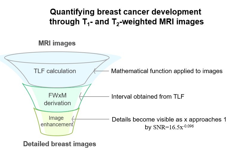
Breast cancer has recently received considerable attention in the field of diagnostic imaging. It can present in various forms, including invasive, in situ, or mixed subtypes. As breast tumors grow faster than other tumor types, non-invasive imaging methods, such as magnetic resonance imaging (MRI), are widely used for their quantitative assessment. This study proposes a novel function that utilizes specific mathematical relationships between relaxation times in MRI to generate maps by defining alpha star. We introduced transverse–longitudinal function (TLF), incorporating T1, T2, and alpha parameters. The function equals zero for a given assumed alpha value. Then, when plotting the TLF, a maximum amount was introduced as a percentage of the maximum width at the x-value. By calculating the inverse of the TLF, the full width at × maximum (FW×M)—the difference between the maximum and minimum alpha stars—was obtained for each image pixel. If this parameter were estimated for the entire image, only one FW×M would be obtained. The derived maps demonstrated breast tumor growth and predictive potential, with a reasonable signal-to-noise ratio of 16.5×−0.096. While the x-value approached 1, more details in the entire breast image became visible. The resulting images with the index value of −0.096 revealed breast structures and other information at different stages, potentially facilitating the quantitative assessment of tumor characteristics and progression.
- Orel SG, Schnall MD. MR imaging of the breast for the detection, diagnosis, and staging of breast cancer. Radiology. 2001;220(1):13-30. doi: 10.1148/radiology.220.1.r01jl3113
- Esserman L, Wolverton D, Hylton N. Magnetic resonance imaging for primary breast cancer management: Current role and new applications. Endocr Relat Cancer. 2002;9:141-153. doi: 10.1677/erc.0.0090141
- Van Goethem M, Schelfout K, Dijckmans L, et al. MR mammography in the pre-operative staging of breast cancer in patients with dense breast tissue: Comparison with mammography and ultrasound. Eur Radiol. 2004;14(5): 809-816. doi: 10.1007/s00330-003-2146-7
- Roganovic D, Djilas D, Vujnovic S, Pavic D, Stojanov D. Breast MRI, digital mammography and breast tomosynthesis: Comparison of three methods for early detection of breast cancer. Bosn J Basic Med Sci. 2015;15(4):64-68. doi: 10.17305/bjbms.2015.616
- Shahbazi-Gahrouei D, Aminolroayaei F, Nematollahi H, Ghaderian M, Gahrouei SS. Advanced magnetic resonance imaging modalities for breast cancer diagnosis: An overview of recent findings and perspectives. Diagnostics. 2022;12(11):2741. doi: 10.3390/diagnostics12112741
- Ashoor M, Khorshidi A. Improving signal-to-noise ratio by maximal convolution of longitudinal and transverse magnetization components in MRI: Application to the breast cancer detection. Med Biol Eng Comput. 2024;62(3):941-954. doi: 10.1007/s11517-023-02994-w
- Daimiel Naranjo I, Lo Gullo R, Saccarelli C, et al. Diagnostic value of diffusion-weighted imaging with synthetic b-values in breast tumors: Comparison with dynamic contrast-enhanced and multiparametric MRI. Eur Radiol. 2021;31:356-367. doi: 10.1007/s00330-020-07094-z
- Alshorman J, Karaminasian M, Sadeghi S, Safari E, Abbaspourtabari H, Altahla R. Prevalence of somatic symptom disorder in Syrian patients with breast cancer: A cross-sectional study. J Clin Basic Psychosom. 2025:025150023. doi: 10.36922/JCBP025150023
- Malik S, Malik A, Islam J, et al. Breaking the metabolic code in triple-negative breast cancer: Mechanistic insights into glycolytic enzyme inhibitors for suppressing metastasis and tumor growth. Cancer Plus. 2025;7(2):25-45. doi: 10.36922/cp.8363
- Khorshidi A, Shams-Abadi M. Simulation of 52m + gMn production yield via chromium target under low-energy proton irradiation from mashhad small cyclotron. Phys Part Nuclei Lett. 2025;22:586-595. doi: 10.1134/S1547477125700219
- Ashoor M, Khorshidi A. Estimation of the number of compartments associated with the apparent diffusion coefficient in MRI: The theoretical and experimental investigations. Am J Roentgenol. 2016;206(3):455-462. doi: 10.2214/AJR.15.14497
- Zanotelli MR, Chada NC, Johnson CA, Reinhart-King CA. The physical microenvironment of tumors: Characterization and clinical impact. Biophys Rev Lett. 2020;15(2):51-82. doi: 10.1142/S1793048020300029
- Huang YL, Shiau C, Wu C, Segall JE, Wu M. The architecture of co-culture spheroids regulates tumor invasion within a 3D extracellular matrix. Biophys Rev Lett. 2020;15(3):131-141. doi: 10.1142/S1793048020500034
- Li X, Thirumalai D. Cooperation among tumor cell subpopulations leads to intratumor heterogeneity. Biophys Rev Lett. 2020;15(2):99-119. doi: 10.1142/S1793048020300042
- Dingli D, Chalub FA, Santos FC, Van Segbroeck S, Pacheco JM. Cancer phenotype as the outcome of an evolutionary game between normal and malignant cells. Br J Cancer. 2009;101(7):1130-1136. doi: 10.1038/sj.bjc.6605288
- Li X, Thirumalai D. Share, but unequally: A plausible mechanism for emergence and maintenance of intratumour heterogeneity. J R Soc Interface. 2019;16(150):20180820. doi: 10.1098/rsif.2018.0820
- Archetti M, Pienta KJ. Cooperation among cancer cells: Applying game theory to cancer. Nat Rev Cancer. 2019;19:110-117. doi: 10.1038/s41568-018-0083-7
- Rockne R, Alvord JREC, Reed PJ, Swanson KR. Modeling the growth and invasion of gliomas, from simple to complex: The goldie locks paradigm. Biophys Rev Lett. 2008;3(1- 2):111-123. doi: 10.1142/S1793048008000642
- Thomas SC, Madaan T, Kamble NS, Siddiqui NA, Pauletti GM, Kotagiri N. Engineered bacteria enhance immunotherapy and targeted therapy through stromal remodeling of tumors. Adv Healthc Mater. 2022;11(2):e2101487. doi: 10.1002/adhm.202101487
- Khalifa MK, Nageeb AM, Mohamed MM, Ezz El Arab LR, Swellam M. Haplotype analysis and linkage disequilibrium of BRCA genes in glioblastoma: Impact on treatment response. Tumor Discov. 2024;3(1):1480. doi: 10.36922/td.1480
- Wang S, Yang D, Mo J, Chen M, Zhang R. The role of pyroptosis-related genes in breast cancer progression. Tumor Discov. 2024;3(3):3469. doi: 10.36922/td.3469
- Zhang Y, Chen B, Wu J, Chen C. Efficacy of pyrotinib and capecitabine in recurrent breast cancer with a HER2-negative genetic switch following systemic therapy: A case report and literature review. Tumor Discov. 2025;4(1):113-119. doi: 10.36922/td.4093
- Okwor VC, Okwor CJ, Musayayi SA, et al. Immune modulation and epigenetic therapies for enhanced outcome of treatment in triple-negative breast cancer. Tumor Discov. 2024;3(3):3383. doi: 10.36922/td.3383
- Zhang Y, Chen B, Lin S, Zhang R, Wu J, Chen C. Emerging immunomodulatory effects of CDK4/6 inhibitors in breast cancer therapy: A comprehensive review. Tumor Discov. 2025;4(3):025190037. doi: 10.36922/TD025190037
- Aye SM, Zu WWM. Characterizing breast cancer in Myanmar: Insights from receptor status and tumor staging. Tumor Discov. 2025:025250051. doi: 10.36922/TD025250051
- Gao H, Lei J, Gui Z, Wang S. Comparative efficacy and safety of Zercepac® plus pyrotinib versus Zercepac® plus pertuzumab in combination with chemotherapy as neoadjuvant therapy for HER2-positive breast cancer: A retrospective study. Eurasian J Med Oncol. 2025;9:025100044. doi: 10.36922/EJMO025100044
- Razem B, Ilhami O, Hamid SE, Oukerroum A, Slimani F. Malignant proliferating trichilemmal tumor post-chemotherapy: A case report. Tumor Discov. 2024;3(2):2344. doi: 10.36922/td.2344
- Aref MH, Aboughaleb IH, Hussein AA, Farag AM, El-Ghaffar SA, El-Sharkawy YH. Malignant versus normal breast tissue: Optical differentiation exploiting hyperspectral imaging system. Tumor Discov. 2023;2(1):258. doi: 10.36922/td.258
- Sosu EK. Optimization of Radiological Protection of Patients Undergoing Mammography Examination in Ghana. PhD Dissertion. University of Cape Coast; 2018. Available from: http://ir.ucc.edu.gh/jspui/handle/123456789/3432 [Last accessed on 2022 May 12].
- Poma GE. Development and Tomographic Reconstruction of an Innovative MBI Device for the Early Breast Cancer Diagnosis. PhD Dissertion, Università Degli Studi di Catania; 2020. Available from: https://hdl.handle.net/20.500.14242/75383 [Last accessed on 2020 Jul 07].
- Khorshidi A, Ashoor M, Abdollahi A. Optimization of breast treatment planning towards lower dose rate: A Monte Carlo simulation study. Inform Med Unlocked. 2023;38:101220. doi: 10.1016/j.imu.2023.101220
- Brix G, Kolem H, Nitz WR, et al. Basics of magnetic resonance imaging and magnetic resonance spectroscopy. In: Reiser M, Semmler W, Hricak H, editors. Magnetic Resonance Tomography. Berlin, Heidelberg: Springer; 2008. doi: 10.1007/978-3-540-29355-2_2
- Abele M. Solid-State NMR Spectroscopy for Structural Investigations in Materials Science. PhD Dissertion. Institute of Physical Chemistry, University of Stuttgart, Stuttgart; 2012. Available from: https://www.ipc.uni-stuttgart.de/ roduner/phd-theses [Last accessed on 2008 Jan 01].
- Landini L, Positano V, Santarelli MF, editors. Advanced Image Processing in Magnetic Resonance Imaging. London: CRC Press; 2018.
- Bloch F, Hansen WW, Packard M. The nuclear induction experiment. Phys Rev. 1946;70:474. doi: 10.1103/PhysRev.70.474
- Liang ZP, Lauterbur PC. Principles of Magnetic Resonance Imaging: A Signal Processing Perspective. Washington, DC: The Institute of Electrical and Electronics Engineers Press; 2000.
- Han SH, An YY, Kang BJ, Kim SH, Lee EJ. Takeaways from pre-contrast T1 and T2 breast magnetic resonance imaging in women with recently diagnosed breast cancer. Iran J Radiol. 2016;13(4):e36271. doi: 10.5812/iranjradiol.36271
- Byrne H. Dissecting cancer through mathematics: from the cell to the animal model. Nat Rev Cancer. 2010;10:221-230. doi: 10.1038/nrc2808
- AlBuainain RY, Bunajem FY, Abdulla HA. Assessment of tumor response to neoadjuvant chemotherapy in breast cancer using MRI and 18F-FDG PET/CT. Eur J Breast Health. 2025;21(1):46-51. doi: 10.4274/ejbh.galenos.2024.2024-8-2
- Qadir A, Singh N, Moe AAK, et al. Potential of MRI in assessing treatment response after neoadjuvant radiation therapy treatment in breast cancer patients: A scoping review. Clin Breast Cancer. 2025;25(1):e1-e9.e2. doi: 10.1016/j.clbc.2024.05.010
- Lorenzon M, Zuiani C, Londero V, Linda A, Furlan A, Bazzocchi M. Assessment of breast cancer response to neoadjuvant chemotherapy: Is volumetric MRI a reliable tool? Eur J Radiol. 2009;71(1):82-88. doi: 10.1016/j.ejrad.2008.03.021
- Mei L, Wang K, Gu Y. Improved fuzzy C-means clustering algorithm-based dynamic contrast-enhanced magnetic resonance imaging features in the diagnosis of invasive breast carcinoma before and after menopause. Comput Math Methods Med. 2022;2022(1):2917844. doi: 10.1155/2022/2917844
- Kang J, Jiang N, Shataer M, Tuersong T. Quantitative bibliometric insights into cisplatin resistance in breast cancer (2010–2024): Implications for drug development. Cancer Plus. 2025;7:025220037. doi: 10.36922/CP025220037
- Greenberg S, Rugo HS. Triple-negative breast cancer: Role of antiangiogenic agents. Cancer J. 2010;16(1):33-38. doi: 10.1097/PPO.0b013e3181d38514
- Steinhauer V, Sergeev NI. Radiomics: Phases of osteoclastic metastasis status in breast cancer identified by morphologic markers. Cancer Plus. 2025;7(1):109-115. doi: 10.36922/cp.6649
- Stewart DA, Winnike JH, McRitchie SL, Clark RF, Pathmasiri WW, Sumner SJ. Metabolomics analysis of hormone-responsive and triple-negative breast cancer cell responses to paclitaxel identify key metabolic differences. J Proteome Res. 2016;15(9):3225-3240. doi: 10.1021/acs.jproteome.6b00430
- Crociani O, Becchetti A, Fanelli D, Arcangeli A. Adhesion-mediated signalling in cancer: Recent advances and mathematical modelling. Biophys Rev Lett. 2014;9(3):285-300. doi: 10.1142/S1793048014300047
- Engelke H. Physics of the extracellular matrix and biology of tumors - a close relationship. Biophys Rev Lett. 2020;15(3):121-130. doi: 10.1142/S1793048020300030
- Margarit DH, Romanelli L, Fendrik AJ. A 3D cellular automaton for cell differentiation in a solid tumor with plasticity. Biophys Rev Lett. 2018;13(1):19-28. doi: 10.1142/S1793048018500029
- Cabuk G, Duce MN, Özgür A, Apaydin FD, Polat A, Orekici G. The diagnostic value of diffusion-weighted imaging and the apparent diffusion coefficient values in the differentiation of benign and malignant breast lesions. J Med Imaging Radiat Oncol. 2015;59(2):141-148. doi: 10.1111/1754-9485.12273
- Oohashi M, Mizuhashi F, Sugawara Y, Saegusa H, Ogura I. Diffusion-weighted magnetic resonance imaging in the palatal tumors: Usefulness of apparent diffusion coefficient value for characterization of benign and malignant tumors. Oral Sci Int. 2020;17(3):142-146. doi: 10.1002/osi2.1058
- Chan SW, Lin CA, Ouyang YC, et al. Characterizing breast tumor heterogeneity through IVIM-DWI parameters and signal decay analysis. Diagnostics (Basel). 2025;15(12):1499. doi: 10.3390/diagnostics15121499
- Kurt N, Kurt BB, Gulsaran U, et al. Diffusion tensor imaging and diffusion-weighted imaging on axillary lymph node status in breast cancer patients. Diagn Interv Radiol. 2022;28(4):329-336. doi: 10.5152/dir.2022.21460
- Mao C, Jiang W, Huang J, et al. Quantitative parameters of diffusion spectrum imaging: HER2 status prediction in patients with breast cancer. Front Oncol. 2022;12:817070. doi: 10.3389/fonc.2022.817070
- Ashoor M, Khorshidi A, Pirouzi A, Abdollahi A, Mohsenzadeh M, Barzi SMZ. Estimation of Reynolds number on microvasculature capillary bed using diffusion and perfusion MRI: The theoretical and experimental investigations. Eur Phys J Plus. 2021;136:152. doi: 10.1140/epjp/s13360-021-01145-0
- Ashoor M, Khorshidi A, Sarkhosh L. Estimation of microvascular capillary physical parameters using MRI assuming a pseudo liquid drop as model of fluid exchange on the cellular level. Rep Pract Oncol Radiother. 2019;24(1):3-11. doi: 10.1016/j.rpor.2018.09.007
- López MT, Feltri AP, Isabe GF, Guida V, Fernandes A, Blanch R. Risk and protective factors associated with breast cancer. Cancer Plus. 2020;2(3):1-6. doi: 10.18063/cp.v2i3.351
- Elbiad O, Mazour O, Ennibi K, Badaoui B, Laraqui A. Variants of unknown significance in BRCA1 and BRCA2 among breast cancer patients in Middle Eastern and North African populations: A systematic review. Eurasian J Med Oncol. 2025;9(1):16-45. doi: 10.36922/ejmo.5800
- Monzon AZ, Franco-López Á, Culebras JM. Benefits and harms of screening: Overdiagnosis and anticipatory medicine – A secondary publication. Tumor Discov. 2022;1(2):228. doi: 10.36922/td.v1i2.228
- Humayun S, Mahmood T. Artificial intelligence for early diagnosis of breast cancer in women: A systematic literature review. Artif Intell Health. 2025;2(2):100-116. doi: 10.36922/aih.4197
- Colman RD, Kotera Y. Addressing the psychological impact of infertility risk arising from breast cancer treatment: Education and self-compassion interventions. Int J Popul Stud 2025;11(5):31-37. doi: 10.36922/ijps.1724
- Lu LJW, Nishino TK, Johnson RF, et al. Comparison of breast tissue measurements using magnetic resonance imaging, digital mammography and a mathematical algorithm. Phys Med Biol. 2012;57:6903. doi: 10.1088/0031-9155/57/21/6903
- Jarrett AM, Kazerouni AS, Wu C, et al. Quantitative magnetic resonance imaging and tumor forecasting of breast cancer patients in the community setting. Nat Protoc. 2021;16:5309-5338. doi: 10.1038/s41596-021-00617-y
- Patel RJS, Wu C, Stowers CE, et al. MRI-based mathematical modeling to predict the response of I-SPY 2 breast cancer patients to neoadjuvant therapy. Clin Cancer Res. 2025. doi: 10.1158/1078-0432.CCR-25-0668
- Ortiz-Abellán C, Aguado-Sarrió E, Prats-Montalbán JM, Camps-Herrero J, Ferrer A. New breast cancer biomarkers from diffusion magnetic resonance imaging based on the Diffusion Tensor using multivariate curve resolution (MCR) models. Chemometr Intell Lab Syst. 2024;251:105171. doi: 10.1016/j.chemolab.2024.105171
- Davenport AA, Lu Y, Gallegos CA, et al. Mathematical model of triple-negative breast cancer in response to combination chemotherapies. Bull Math Biol. 2023;85:7. doi: 10.1007/s11538-022-01108-1
- Campana A, Gandomkar Z, Giannotti N, Reed W. The use of radiomics in magnetic resonance imaging for the pre-treatment characterisation of breast cancers: A scoping review. J Med Radiat Sci. 2023;70(4):462-478. doi: 10.1002/jmrs.709
- Nafiss N, Heiranizadeh N, Shirinzadeh-Dastgiri A, et al. The application of artificial intelligence in breast cancer. Eurasian J Med Oncol. 2024;8(3):235-244. doi: 10.14744/ejmo.2024.45903
- Haider S. Catalyzing breast cancer diagnosis: Ai advancements in mammography. Eurasian J Med Oncol. 2023;7(4):402-403. doi: 10.14744/ejmo.2023.93033
- Botlagunta M, Botlagunta M, Venkata MD, Kanakapudi C, Khan Z. Correlation-based comparative machine learning analysis for the classification of metastatic breast cancer using blood profile. Eurasian J Med Oncol. 2024;8(2):152-164. doi: 10.14744/ejmo.2024.52521
- Bandyopadhyay A, Mondal HS, Dam B, Patranabis DC, Pal B. Innovative infrared imaging approach for breast cancer screening: Integrating rotational thermography and machine learning analysis. Artif Intell Health. 2024;1(3):64-79. doi: 10.36922/aih.3312
- Leong HJY, Tan HD, Yap WH, Chia AYY, Zacchigna S, Tang YQ. Identification of potentially therapeutic target genes in metastatic breast cancer via integrative network analysis. Eurasian J Med Oncol. 2023;7(4):371-387. doi: 10.14744/ejmo.2023.80452
- Ashoor M, Khorshidi A. Extraction of hybrid data using intrinsic interactions of relaxation times in MRI. Int J Biomath. 2025. doi: 10.1142/S1793524525500159
- Liu X, Hong Z, Ding H, et al. Assessment of cerebral venous sinus: Anatomical and functional diagnostic performance of three-dimensional reconstruction models based on venous sinus MRI and CT images. Brain Heart. 2024;2(2):2756. doi: 10.36922/bh.2756
- Ashoor M, Khorshidi A. Introducing a parametric function on relaxation times in magnetic resonance imaging. Multimed Tools Appl. 2025;84:18247-18262. doi: 10.1007/s11042-024-19789-2
- Khorshidi A. Examining neural correlates of sexual preferences between Persian homo- and heterosexual males using psychological assessments and functional magnetic resonance imaging in specifying cognitive map: A limited and cross-sectional study. J Pediatr Neurol. 2024;21:1-16. doi: 10.1055/s-0044-1788630
- Khorshidi A, Ashoor M. New deformity outline on the breast radiation therapy for diminishing absorbed dose ratio. Braz J Radiat Sci. 2023;11(3):1-12. doi: 10.15392/2319-0612.2023.2281
- Jensen JR, Do D, Chang YP, et al. CoreView imaging on needle: Rapid core-needle biopsy imaging for point-of-care breast cancer diagnosis. Global Transl Med. 2025;4:025170039. doi: 10.36922/GTM025170039
- Khorshidi A. Segmentation of tumor region in respiratory disease by extended algorithm. Int J Mod Phys C. 2023;34(12):2350164. doi: 10.1142/S0129183123501644
- Puvvula PK, Puvvula RS. Clinical advancements in breast cancer research: A comprehensive review. Tumor Discov. 2025:025090016. doi: 10.36922/TD025090016