

Tracking neurodegeneration in multiple sclerosis: the role of optical coherence tomography, optical coherence tomography angiography, and artificial intelligence-based imaging

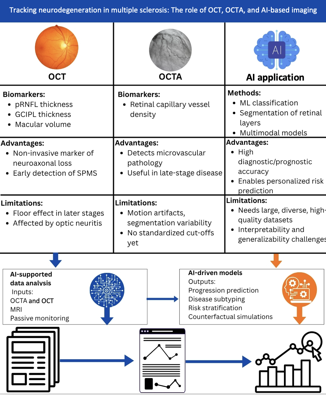
Multiple sclerosis (MS) represents a chronic autoimmune disease involving the central nervous system through inflammation and progressive neurodegeneration. Monitoring disease progression, specifically in terms of neuroaxonal loss, has critical importance in guiding treatment and improving patient outcomes. This review discusses the evolving role of optical coherence tomography (OCT) and artificial intelligence (AI) in monitoring MS progression, highlighting their efficiency in assessing neuroaxonal damage, predicting disability, and complementing conventional imaging techniques. We synthesize current evidence on OCT and OCT angiography (OCTA) metrics, including peripapillary retinal nerve fiber layer (pRNFL) and ganglion cell/inner plexiform layer (GCIPL) thickness, and their correlations with disability, cognitive impairment, magnetic resonance imaging (MRI) findings, and clinical progression. We further review machine learning and deep learning applications for OCT-based classification, segmentation, and prognosis in MS. Thinning of pRNFL and GCIPL correlates with higher expanded disability status scale, brain atrophy, and impaired cognition, often preceding clinical signs of progression. OCTA reveals reduced retinal capillary density in MS, supporting a vascular component in disease pathology. AI tools enhance OCT analysis through automated layer segmentation and predictive modeling, enabling individualized risk stratification and early detection of secondary progression. Multimodal AI frameworks that combine OCT with MRI and clinical data further enhance prognostic accuracy. In summary, OCT and AI are transforming MS disease progression tracking by providing affordable, non-invasive biomarkers reflecting neurodegeneration and cerebrovascular dysfunction. For achieving their full potential as predictive and therapeutic tools in individualized care for MS patients, standardization, external validation, and integration into clinical practice are essential.
- Lassmann H. Multiple sclerosis pathology. Cold Spring Harb Perspect Med. 2018;8(3):a028936. doi: 10.1101/cshperspect.a028936
- Walton C, King R, Rechtman L, et al. Rising prevalence of multiple sclerosis worldwide: Insights from the Atlas of MS. Mult Scl. 2020;26(14):1816-1821. doi: 10.1177/1352458520970841
- Ramagopalan SV, Sadovnick AD. Epidemiology of multiple sclerosis. Neurol Clin. 2011;29(2):207-217. doi: 10.1016/j.ncl.2010.12.010
- Stadelmann C, Wegner C, Brück W. Inflammation, demyelination, and degeneration -recent insights from MS pathology. Biochim Biophys Acta (BBA) Mol Basis Dis. 2011;1812(2):275-282. doi: 10.1016/j.bbadis.2010.07.007
- Khan G, Hashim MJ. Epidemiology of multiple sclerosis: Global, regional, National and sub-national-level estimates and future projections. J Epidemiol Glob Health. 2025;15(1):21. doi: 10.1007/s44197-025-00353-6
- Calabrese M, Rinaldi F, Grossi P, et al. Basal ganglia and frontal/parietal cortical atrophy is associated with fatigue in relapsing-remitting multiple sclerosis. Mult Scler. 2010;16(10):1220-1228. doi: 10.1177/1352458510376405
- Sousa C, Jacques T, Sá MJ, Alves RA. Cognitive impairment in multiple sclerosis phenotypes: Neuropsychological assessment in a Portuguese sample. Appl Neuropsychol Adult. 2024;31(6):1153-1162. doi: 10.1080/23279095.2022.2112681
- Belbasis L, Bellou V, Evangelou E, Ioannidis JP, Tzoulaki I. Environmental risk factors and multiple sclerosis: An umbrella review of systematic reviews and meta-analyses. Lancet Neurol. 2015;14(3):263-273. doi: 10.1016/S1474-4422(14)70267-4
- Meca-Lallana JE, Casanova B, Rodríguez-Antigüedad A, et al. Consensus on early detection of disease progression in patients with multiple sclerosis. Front Neurol. 2022;13:931014. doi: 10.3389/fneur.2022.931014
- Waubant E. Improving outcomes in multiple sclerosis through early diagnosis and effective management. Prim Care Companion CNS Disord. 2012;14(5):PCC.11016co2cc27363. doi: 10.4088/PCC.11016co2cc
- Meca-Lallana V, Berenguer-Ruiz L, Carreres-Polo J, et al. Deciphering multiple sclerosis progression. Front Neurol. 2021;12:608491. doi: 10.3389/fneur.2021.608491
- Criste G, Trapp B, Dutta R. Axonal loss in multiple sclerosis: Causes and mechanisms. Handb Clinical Neurol. 2014;122:101-113. doi: 10.1016/B978-0-444-52001-2.00005-4
- Rocca MA, Preziosa P, Barkhof F, et al. Current and future role of MRI in the diagnosis and prognosis of multiple sclerosis. Lancet Reg Health Eur. 2024;44:100978. doi: 10.1016/j.lanepe.2024.100978.
- Rjeily NB, Solomon AJ. Misdiagnosis of multiple sclerosis: Past, present, and future. Curr Neurol Neurosci Rep. 2024;24(11):547-557. doi: 10.1007/s11910-024-01371-w
- Amezcua L. Progressive multiple sclerosis. Continuum (Minneap Minn). 2022;28(4):1083-1103. doi: 10.1212/CON.0000000000001157
- Kurtzke JF. Rating neurologic impairment in multiple sclerosis: An expanded disability status scale (EDSS). Neurology. 1983;33(11):1444-1452. doi: 10.1212/wnl.33.11.1444
- Cohen M, Bresch S, Thommel Rocchi O, et al. Should we still only rely on EDSS to evaluate disability in multiple sclerosis patients? A study of inter and intra rater reliability. Mult Scler Relat Disord. 2021;54:103144. doi: 10.1016/j.msard.2021.103144
- Benedict RH, DeLuca J, Phillips G, et al. Validity of the symbol digit modalities test as a cognition performance outcome measure for multiple sclerosis. Mult Scler. 2017;23(5):721-733. doi: 10.1177/1352458517690821
- Filippi M, Rocca MA, Ciccarelli O, et al. MRI criteria for the diagnosis of multiple sclerosis: MAGNIMS consensus guidelines. Lancet Neurol. 2016;15(3):292-303. doi: 10.1016/S1474-4422(15)00393-2
- Bakshi R, Thompson AJ, Rocca MA, et al. MRI in multiple sclerosis: Current status and future prospects. Lancet Neurol. 2008;7(7):615-625. doi: 10.1016/S1474-4422(08)70137-6
- Neema M, Stankiewicz J, Arora A, Guss ZD, Bakshi R. MRI in multiple sclerosis: What’s inside the toolbox? Neurotherapeutics. 2007;4(4):602-617. doi: 10.1016/j.nurt.2007.08.001
- Tomassini V, Sinclair A, Sawlani V, et al. Diagnosis and management of multiple sclerosis: MRI in clinical practice. J Neurol. 2020;267(10):2917-2925. doi: 10.1007/s00415-020-09930-0
- Schmierer K, Campion T, Sinclair A, et al. Towards a standard MRI protocol for multiple sclerosis across the UK. Br J Radiol. 2019;92(1101):20180926. doi: 10.1259/bjr.20180926
- Petzold A, De Boer JF, Schippling S, et al. Optical coherence tomography in multiple sclerosis: A systematic review and meta-analysis. Lancet Neurol. 2010;9(9):921-932. doi: 10.1016/S1474-4422(10)70168-X
- Petzold A, Balcer LJ, Calabresi PA, et al. Retinal layer segmentation in multiple sclerosis: A systematic review and meta-analysis. Lancet Neurol. 2017;16(10):797-812. doi: 10.1016/S1474-4422(17)30278-8
- Martinez-Lapiscina EH, Arnow S, Wilson JA, et al. Retinal thickness measured with optical coherence tomography and risk of disability worsening in multiple sclerosis: A cohort study. Lancet Neurol. 2016;15(6):574-584. doi: 10.1016/S1474-4422(16)00068-5
- Oberwahrenbrock T, Schippling S, Ringelstein M, et al. Retinal damage in multiple sclerosis disease subtypes measured by high-resolution optical coherence tomography. Mult Scler Int. 2012;2012(1):530305. doi: 10.1155/2012/530305
- Mirmosayyeb O, Yazdan Panah M, Moases Ghaffary E, et al. The relationship between optical coherence tomography and magnetic resonance imaging measurements in people with multiple sclerosis: A systematic review and meta-analysis. J Neurol Sci. 2025;470:123401. doi: 10.1016/j.jns.2025.123401
- Mirmosayyeb O, Yazdan Panah M, Mokary Y, et al. Optical coherence tomography (OCT) measurements and disability in multiple sclerosis (MS): A systematic review and meta-analysis. J Neurol Sci. 2023;454:120847. doi: 10.1016/j.jns.2023.120847
- Mirmosayyeb O, Zivadinov R, Weinstock-Guttman B, Benedict RHB, Jakimovski D. Optical coherence tomography (OCT) measurements and cognitive performance in multiple sclerosis: A systematic review and meta-analysis. J Neurol. 2023;270(3):1266-1285. doi: 10.1007/s00415-022-11449-5
- Ortiz M, Mallen V, Boquete L, et al. Diagnosis of multiple sclerosis using optical coherence tomography supported by artificial intelligence. Mult Scler Relat Disord. 2023;74:104725. doi: 10.1016/j.msard.2023.104725
- Balyen L, Peto T. Promising artificial intelligence-machine learning-deep learning algorithms in ophthalmology. Asia- Pac J Ophthalmol (Phila). 2019;8(3):264-272. doi: 10.22608/APO.2018479
- Perdomo O, Rios H, Rodríguez FJ, et al. Classification of diabetes-related retinal diseases using a deep learning approach in optical coherence tomography. Comput Methods Prog Biomed. 2019;178:181-189. doi: 10.1016/j.cmpb.2019.06.016
- He Y, Carass A, Liu Y, et al. Structured layer surface segmentation for retina OCT using fully convolutional regression networks. Med Image Anal. 2021;68:101856. doi: 10.1016/j.media.2020.101856
- Andorra M, Freire A, Zubizarreta I, et al. Predicting disease severity in multiple sclerosis using multimodal data and machine learning. J Neurol. 2024;271(3):1133-1149. doi: 10.1007/s00415-023-12132-z
- Fujimoto JG, Pitris C, Boppart SA, Brezinski ME. Optical coherence tomography an emerging technology for biomedical imaging and optical biopsy. Neoplasia. 2000;2:9-25. doi: 10.1038/sj.neo.7900071
- Wojtkowski M. High-speed optical coherence tomography: Basics and applications. Appl Opt. 2010;49(16):D30-D61. doi: 10.1364/AO.49.000D30
- Noval S, Contreras I, Muñoz S, Oreja-Guevara C, Manzano B, Rebolleda G. Optical coherence tomography in multiple sclerosis and neuromyelitis optica: An update. Mult Scler Int. 2011;2011(1):472790. doi: 10.1155/2011/472790
- Bagci AM, Shahidi M, Ansari R, Blair M, Blair NP, Zelkha R. Thickness profiles of retinal layers by optical coherence tomography image segmentation. Am J Ophthalmol. 2008;146(5):679-687.e1. doi: 10.1016/j.ajo.2008.06.010
- Suh A, Hampel G, Vinjamuri A, et al. Oculomics analysis in multiple sclerosis: Current ophthalmic clinical and imaging biomarkers. Eye. 2024;38(14):2701-2710. doi: 10.1038/s41433-024-03132-y
- Vidal-Jordana A, Pareto D, Cabello S, et al. Optical coherence tomography measures correlate with brain and spinal cord atrophy and multiple sclerosis disease-related disability. Eur J Neurol. 2020;27(11):2225-2232. doi: 10.1111/ene.14421
- El Ayoubi NK, Ismail A, Fahd F, Younes L, Chakra NA, Khoury SJ. Retinal optical coherence tomography measures in multiple sclerosis: A systematic review and meta-analysis. Ann Clin Transl Neurol. 2024;411(9):2236-2253. doi: 10.1002/acn3.52165
- Lublin FD, Reingold SC, Cohen JA, et al. Defining the clinical course of multiple sclerosis. Neurology. 2014;83(3):278-286. doi: 10.1212/WNL.0000000000000560
- Ford H. Clinical presentation and diagnosis of multiple sclerosis. Clin Med. 2020;20(4):380-383. doi: 10.7861/clinmed.2020-0292
- Lorscheider J, Buzzard K, Jokubaitis V, et al. Defining secondary progressive multiple sclerosis. Brain. 2016;139(9):2395-2405. doi: 10.1093/brain/aww173
- Thompson AJ, Banwell BL, Barkhof F, et al. Diagnosis of multiple sclerosis: 2017 revisions of the McDonald criteria. Lancet Neurol. 2018;17(2):162-173. doi: 10.1016/S1474-4422(17)30470-2
- Sormani MP, De Stefano N. Defining and scoring response to IFN-β in multiple sclerosis. Nat Rev Neurol. 2013;9(9):504-512. doi: 10.1038/nrneurol.2013.146
- Cutter GR, Baier ML, Rudick RA, et al. Development of a multiple sclerosis functional composite as a clinical trial outcome measure. Brain. 1999;122(5):871-882. doi: 10.1093/brain/122.5.871
- Kappos L, Wolinsky JS, Giovannoni G, et al. Contribution of relapse-independent progression vs relapse-associated worsening to overall confirmed disability accumulation in typical relapsing multiple sclerosis in a pooled analysis of 2 randomized clinical trials. JAMA Neurol. 2020;77(9):1132-1140. doi: 10.1001/jamaneurol.2020.1568
- Zivadinov R, Cox JL. Neuroimaging in multiple sclerosis. Int Rev Neurobio. 2007;79:449-474. doi: 10.1016/S0074-7742(07)79020-7
- Montalban X, Hauser SL, Kappos L, et al. Ocrelizumab versus placebo in primary progressive multiple sclerosis. N Engl J Med. 2017;376(3):209-220. doi: 10.1056/NEJMoa1606468
- Kappos L, Bar-Or A, Cree BAC, et al. Siponimod versus placebo in secondary progressive multiple sclerosis (EXPAND): A double-blind, randomized, phase 3 study. Lancet. 2018;391(10127):1263-1273. doi: 10.1016/S0140-6736(18)30475-6
- Hauser SL, Bar-Or A, Comi G, et al. Ocrelizumab versus interferon beta-1a in relapsing multiple sclerosis. N Engl J Med. 2017;376(3):221-234. doi: 10.1056/NEJMoa1601277
- Confavreux C, Vukusic S, Adeleine P. Early clinical predictors and progression of irreversible disability in multiple sclerosis: An amnesic process. Brain. 2003;126(4):770-782. doi: 10.1093/brain/awg081
- Fisniku LK, Brex PA, Altmann DR, et al. Disability and T2 MRI lesions: A 20-year follow-up of patients with relapse onset of multiple sclerosis. Brain. 2008;131(3):808-817. doi: 10.1093/brain/awm329
- Absinta M, Sati P, Masuzzo F, et al. Association of chronic active multiple sclerosis lesions with disability in vivo. JAMA Neurol. 2019;76(12):1474-1483. doi: 10.1001/jamaneurol.2019.2399
- Alonso R, Gonzalez-Moron D, Garcea O. Optical coherence tomography as a biomarker of neurodegeneration in multiple sclerosis: A review. Mult Scler Relat Disord. 2018;22:77-82. doi: 10.1016/j.msard.2018.03.007
- Swinnen S, De Wit D, Van Cleemput L, Cassiman C, Dubois B. Optical coherence tomography as a prognostic tool for disability progression in MS: A systematic review. J Neurol. 2023;270(2):1178-1186. doi: 10.1007/s00415-022-11474-4
- Toscano S, Chisari CG, Biondi A, Patti F. Early reduction of retinal thickness predicts physical and cognitive disability in newly diagnosed multiple sclerosis patients: results from a cross-sectional study. Neurol Sci. 2024;45(11):5385-5394. doi: 10.1007/s10072-024-07664-9
- Dreyer-Alster S, Gal A, Achiron A. Optical coherence tomography is associated with cognitive impairment in multiple sclerosis. J Neuroophthalmol. 2022;42(1):e14-e21. doi: 10.1097/WNO.0000000000001326
- Tsoukaki N, Anagnostopoulou A, Kartsidis P, et al. The Mediating Role of Trait Mental Fatigue in Cognitive Decline among PwMS: Implications for Verbal Memory and Information Processing Speed. [Preprint]; 2024. doi: 10.21203/rs.3.rs-5515189/v1
- Rocca MA, Mesaros S, Preziosa P, et al. Wallerian and trans-synaptic degeneration contribute to optic radiation damage in multiple sclerosis: A diffusion tensor MRI study. Mult Scler J. 2013;19(12):1610-1617. doi: 10.1177/1352458513485146
- Simon JH, Kinkel RP, Jacobs L, Bub L, Simonian N. A wallerian degeneration pattern in patients at risk for MS. Neurology. 2000;54(5):1155-1160. doi: 10.1212/wnl.54.5.1155
- Papadopoulou A, Gaetano L, Pfister A, et al. Damage of the lateral geniculate nucleus in MS: Assessing the missing node of the visual pathway. Neurology. 2019;92(19):e2240-e2249. doi: 10.1212/WNL.0000000000007450
- Shenoy N, Liu F, Narayanan SP. Loss of cells in the retinal ganglion cell layer as an early indication of neurodegeneration in multiple sclerosis. J Integr Neurosci. 2024;23(7):129. doi: 10.31083/j.jin2307129
- Klistorner A, Sriram P, Vootakuru N, et al. Axonal loss of retinal neurons in multiple sclerosis associated with optic radiation lesions. Neurology. 2014;82(24):2165-2172. doi: 10.1212/WNL.0000000000000522
- Yap TE, Balendra SI, Almonte MT, Cordeiro MF. Retinal correlates of neurological disorders. Ther Adv Chronic Dis. 2019;10:1-32. doi: 10.1177/2040622319882205
- Lambe J, Fitzgerald KC, Murphy OC, et al. Association of spectral-domain OCT with long-term disability worsening in multiple sclerosis. Neurology. 2021;96(16):e2058-e2069. doi: 10.1212/WNL.0000000000011788
- El Ayoubi NK, Sabbagh HM, Bou Rjeily N, Hannoun S, Khoury SJ. Rate of retinal layer thinning as a biomarker for conversion to progressive disease in multiple sclerosis. Neurol Neuroimmunol Neuroinflamm. 2022;9(6):e200030. doi: 10.1212/NXI.0000000000200030
- Bsteh G, Hegen H, Krajnc N, et al. Retinal thinning differentiates treatment effects in relapsing multiple sclerosis below the clinical threshold. Ann Clin Transl Neurol. 2025;12(2):345-354. doi: 10.1002/acn3.52279
- Berek K, Hegen H, Hocher J, et al. Retinal layer thinning as a biomarker of long-term disability progression in multiple sclerosis. Mult Scler. 2022;28(12):1871-1880. doi: 10.1177/13524585221097566
- Petzold A, Schippling S, Hanson JVM. OCT and Multiple Sclerosis. OCT and Imaging in Central Nervous System Diseases: The Eye as a Window to the Brain. Vol. 30. Berlin: Springer; 2025. p. 249.
- Balk LJ, Coric D, Knier B, et al. Retinal inner nuclear layer volume reflects inflammatory disease activity in multiple sclerosis; A longitudinal OCT study. Mult Scler J Exp Transl Clin. 2019;5(3):1-11. doi: 10.1177/2055217319871582
- Costello F, Burton JM. Retinal imaging with optical coherence tomography: A biomarker in multiple sclerosis? Eye Brain. 2018;10:47-63. doi: 10.2147/EB.S139417
- Green AJ, McQuaid S, Hauser SL, Allen IV, Lyness R. Ocular pathology in multiple sclerosis: Retinal atrophy and inflammation irrespective of disease duration. Brain. 2010;133(6):1591-1601. doi: 10.1093/brain/awq080
- Sethi V, Yousry T, Muhlet N, et al. A longitudinal study of cortical grey matter lesion subtypes in relapse-onset multiple sclerosis. J Neurol Neurosurg Psychiatr. 2016;87(7):750-753. doi: 10.1136/jnnp-2015-311102
- Pulido-Valdeolivas I, Andorrà M, Gómez-Andrés D, et al. Retinal and brain damage during multiple sclerosis course: Inflammatory activity is a key factor in the first 5 years. Sci Rep. 2020;10(1):13333. doi: 10.1038/s41598-020-70255-z
- Riboni Verri G, Chen BS, McMurran CE, et al. Visual outcome measures of remyelination and neuroprotection in multiple sclerosis. BMJ Neurol Open. 2024;6:e000560. doi: 10.1136/bmjno-2023-000560
- Rocholz R, Corvi F, Weichsel J, Schmidt S, Staurenghi G. OCT Angiography (OCTA) in retinal diagnostics. In: Bille JF, editor. High Resolution Imaging in Microscopy and Ophthalmology: New Frontiers in Biomedical Optics. Cham: Springer International Publishing; 2019. p. 135-160.
- Choi WJ. Imaging motion: A comprehensive review of optical coherence tomography angiography. Adv Exp Med Biol. 2021;1310:343-365. doi: 10.1007/978-981-33-6064-8_12
- Lavia C, Bonnin S, Maule M, Erginay A, Tadayoni R, Gaudric A. Vessel density of superficial, intermediate, and deep capillary plexuses using optical coherence tomography angiography. Retina. 2019;39(2):247-258. doi: 10.1097/IAE.0000000000002413
- Kleerekooper I, Houston S, Dubis AM, Trip SA, Petzold A. Optical coherence tomography angiography (OCTA) in multiple sclerosis and neuromyelitis optica spectrum disorder. Front Neurol. 2020;11:604049. doi: 10.3389/fneur.2020.604049
- Ciccarelli O, Barkhof F, Bodini B, et al. Pathogenesis of multiple sclerosis: Insights from molecular and metabolic imaging. Lancet Neurol. 2014;13(8):807-822. doi: 10.1016/S1474-4422(14)70101-2
- Wang L, Murphy O, Caldito NG, Calabresi PA, Saidha S. Emerging applications of optical coherence tomography angiography (OCTA) in neurological research. Eye Vis (Lond). 2018;5(1):11. doi: 10.1186/s40662-018-0104-3.
- Arya M, Rashad R, Sorour O, Moult EM, Fujimoto JG, Waheed NK. Optical coherence tomography angiography (OCTA) flow speed mapping technology for retinal diseases. Expert Rev Med Dev. 2018;15(12):875-882. doi: 10.1080/17434440.2018.1548932
- Bostan M, Chua J, Sim YC, et al. Microvascular changes in the macular and parafoveal areas of multiple sclerosis patients without optic neuritis. Sci Rep. 2022;12(1):13366. doi: 10.1038/s41598-022-17344-3
- Pujari A, Bhaskaran K, Sharma P, et al. Optical coherence tomography angiography in neuro-ophthalmology: Current clinical role and future perspectives. Surv Ophthalmol. 2021;66(3):471-481. doi: 10.1016/j.survophthal.2020.10.009
- Lerche RC, Schaudig U, Scholz F, Walter A, Richard G. Structural changes of the retina in retinal vein occlusion- -imaging and quantification with optical coherence tomography. Ophthalmic Surg Lasers. 2001;32(4):272-280.
- Pietroboni AM, Dell’Arti L, Caprioli M, et al. The loss of macular ganglion cells begins from the early stages of disease and correlates with brain atrophy in multiple sclerosis patients. Mult Scler J. 2019;25(1):31-38. doi: 10.1177/1352458517740214
- Liu J, Song S, Gu X, Li H, Yu X. Microvascular impairments detected by optical coherence tomography angiography in multiple sclerosis patients: A systematic review and meta-analysis. Front Neurosci. 2023;16:1121899. doi: 10.3389/fnins.2022.1121899
- Wicklein R, Kreitner L, Wild A, et al. Retinal small vessel pathology is associated with disease burden in multiple sclerosis. Mult Scler. 2024;30(7):812-819. doi: 10.1177/13524585241247775
- Mohammadi S, Gouravani M, Salehi MA, et al. Optical coherence tomography angiography measurements in multiple sclerosis: A systematic review and meta-analysis. J Neuroinflammation. 2023;20(1):85. doi: 10.1186/s12974-023-02763-4
- Spencer JI, Bell JS, DeLuca GC. Vascular pathology in multiple sclerosis: Reframing pathogenesis around the blood-brain barrier. J Neurol Neurosurg Psychiatry. 2018;89(1):42-52. doi: 10.1136/jnnp-2017-316011
- Murphy OC, Kwakyi O, Iftikhar M, et al. Alterations in the retinal vasculature occur in multiple sclerosis and exhibit novel correlations with disability and visual function measures. Mult Scler. 2020;26(7):815-828. doi: 10.1177/1352458519845116
- Li J, Chang Y, Zhang Y, et al. Visual function and disability are associated with microcystic macular edema, macular and peripapillary vessel density in patients with neuromyelitis optica spectrum disorder. Front Neurol. 2022;13:1019959. doi: 10.3389/fneur.2022.1019959
- Tsokolas G, Tsaousis KT, Diakonis VF, Matsou A, Tyradellis S. Optical coherence tomography angiography in neurodegenerative diseases: A review. Eye Brain. 2020;12:73-87. doi: 10.2147/EB.S193026
- Ozcelik S, Kaya E, Guney F, et al. The relationship between retinal layer thickness and cognition in people with multiple sclerosis. J Mult Scler Res. 2025;4(3):59-66. doi: 10.4274/jmsr.galenos.2024.2024-12-1
- Zhang YS, Zhou N, Knoll BM, et al. Parafoveal vessel loss and correlation between peripapillary vessel density and cognitive performance in amnestic mild cognitive impairment and early Alzheimer’s Disease on optical coherence tomography angiography. PLoS One. 2019;14(4):e0214685. doi: 10.1371/journal.pone.0214685
- Sousa DSC. Retinal Vascular Reactivity Study using Optical Coherence Tomography Angiography. Portugal: Universidade de Lisboa; 2021.
- Coscas GJ, Lupidi M, Coscas F, Cagini C, Souied EH. Optical coherence tomography angiography versus traditional multimodal imaging in assessing the activity of exudative age-related macular degeneration: A new diagnostic challenge. Retina. 2015;35(11):2219-2228. doi: 10.1097/IAE.0000000000000766
- Balk LJ, Cruz-Herranz A, Albrecht P, et al. Timing of retinal neuronal and axonal loss in MS: A longitudinal OCT study. J Neurol. 2016;263(7):1323-1331. doi: 10.1007/s00415-016-8127-y
- Wu JH, Moghimi S, Nishida T, et al. Association of macular OCT and OCTA parameters with visual acuity in glaucoma. Br J Ophthalmol. 2023;107(11):1652-1657. doi: 10.1136/bjo-2022-321460.
- Narayanan D, Cheng H, Bonem KN, Saenz R, Tang RA, Frishman LJ. Tracking changes over time in retinal nerve fiber layer and ganglion cell-inner plexiform layer thickness in multiple sclerosis. Mult Scler. 2014;20(10):1331-1341. doi: 10.1177/1352458514523498
- Haider L, Zrzavy T, Hametner S, et al. The topograpy of demyelination and neurodegeneration in the multiple sclerosis brain. Brain. 2016;139(3):807-815. doi: 10.1093/brain/awv398
- Mirmosayyeb O, Yazdan Panah M, Kord R, et al. Optical coherence tomography angiography biomarkers in multiple sclerosis and neuromyelitis optica spectrum disorders: A systematic review. Int J Retina Vitreous. 2025;11(1):71. doi: 10.1186/s40942-025-00698-x
- Bostan M, Pîrvulescu R, Tiu C, Bujor I, Popa-Cherecheanu A. OCT and OCT-A biomarkers in multiple sclerosis - review. Rom J Ophthalmol. 2023;67(2):107-110. doi: 10.22336/rjo.2023.20
- Jiang H, Delgado S, Tan J, et al. Impaired retinal microcirculation in multiple sclerosis. Mult Scler J. 2016;22(14):1812-1820. doi: 10.1177/1352458516631035
- Sampson DM, Dubis AM, Chen FK, Zawadzki RJ, Sampson DD. Towards standardizing retinal optical coherence tomography angiography: A review. Light Sci Appl. 2022;11(1):63. doi: 10.1038/s41377-022-00740-9
- Munk MR, Giannakaki-Zimmermann H, Berger L, et al. OCT-angiography: A qualitative and quantitative comparison of 4 OCT-A devices. PLoS One. 2017;12(5):e0177059. doi: 10.1371/journal.pone.0177059
- Wicklein R, Yam C, Noll C, et al. The OSCAR-MP consensus criteria for quality assessment of retinal optical coherence tomography angiography. Neurol Neuroimmunol Neuroinflamm. 2023;10(6):e200169. doi: 10.1212/NXI.0000000000200169
- Lauermann JL, Woetzel AK, Treder M, et al. Prevalences of segmentation errors and motion artifacts in OCT-angiography differ among retinal diseases. Graefes Arch Clin Exp Ophthalmol. 2018;256(10):1807-1816. doi: 10.1007/s00417-018-4053-2
- Shen Z, Zhang S, Yu W, Yue M, Hong C. Optical coherence tomography angiography: Revolutionizing clinical diagnostics and treatment in central nervous system disease. Aging Dis. 2024;16(1):77-114. doi: 10.14336/AD.2024.0112
- Rzepiński Ł, Kucharczuk J, Tkaczyńska M, Parisi V, Grzybowski A. Swept-source optical coherence tomography thresholds in differentiating clinical outcomes in a real-world cohort of treatment-naïve multiple sclerosis patients. Brain Sci. 2023;13:591. doi: 10.3390/brainsci13040591
- Maloca PM, Lee AY, De Carvalho ER, et al. Validation of automated artificial intelligence segmentation of optical coherence tomography images. PLoS One. 2019;14(8):e0220063. doi: 10.1371/journal.pone.0220063
- Goswami M. A.I. pipeline for accurate retinal layer segmentation using OCT 3D images. Photonics. 2023;10(3):275. doi: 10.3390/photonics10030275
- Bhargava P, Lang A, Al-Louzi O, et al. Applying an open-source segmentation algorithm to different OCT devices in multiple sclerosis patients and healthy controls: Implications for clinical trials. Mult Scler Int. 2015;2015(1):136295. doi: 10.1155/2015/136295
- Ahmed SF, Alam SB, Hassan M, et al. Deep learning modelling techniques: Current progress, applications, advantages and challenges. Artif Intellig Rev. 2023;56(11):13521-13617. doi: 10.1007/s10462-023-10466-8
- Viedma IA, Alonso-Caneiro D, Read SA, Collins MJ. OCT retinal and choroidal layer instance segmentation using mask R-CNN. Sensors. 2022;22:2016. doi: 10.3390/s22052016.
- Shahrian Varnousfaderani E, Wu J, Vogl WD, et al. A novel benchmark model for intelligent annotation of spectral-domain optical coherence tomography scans using the example of cyst annotation. Comput Methods Programs Biomed. 2016;130:93-105. doi: 10.1016/j.cmpb.2016.03.012
- Seon SG. Deep Learning Based Glaucoma Diagnosis Support System. South Korea: Seoul National University Graduate School; 2021. p. 131.
- He Y, Carass A, Jedynak BM, et al. Topology Guaranteed Segmentation of the Human Retina from OCT using Convolutional Neural Networks. [arXiv Preprint]; 2018.
- He Y, Carass A, Liu Y, et al. Deep learning based topology guaranteed surface and MME segmentation of multiple sclerosis subjects from retinal OCT. Biomed Opt Express. 2019;10(10):5042-5058. doi: 10.1364/BOE.10.005042
- Garcia-Martin E, Polo V, Larrosa JM, et al. Retinal layer segmentation in patients with multiple sclerosis using spectral domain optical coherence tomography. Ophthalmology. 2014;121(2):573-579. doi: 10.1016/j.ophtha.2013.09.035.
- Sampson DM, Sampson DD. AI-Driven Innovations in signal/image processing and data analysis for optical coherence tomography in clinical applications. In: Chalyan AT, Sampson DD, editors. Biophotonics and Biosensing A. Netherlands: Elsevier; 2024. p. 417-480.
- Mehta N, Lee CS, Mendonça LSM, et al. Model-to-data approach for deep learning in optical coherence tomography intraretinal fluid segmentation. JAMA Ophthalmol. 2020;138(10):1017-1024. doi: 10.1001/jamaophthalmol.2020.2769
- Montolío A, Martín-Gallego A, Cegoñino J, et al. Machine learning in diagnosis and disability prediction of multiple sclerosis using optical coherence tomography. Comput Biol Med. 2021;133:104416. doi: 10.1016/j.compbiomed.2021.104416
- Ciftci Kavaklioglu B, Erdman L, Goldenberg A, et al. Machine learning classification of multiple sclerosis in children using optical coherence tomography. Mult Scler. 2022;28(14):2253-2262. doi: 10.1177/13524585221112605
- Montolío A, Cegoñino J, Garcia-Martin E, Pérez Del Palomar A. Comparison of machine learning methods using spectralis OCT for diagnosis and disability progression prognosis in multiple sclerosis. Ann Biomed Eng. 2022;50(5):507-528. doi: 10.1007/s10439-022-02930-3
- Cavaliere C, Vilades E, Alonso-Rodríguez MC, et al. Computer-aided diagnosis of multiple sclerosis using a support vector machine and optical coherence tomography features. Sensors. 2019;19(23):5323. doi: 10.3390/s19235323
- Li D, Ran AR, Cheung CY, Prince JL. Deep learning in optical coherence tomography: Where are the gaps? Clin Exp Ophthalmol. 2023;51(8):853-863. doi: 10.1111/ceo.14258
- Aslam N, Khan IU, Bashamakh A, et al. Multiple sclerosis diagnosis using machine learning and deep learning: challenges and opportunities. Sensors. 2022;22:7856. doi: 10.3390/s22207856.
- López-Dorado A, Ortiz M, Satue M, et al. Early diagnosis of multiple sclerosis using swept-source optical coherence tomography and convolutional neural networks trained with data augmentation. Sensors. 2022;22(1):167. doi: 10.3390/s22010167
- Arian R, Arian R, Soltanipour A, Ashtari F, Rabbani H, Kafieh R. Discrimination of multiple sclerosis using scanning laser ophthalmoscopy images with autoencoder-based feature extraction. Mult Scler Relat Disord. 2024;88:105743. doi: 10.1016/j.msard.2024.105743
- Tan JH, Acharya UR, Bhandary SV, Chua KC, Sivaprasad S. Segmentation of optic disc fovea and retinal vasculature using a single convolutional neural network. J Comput Sci. 2017;20:70-79. doi: 10.1016/j.jocs.2017.02.006
- He Y, Carass A, Liu Y, Calabresi PA, Saidha S, Prince JL. Longitudinal deep network for consistent OCT layer segmentation. Biomed Optics Express. 2023;14(5):1874-1893. doi: 10.1364/BOE.487518
- Mishra Z, Wang ZC, Xu E, et al. Recurrent and concurrent prediction of longitudinal progression of stargardt atrophy and geographic atrophy towards comparative performance on optical coherence tomography as on fundus autofluorescence. Appl Sci. 2024;14(17):7773. doi: 10.3390/app14177773
- Montolíoa A, Cegoñinoa J, Garcia-Martinb E, Pérez del Palomara A. LSTM Recurrent Neural Network to Predict the Disability Course. Berlin: Springer; 2020.
- Yaghoubi N, Masumi H, Fatehi MH, et al. Utilizing long short-term memory for detecting multiple sclerosis based on vessel analysis. Int J Optics Photon. 2023;17(1):103-116. doi: 10.61186/ijop.17.1.103
- Sreenivasan AP, Vaivade A, Noui Y, et al. Conformal prediction enables disease course prediction and allows individualized diagnostic uncertainty in multiple sclerosis. NPJ Digit Med. 2025;8(1):224. doi: 10.1038/s41746-025-01616-z
- Wang S, He X, Jian Z, et al. Advances and prospects of multi-modal ophthalmic artificial intelligence based on deep learning: A review. Eye Vis (Lond). 2024;11(1):38. doi: 10.1186/s40662-024-00405-1
- Eshaghi A, Young AL, Wijeratne PA, et al. Identifying multiple sclerosis subtypes using unsupervised machine learning and MRI data. Nat Commun. 2021;12(1):2078. doi: 10.1038/s41467-021-22265-2
- Thabet RM, Shedeed HA, Al-Berry M, Khattab D. Multiple sclerosis classification and segmentation in neuroimaging MRI using different machine and deep learning techniques: A review. Artif Int Rev. 2025;58(8):255. doi: 10.1007/s10462-025-11143-8
- Saidha S, Al-Louzi O, Ratchford JN, et al. Optical coherence tomography reflects brain atrophy in multiple sclerosis: A four‐year study. Ann Neurol. 2015;78(5):801-813. doi: 10.1002/ana.24487
- Campanioni S, Veiga C, Prieto-González JM, et al. Explainable machine learning on baseline MRI predicts multiple sclerosis trajectory descriptors. PLoS One. 2024;19(7):e0306999. doi: 10.1371/journal.pone.0306999
- Yousef H, Malagurski Tortei B, Castiglione F. Predicting multiple sclerosis disease progression and outcomes with machine learning and MRI-based biomarkers: A review. J Neurol. 2024;271(10):6543-6572. doi: 10.1007/s00415-024-12651-3
- Cree BA, Hollenbach JA, Bove R, et al. Silent progression in disease activity-free relapsing multiple sclerosis. Ann Neurol. 2019;85(5):653-666. doi: 10.1002/ana.25463
- Dahrouj M, Miller JB. Artificial intelligence (AI) and retinal optical coherence tomography (OCT). In: Seminars in Ophthalmology. London: Taylor and Francis; 2021.
- Yeh PH, Tan O, Silbermann E, et al. Differentiating multiple sclerosis and glaucoma with sectoral pattern analysis of peripapillary nerve fiber layer. Transl Vis Sci Technol. 2024;13(11):11-11. doi: 10.1167/tvst.13.11.11
- Leandro I, Lorenzo B, Aleksandar M, et al. OCT-based deep-learning models for the identification of retinal key signs. Sci Rep. 2023;13(1):14628. doi: 10.1038/s41598-023-41362-4
- Doustar J, Torbati T, Black KL, Koronyo Y, Koronyo- Hamaoui M. Optical coherence tomography in Alzheimer’s disease and other neurodegenerative diseases. Front Neurol. 2017;8:701. doi: 10.3389/fneur.2017.00701
- Suh A, Ong J, Kamran SA, et al. Retina oculomics in neurodegenerative disease. Ann Biomed Eng. 2023;51(12):2708-2721. doi: 10.1007/s10439-023-03365-0
- Praet J, Anderhalten L, Comi G, et al. RECLAIM-a retrospective multicenter observational study aimed at enabling the development of artificial intelligence-driven prognostic models for disease progression in multiple sclerosis. Front Neurol. 2025;16:1557947. doi: 10.3389/fneur.2025.1557947
- Naji Y, Mahdaoui M, Klevor R, Kissani N. Artificial intelligence and multiple sclerosis: Up-to-date review. Cureus. 2023;15(9):e45412. doi: 10.7759/cureus.45412