
Influence of ursodeoxycholic acid on hepatic lipid deposition, blood glucose, and insulin levels in high-fat -fed Sprague–Dawley rats

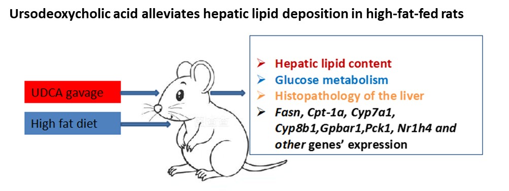
Background: Metabolic dysfunction-associated steatotic liver disease (MASLD) is becoming a growing public health concern. MASLD often coexists with abnormal glucose metabolism. Currently, there is no standard treatment for MASLD. Aim: To elucidate the influence of ursodeoxycholic acid (UDCA) on hepatic lipid deposition, blood glucose, and insulin levels in high-fat diet-induced Sprague–Dawley (SD) rats. Methods: SD rats were randomly assigned to three groups: Group A, which comprised a normal-diet control group; Group B, which comprised a high-fat-fed group; and Group C, which comprised rats treated with UDCA combined with a high-fat diet. At the end of week 4, we performed oral glucose tolerance tests, measured liver enzymes, triglyceride (TG) and total cholesterol (TC) contents, identified morphological changes of the liver, and quantified the expression of major genes in fatty acid, glucose, and bile acid metabolic pathways using real-time polymerase chain reaction. Results: In comparison to Group A, Group B had higher postprandial glucose, insulin, C-peptide, and hepatic TG and TC concentrations (all p < 0.05); metabolism-related genes were differentially expressed in Group B. In comparison to Group B, the values of these indices were all decreased in Group C (all p < 0.05); UDCA reversed the mRNA expression of metabolism-related genes in Group C (all p < 0.05). Conclusion: UDCA administration decreased hepatic lipid content, blood glucose, and insulin levels in SD rats. Relevance for patients: UDCA may be helpful in the treatment of MASLD and its related comorbidities in clinical practice.
- Huby T, Gautier EL. Immune cell-mediated features of non-alcoholic steatohepatitis. Nat Rev Immunol. 2022;22(7):429-443. doi: 10.1038/s41577-021-00639-3
- Riazi K, Azhari H, Charette JH, et al. The prevalence and incidence of NAFLD worldwide: a systematic review and meta-analysis. Lancet Gastroenterol Hepatol. 2022;7(9):851-861. doi: 10.1016/S2468-1253(22)00165-0
- Eslam M, Sanyal AJ, George J. International Consensus Panel MAFLD: a consensus-driven proposed nomenclature for metabolic associated fatty liver disease. Gastroenterology. 2020;158(7):1999-2014.e1. doi: 10.1053/j.gastro.2019.11.312
- Friedman SL, Neuschwander-Tetri BA, Rinella M, Sanyal AJ. Mechanisms of NAFLD development and therapeutic strategies. Nat Med. 2018;24(7):908-922. doi: 10.1038/s41591-018-0104-9
- Ter Horst KW, Serlie MJ. Fructose Consumption, Lipogenesis, and Non-Alcoholic Fatty Liver Disease. Nutrients. 2017;9(9):981. doi: 10.3390/nu9090981
- Ipsen DH, Lykkesfeldt J, Tveden-Nyborg P. Molecular mechanisms of hepatic lipid accumulation in non-alcoholic fatty liver disease. Cell Mol Life Sci. 2018;75(18):3313-3327. doi: 10.1007/s00018-018-2860-6
- Dorn C, Riener MO, Kirovski G, et al. Expression of fatty acid synthase in nonalcoholic fatty liver disease. Int J Clin Exp Pathol. 2010;3(5):505-514.
- Ishii S, Iizuka K, Miller BC, Uyeda K. Carbohydrate response element binding protein directly promotes lipogenic enzyme gene transcription. Proc Natl Acad Sci USA. 2004;101(44):15597-15602. doi: 10.1073/pnas.0405238101
- Schlaepfer IR, Joshi M. CPT1A-mediated Fat Oxidation, Mechanisms, and Therapeutic Potential. Endocrinology. 2020;161(2):bqz046. doi: 10.1210/endocr/bqz046
- Alshawsh MA, Alsalahi A, Alshehade SA, et al. A Comparison of the Gene Expression Profiles of Non-Alcoholic Fatty Liver Disease between Animal Models of a High-Fat Diet and Methionine-Choline-Deficient Diet. Molecules. 2022;27(3):858. doi: 10.3390/molecules27030858
- Chiang JY. Bile acid metabolism and signaling. Compr Physiol. 2013;3(3):1191-1212. doi: 10.1002/j.2040-4603.2013.tb00517.x
- Di Ciaula A, Garruti G, Lunardi Baccetto R, et al. Bile Acid Physiology. Ann Hepatol. 2017;16:s4-s14. doi: 10.5604/01.3001.0010.5493
- Gottlieb A, Canbay A. Why Bile Acids Are So Important in Non-Alcoholic Fatty Liver Disease (NAFLD) Progression. Cells. 2019;8(11):1358. doi: 10.3390/cells8111358
- Younossi ZM, Loomba R, Anstee QM, et al. Diagnostic modalities for nonalcoholic fatty liver disease, nonalcoholic steatohepatitis, and associated fibrosis. Hepatology. 2018;68(1):349-360. doi: 10.1002/hep.29721
- Subudhi S, Drescher HK, Dichtel LE, et al. Distinct Hepatic Gene-Expression Patterns of NAFLD in Patients With Obesity. Hepatol Commun. 2022;6(1):77-89. doi: 10.1002/hep4.1789
- Ishizaki K, Imada T, Tsurufuji M. Hepatoprotective bile acid ‘ursodeoxycholic acid (UDCA)’ Property and difference as bile acids. Hepatol Res. 2005;33(2):174-177. doi: 10.1016/j.hepres.2005.09.029
- Beuers U. Drug insight: mechanisms and sites of action of ursodeoxycholic acid in cholestasis. Nat Clin Pract Gastroenterol Hepatol. 2006;3(6):318-328. doi: 10.1038/ncpgasthep0521
- Bai XP, Du WJ, Xing HB, Yang GH, Bai R. Influence of ursodeoxycholic acid on blood glucose, insulin and GLP-1 in rats with liver fibrosis induced by bile duct ligation. Diabetol Metab Syndr. 2023;15(1):18. doi: 10.1186/s13098-023-00989-z
- Castro RE, Ferreira DM, Afonso MB, et al. miR-34a/SIRT1/p53 is suppressed by ursodeoxycholic acid in the rat liver and activated by disease severity in human non-alcoholic fatty liver disease. J Hepatol. 2013;58(1):119-125. doi: 10.1016/j.jhep.2012.08.008
- Yang W, Liu J, Shan Z, et al. Acarbose compared with metformin as initial therapy in patients with newly diagnosed type 2 diabetes: an open-label, non-inferiority randomised trial. Lancet Diabetes Endocrinol. 2014;2(1):46-55. doi: 10.1016/S2213-8587(13)70021-4
- Bai XP, Li TT, Guo LL, Wang J, Dong F. The Influence of Hyperglycemia on Liver Triglyceride Deposition in Partially Pancreatectomized Rats. Horm Metab Res. 2024;56(2):159-166. doi: 10.1055/a-2198-1132
- Subramanian P, Hampe J, Tacke F, Chavakis T. Fibrogenic Pathways olic Dysfunction Associated Fatty Liver Disease (MAFLD). Int J Mol Sci. 2022;23(13):6996. doi: 10.3390/ijms23136996
- Sauerbruch T, Hennenberg M, Trebicka J, Beuers U. Bile Acids, Liver Cirrhosis, and Extrahepatic Vascular Dysfunction. Front Physiol. 2021;12:718783. doi: 10.3389/fphys.2021.718783
- Nadinskaia M, Maevskaya M, Ivashkin V, et al. Ursodeoxycholic acid as a means of preventing atherosclerosis, steatosis and liver fibrosis in patients with nonalcoholic fatty liver disease. World J Gastroenterol. 2021;27(10):959-975. doi: 10.3748/wjg.v27.i10.959
- Li H, Wang Q, Chen P, Zhou C, Zhang X, Chen Li. Ursodeoxycholic Acid Treatment Restores Gut Microbiota and Alleviates Liver Inflammation in Non-Alcoholic Steatohepatitic Mouse Model. Front Pharmacol. 2021;12:788558. doi: 10.3389/fphar.2021.788558
- Mueller M, Thorell A, Claudel T, et al. Ursodeoxycholic acid exerts farnesoid X receptor-antagonistic effects on bile acid and lipid metabolism in morbid obesity. J Hepatol. 2015;62(6):1398-1404. doi: 10.1016/j.jhep.2014.12.034
- Matsukawa T, Yagi T, Uchida T, et al. Hepatic FASN deficiency differentially affects nonalcoholic fatty liver disease and diabetes in mouse obesity models. JCI Insight. 2023;8(17):e161282. doi: 10.1172/jci.insight.161282
- Chen YS, Liu HM, Lee TY. Ursodeoxycholic Acid Regulates Hepatic Energy Homeostasis and White Adipose Tissue Macrophages Polarization in Leptin-Deficiency Obese Mice. Cells. 2019;8(3):253. doi: 10.3390/cells8030253
- Mouzaki M, Wang AY, Bandsma R, et al. Bile Acids and Dysbiosis in Non-Alcoholic Fatty Liver Disease. PLoS ONE. 2016;11(5):e0151829. doi: 10.1371/journal.pone.0151829
- Allen K, Jaeschke H, Copple BL. Bile acids induce inflammatory genes in hepatocytes: a novel mechanism of inflammation during obstructive cholestasis. Am J Pathol. 2011;178(1):175-186. doi: 10.1016/j.ajpath.2010.11.026
- Taylor R, Basaly V, Kong B, et al. Effects of therapeutically approved individual bile acids on the development of metabolic dysfunction-associated steatohepatitis a low bile acid mouse model. Toxicol Sci. 2024;202(2):179-195. doi: 10.1093/toxsci/kfae110
- Mak KM, Chen LL, Lee TF. Codistribution of collagen type IV and laminin in liver fibrosis of elderly cadavers: immunohistochemical marker of perisinusoidal basement membrane formation. Anat Rec. 2013;296(6):953-964. doi: 10.1002/ar.22694
- Liu Y, Zhang J, Chen Y, et al. The correlation and role analysis of COL4A1 and COL4A2 in hepatocarcinogenesis. Aging. 2020;12(1):204-223. doi: 10.18632/aging.102610
- Elhini SH, Wahsh EA, Elberry AA, et al. The Impact of an SGLT2 Inhibitor versus Ursodeoxycholic Acid on Liver Steatosis in Diabetic Patients. Pharmaceuticals. 2022;15(12):1516. doi: 10.3390/ph15121516
- Chiang JYL, Ferrell JM. Bile acid receptors FXR and TGR5 signaling in fatty liver diseases and therapy. Am J Physiol Gastrointest Liver Physiol. 2020;318(3):G554-573. doi: 10.1152/ajpgi.00223.2019
- Brevini T, Maes M, Webb GJ, et al. FXR inhibition may protect from SARS-CoV-2 infection by reducing ACE2. Nature. 2023;615(7950):134-142. doi: 10.1038/s41586-022-05594-0
- Hatting M, Tavares CDJ, Sharabi K, Rines AK, Puigserver P. Insulin regulation of gluconeogenesis. Ann N Y Acad Sci. 2018;1411(1):21-35. doi: 10.1111/nyas.13435
- Renga B, Mencarelli A, D’Amore C, et al. Glucocorticoid receptor mediates the gluconeogenic activity of the farnesoid X receptor in the fasting condition. FASEB J. 2012;26(7):3021-3031. doi: 10.1096/fj.11-195701
- Wang XX, Xie C, Libby AE, et al. The role of FXR and TGR5 in reversing and preventing progression of Western diet-induced hepatic steatosis, inflammation, and fibrosis in mice. J Biol Chem. 2022;298(11):102530. doi: 10.1016/j.jbc.2022.102530
- Pathak P, Liu H, Boehme S, et al. Farnesoid X receptor induces Takeda G-protein receptor 5 cross-talk to regulate bile acid synthesis and hepatic metabolism. J Biol Chem. 2017;292(26):11055-11069. doi: 10.1074/jbc.M117.784322
- Spatz M, Ciocan D, Merlen G, et al. Bile acid-receptor TGR5 deficiency worsens liver injury in alcohol-fed mice by inducing intestinal microbiota dysbiosis. JHEP Rep. 2021;3(2):100230. doi: 10.1016/j.jhepr.2021.100230
- Yoon S, Lee H, Ji SC, Yoon SH, Cho JY, Chung JY. Pharmacokinetics and Pharmacodynamics of Ursodeoxycholic Acid in an Overweight Population With Abnormal Liver Function. Clin Pharmacol Drug Dev. 2021;10(1):68-77. doi: 10.1002/cpdd.790