

Computed tomography-based evaluation of anatomical variations in the sinonasal region in symptomatic patients: A cross-sectional study

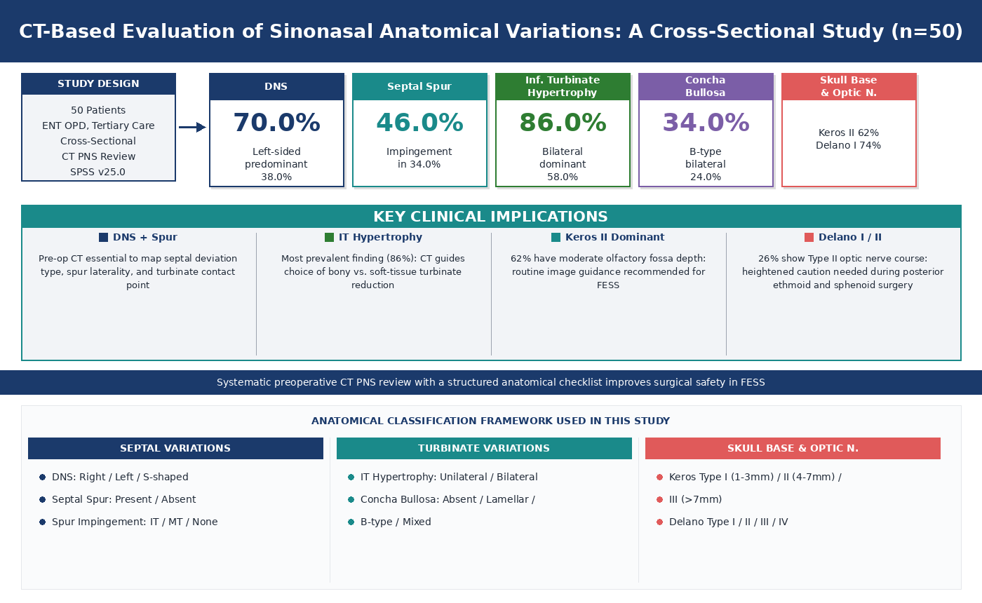
Background: Sinonasal anatomical variations are common structural alterations that influence nasal airflow, sinus drainage, and the safety of functional endoscopic sinus surgery. Computed tomography of the paranasal sinuses (CT PNS) is the gold standard for preoperative assessment of these variations; however, region-specific prevalence data from South Indian cohorts remain limited. Aim: This hospital-based descriptive cross-sectional study aimed to systematically evaluate the prevalence and patterns of key sinonasal anatomical variations in symptomatic patients. Methods: Fifty patients aged 18 years and above who presented to the ear, nose, and throat (ENT) outpatient department of a tertiary care hospital and had undergone CT PNS imaging were enrolled through convenience sampling over a three-month period. Patients with prior sinonasal surgery or recent facial trauma were excluded. CT images were reviewed using standardized high-resolution parameters by an experienced radiologist in collaboration with the ENT team. Data were summarized using descriptive statistics, including frequencies, percentages, means, and standard deviations. Results: Among the 50 patients (mean age, 33.0 ± 14.0 years; 58% female), 70% had a deviated nasal septum, with left-sided deviation being the most common pattern (38.0% of all patients). Septal spurs were present in 46.0%, with spur-induced turbinate impingement in 34.0% of all patients. Inferior turbinate hypertrophy was the most prevalent finding (86.0%), predominantly bilateral (58.0%). Middle turbinate pneumatization (concha bullosa) was identified in 34.0%. Keros type II (62.0%) and Delano type I (74.0%) were the dominant skull base and optic nerve configurations, respectively. Conclusion: Common anatomical variations were frequently identified in symptomatic South Indian patients. Relevance for patients: Systematic CT PNS evaluation is essential for preoperative planning, and region-specific normative data are recommended to enhance surgical safety.
- Stammberger HR, Kennedy DW. Paranasal sinuses: anatomic terminology and nomenclature. Ann Otol Rhinol Laryngol. 1995;104(10_suppl):7-16. doi: 10.1177/000348949510410s01
- Zinreich SJ. Imaging of the nasal cavity and paranasal sinuses. Curr Opin Radiol. 1992;4(1):112-116.
- Van Alyea OE. Nasal Sinuses: An Anatomic and Clinical Consideration. Baltimore: Williams & Wilkins; 1942.
- Lund VJ, Mackay IS. Staging in rhinosinusitis. Rhinology. 1993;31(4):183-184.
- Earwaker J. Anatomic variants in sinonasal CT. RadioGraphics. 1993;13(2):381-415. doi: 10.1148/radiographics.13.2.8460226
- Bolger WE, Butzin CA, Parsons DS. Paranasal sinus bony anatomic variations and mucosal abnormalities: CT analysis for endoscopic sinus surgery. Laryngoscope. 1991;101(1):56-64. doi: 10.1288/00005537-199101000-00010
- DeLano MC, Fun FY, Zinreich SJ. Relationship of the optic nerve to the posterior paranasal sinuses: a CT anatomic study. Am J Neuroradiol. 1996;17(4):669-675.
- Rao VM, Sharma D, Madan A. Imaging of frontal sinus disease: concepts, interpretation, and technology. Otolaryngol Clin North Am. 2001;34(1):23-39. doi: 10.1016/s0030-6665(05)70292-9
- Stallman JS, Lobo JN, Som PM. The incidence of concha bullosa and its relationship to nasal septal deviation and paranasal sinus disease. Am J Neuroradiol. 2004;25(9):1613-1618.
- Keros P. Über die praktische Bedeutung der Niveauunterschiede der Lamina cribrosa des Ethmoids [On the practical significance of the level differences of the lamina cribrosa of the ethmoid]. Z Laryngol Rhinol Otol. 1962;41:809-813. [In German]
- Ramadan HH. Surgical causes of failure in endoscopic sinus surgery. Laryngoscope. 1999;109(1):27-29. doi: 10.1097/00005537-199901000-00006
- Kumar P, Rakesh BS, Prasad R. Anatomical variations of sinonasal region, a coronal CT scan study. Int J Contemporary Med Res. 2016;3(9):2601-2604.
- Gouripur K, Kumar U, Janagond AB, Elangovan S, Srinivasa V. Incidence of sinonasal anatomical variations associated with chronic sinusitis by CT scan in Karaikal, South India. Int J Otorhinolaryngol Head Neck Surg. 2017;3(3):576-580. doi: 10.18203/issn.2454-5929.ijohns20172291
- Kennedy DW. Prognostic factors, outcomes and staging in ethmoid sinus surgery. Laryngoscope. 1992;102(12 Pt 2 Suppl 57):1-18.
- Harar RP, Chadha NK, Rogers G. The role of septal deviation in adult chronic rhinosinusitis: a study of 500 patients. Rhinology. 2004;42(3):126-130.
- Badia L, Lund VJ, Wei W, Ho WK. Ethnic variation in sinonasal anatomy on CT-scanning. Rhinology. 2005;43(3):210-214.
- Mokhasanavisu VJ, Singh R, Balakrishnan R, Kadavigere R. Ethnic variation of sinonasal anatomy on CT scan and volumetric analysis. Indian J Otolaryngol Head Neck Surg. 2019;71(Suppl 3):2157-2164.
- Orhan I, Aydin S, Ormeci T, Yilmaz F. A radiological analysis of inferior turbinate in patients with deviated nasal septum by using computed tomography. Am J Rhinol Allergy. 2014;28(1):e68-e72. doi: 10.2500/ajra.2014.28.4007
- Mygind N, Lilholdt T. Nasal polyps treatment: medical management. Allergy Asthma Proc. 1996;17(5):275-282. doi: 10.2500/108854196778662228
- Wormald PJ. The agger nasi cell: the key to understanding the anatomy of the frontal recess. Otolaryngol Head Neck Surg. 2003;129(5):497-507. doi: 10.1016/S0194-5998(03)01581-X
- Naidoo Y, Bassiouni A, Keen M, Wormald PJ. Long-term outcomes for the endoscopic modified Lothrop/Draf III procedure: a 10-year review. Laryngoscope. 2014;124(1):43- 49. doi: 10.1002/lary.24258
- Papadopoulou AM, Chrysikos D, Samolis A, Tsakotos G, Troupis T. Anatomical variations of the nasal cavities and paranasal sinuses: a systematic review. Cureus. 2021;13(1):e12727. doi: 10.7759/cureus.12727
- Hatipoğlu HG, Cetin MA, Yüksel E. Concha bullosa types: their relationship with sinusitis, ostiomeatal and frontal recess disease. Diagn Interv Radiol. 2005;11(3):145-149.
- Alrumaih RA, Ashoor MM, Obidan AA, Al-Khater KM, Al-Jubran SA. Radiological sinonasal anatomy: exploring the Saudi population. Saudi Med J. 2016;37(5):521-526. doi: 10.15537/smj.2016.5.13904
- Maru YK, Gupta Y. Concha bullosa: frequency and appearances on sinonasal CT. Indian J Otolaryngol Head Neck Surg. 1999;52(1):40-44. doi: 10.1007/BF02996431
- Smith KD, Edwards PC, Saini TS, Norton NS. The prevalence of concha bullosa and nasal septal deviation and their relationship to maxillary sinusitis by volumetric tomography. Int J Dent. 2010;2010:1-5. doi: 10.1155/2010/404982
- Uygur K, Tüz M, Dogru H. The correlation between septal deviation and concha bullosa. Otolaryngol Head Neck Surg. 2003;129(1):33-36. doi: 10.1016/S0194-59980300479-0
- Fadda GL, Rosso S, Aversa S, Petrelli A, Ondolo C, Succo G. Multiparametric statistical correlations between paranasal sinus anatomic variations and chronic rhinosinusitis. Acta Otorhinolaryngol Ital. 2012;32(4):244-251.
- Nouraei SA, Elisay AR, Dimarco A, et al. Variations in paranasal sinus anatomy: implications for the pathophysiology of chronic rhinosinusitis and safety of endoscopic sinus surgery. J Otolaryngol Head Neck Surg. 2009;38(1):32-37. doi: 10.2310/7070.2008.070266
- Ozcan KM, Selcuk A, Ozcan I, Akdogan O, Dere H. Anatomical variations of nasal turbinates. J Craniofac Surg. 2008;19(6):1678-1682. doi: 10.1097/SCS.0b013e318188a29d
- Stammberger H, Shankar L, Hawke M, Evans K. An Atlas of Imaging of the Paranasal Sinuses. London, UK: Martin Dunitz; 1994:41-81. doi: 10.1201/9781003076438-1
- Calhoun KH, Waggenspack GA, Simpson CB, Hokanson JA, Bailey BJ. CT evaluation of the paranasal sinuses in symptomatic and asymptomatic populations. Otolaryngol Head Neck Surg. 1991;104(4):480-483. doi: 10.1177/019459989110400409
- Kantarci M, Karasen RM, Alper F, Onbas O, Okur A, Karaman A. Remarkable anatomic variations in paranasal sinus region and their clinical importance. Eur J Radiol. 2004;50(3):296-302. doi: 10.1016/j.ejrad.2003.08.012
- Lund VJ, Kennedy DW. Quantification for staging sinusitis. Ann Otol Rhinol Laryngol Suppl. 1995;167:17-21. doi: 10.1177/000348949510410s02
- Adeel M, Rajput MS, Akhter S, Ikram M, Arain A, Khattak YJ. Anatomical variations of nose and para-nasal sinuses; CT scan review. J Pak Med Assoc. 2013;63(3):317-319. doi: 10.1016/j.jasi.2015.04.017
- Weinberger DG, Anand VK, Al-Rawi M, Cheng HJ, Messina AV. Surgical anatomy and variations of the Onodi cell. Am J Rhinol. 1996;10(6):365-370. doi: 10.2500/105065896781794851
- Thakur P, Sharma M, Kotwal S, et al. Inter observer agreement among radiologist and otorhinolaryngologists on paranasal sinus computed tomography scans in chronic rhinosinusitis. Indian J Otolaryngol Head Neck Surg. 2022;74(4):501-509. doi: 10.1007/s12070-021-03016-7
- Devaraja K, Doreswamy SM, Pujary K, Ramaswamy B, Pillai S. Anatomical variations of the nose and paranasal sinuses: a computed tomographic study. Indian J Otolaryngol Head Neck Surg. 2019;71(Suppl 3):2231-2240. doi: 10.1007/s12070-019-01716-9
- Stammberger H. Functional Endoscopic Sinus Surgery: The Messerklinger Technique. Philadelphia: BC Decker; 1991. ISBN: 9780941158961.
- Sethi DS, Stanley RE, Pillay PK. Endoscopic anatomy of the sphenoid sinus and sella turcica. J Laryngol Otol. 1995;109(10):951-955. doi: 10.1017/s0022215100131743