

Lipid metabolism dysregulation in Parkinson’s disease: Mechanistic insights and therapeutic implications

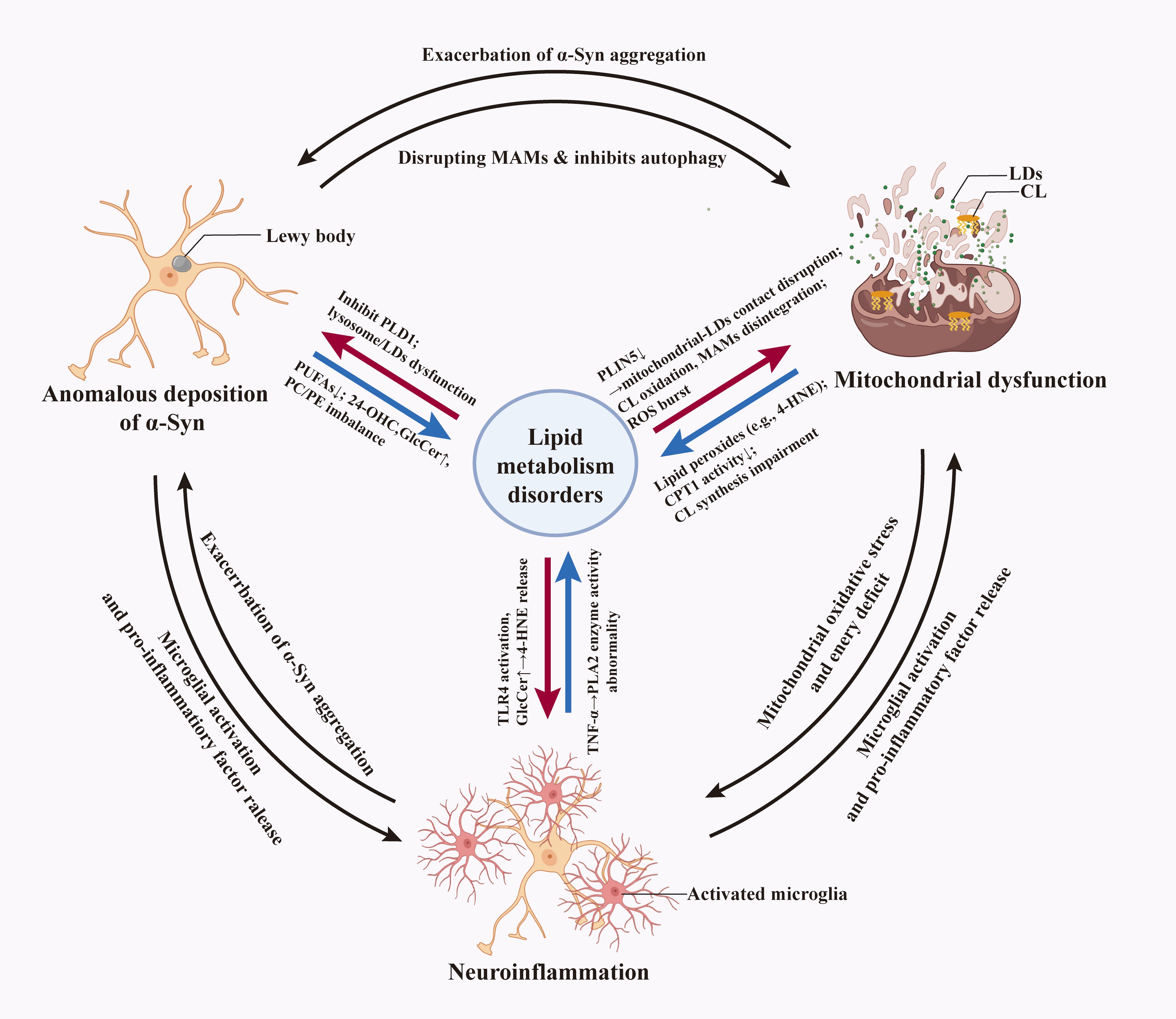
Parkinson’s disease (PD) is a progressive neurodegenerative disorder characterized by the selective degeneration of nigrostriatal dopaminergic neurons and pathological accumulation of α-synuclein (α-Syn) aggregates. Emerging evidence indicates the important role of lipid metabolism dysregulation in driving these pathological features. As major structural components of brain tissue and critical regulators of neuronal function, lipids are involved in diverse biological processes, including cell membrane formation, intercellular signaling, energy storage, and homeostasis. Their dysregulation directly affects neural functions, such as synaptic transmission, antioxidant defense, and inflammatory modulation. PD is recognized not only as a “proteinopathy” but also as an “organelle communication disorder,” involving dysfunction of membrane contact sites across mitochondria, endoplasmic reticulum, lysosomes, and lipid droplets (LDs)—a process that may constitute an early pathogenic event. It is noteworthy that several proteins mediating LDs–organelle contacts are disease-related factors encoded by mutated genes in inherited neurological and metabolic disorders. Despite the extensive communication between intracellular LDs and other organelles through these contact sites, the systematic integration of lipid metabolism dysregulation into core PD pathogenesis remains elusive. This review provides a comprehensive overview of the mechanisms underlying lipid–organelle interactions in PD pathogenesis, with a specific focus on the triangular interplay among the three core pathological hallmarks: α-Syn aggregation, mitochondrial dysfunction, and neuroinflammation, and their convergence with the lipid metabolic network. By analyzing molecular mechanisms and clinical implications, with particular focus on lipid-related biomarkers and therapeutic strategies targeting organelle communication pathways, this review aims to provide new insights into the role of lipid dyshomeostasis in PD pathogenesis and identify feasible therapeutic targets.
- Björklund A, Björklund T, Kirik D. Gene therapy for dopamine replacement in Parkinson’s disease. Sci Transl Med. 2009;1(2):2ps2. doi: 10.1126/scitranslmed.3000350
- Mollenhauer B, Von Arnim CAF. Toward preventing Parkinson’s disease. Science. 2022;377(6608):818-819. doi: 10.1126/science.add7162
- Ben-Shlomo Y, Darweesh S, Llibre-Guerra J, Marras C, San Luciano M, Tanner C. The epidemiology of Parkinson’s disease. Lancet. 2024;403(10423):283-292. doi: 10.1016/S0140-6736(23)01419-8
- Wu KM, Xu QH, Liu YQ, et al. Neuronal FAM171A2 mediates α-synuclein fibril uptake and drives Parkinson’s disease. Science. 2025; 387(6736):892-900. doi: 10.1126/science.adp3645
- Schmidt S, Luecken MD, Trümbach D, et al. Primary cilia and SHH signaling impairments in human and mouse models of Parkinson’s disease. Nat Commun. 2022;13(1):4819. doi: 10.1038/s41467-022-32229-9
- Sampson TR, Debelius JW, Thron T, et al. Gut microbiota regulate motor deficits and neuroinflammation in a model of Parkinson’s disease. Cell. 2016;167(6):1469-1480.e12. doi: 10.1016/j.cell.2016.11.018
- Burtscher J, Moraud EM, Malatesta D, Millet GP, Bally JF, Patoz A. Exercise and gait/movement analyses in treatment and diagnosis of Parkinson’s disease. Ageing Res Rev. 2024;93:102147. doi: 10.1016/j.arr.2023.102147
- Armstrong MJ, Okun MS. Diagnosis and treatment of Parkinson disease: A review. JAMA. 2020;323(6):548-560. doi: 10.1001/jama.2019.22360
- Osetrova M, Tkachev A, Mair W, et al. Lipidome atlas of theadult human brain. Nat Commun. 2024;15(1):4455. doi: 10.1038/s41467-024-48734-y
- Yoon JH, Seo Y, Jo YS, et al. Brain lipidomics: From functional landscape to clinical significance. Sci Adv. 2022;8(37):eadc9317. doi: 10.1126/sciadv.adc9317
- Hussain G, Haseeb A, Azhar R, et al. Lipids as biomarkers of brain disorders. Crit Rev Food Sci Nutr. 2020;60(3):351-374. doi: 10.1080/10408398.2018.1529653
- Qin B, Fu Y, Raulin AC, et al. Lipid metabolism in health and disease: Mechanistic and therapeutic insights for Parkinson’s disease. Chin Med J. 2025;138(12):1411-1423. doi: 10.1097/cm9.0000000000003627
- Pizzuto M, Pelegrin P. Cardiolipin in immune signaling and cell death. Trends Cell Biol. 2020;30(11):892-903. doi: 10.1016/j.tcb.2020.09.004
- Xiao M, Zhong H, Xia L, Tao Y, Yin H. Pathophysiology of mitochondrial lipid oxidation: Role of 4-hydroxynonenal (4-HNE) and other bioactive lipids in mitochondria. Free Radic Biol Med. 2017;111:316-327. doi: 10.1016/j.freeradbiomed.2017.04.363
- Parida PK, Marquez-Palencia M, Ghosh S, et al. Limiting mitochondrial plasticity by targeting DRP1 induces metabolic reprogramming and reduces breast cancer brain metastases. Nat Cancer. 2023;4(6):893-907. doi: 10.1038/s43018-023-00563-6
- Xiong X, Hasani S, Young LEA, et al. Activation of Drp1 promotes fatty acids-induced metabolic reprograming to potentiate Wnt signaling in colon cancer. Cell Death Differ. 2022;29(10):1913-1927. doi: 10.1038/s41418-022-00974-5
- Wedan RJ, Longenecker JZ, Nowinski SM. Mitochondrial fatty acid synthesis is an emergent central regulator of mammalian oxidative metabolism. Cell Metab. 2024;36(1):36-47. doi: 10.1016/j.cmet.2023.11.017
- Choi ML, Chappard A, Singh BP, et al. Pathological structural conversion of α-synuclein at the mitochondria induces neuronal toxicity. Nat Neurosci. 2022;25(9):1134-1148. doi: 10.1038/s41593-022-01140-3
- Conde MA, Alza NP, Iglesias González PA, et al. Phospholipase D1 downregulation by α-synuclein: Implications for neurodegeneration in Parkinson’s disease. Biochim Biophys Acta Mol Cell Biol Lipids. 2018;1863(6):639-650. doi: 10.1016/j.bbalip.2018.03.006
- Bagh MB, Peng S, Chandra G, et al. Misrouting of v-ATPase subunit V0a1 dysregulates lysosomal acidification in a neurodegenerative lysosomal storage disease model. Nat Commun. 2017;8:14612. doi: 10.1038/ncomms14612
- Hu M, Li P, Wang C, et al. Parkinson’s disease-risk protein TMEM175 is a proton-activated proton channel in lysosomes. Cell. 2022;185(13):2292-2308.e20. doi: 10.1016/j.cell.2022.05.021
- Jin H, Wang L, Shen Y, Mao C, Chen J, Liu C. Inflammation-associated peripheral blood cells and serum lipid levels are associated with Parkinson’s disease. Chin Med J (Engl). 2023;136(5):621-622. doi: 10.1097/CM9.0000000000002397
- Ioannou MS, Jackson J, Sheu SH, et al. Neuron-astrocyte metabolic coupling protects against activity-induced fatty acid toxicity. Cell. 2019;177(6):1522-1535.e14. doi: 10.1016/j.cell.2019.04.001
- Cantuti-Castelvetri L, Fitzner D, Bosch-Queralt M, et al. Defective cholesterol clearance limits remyelination in the aged central nervous system. Science. 2018;359(6376):684-688. doi: 10.1126/science.aan4183
- Hamilton LK, Dufresne M, Joppé SE, et al. Aberrant lipid metabolism in the forebrain niche suppresses adult neural stem cell proliferation in an animal model of Alzheimer’s disease. Cell Stem Cell. 2015;17(4):397-411. doi: 10.1016/j.stem.2015.08.001
- Glajch KE, Moors TE, Chen Y, et al. Wild-type GBA1 increases the α-synuclein tetramer-monomer ratio, reduces lipid-rich aggregates, and attenuates motor and cognitive deficits in mice. Proc Natl Acad Sci U S A. 2021;118(31):e2103425118. doi: 10.1073/pnas.2103425118
- Paillusson S, Gomez-Suaga P, Stoica R, et al. α-Synuclein binds to the ER-mitochondria tethering protein VAPB to disrupt Ca2+ homeostasis and mitochondrial ATP production. Acta Neuropathol. 2017;134(1):129-149. doi: 10.1007/s00401-017-1704-z
- Zhang L, Zhou Y, Yang Z, et al. Lipid droplets in central nervous system and functional profiles of brain cells containing lipid droplets in various diseases. J Neuroinflammation. 2025;22(1):7. doi: 10.1186/s12974-025-03334-5
- Guardia-Laguarta C, Area-Gomez E, Rüb C, et al. α-Synuclein is localized to mitochondria-associated ER membranes. J Neurosci. 2014;34(1):249-259. doi: 10.1523/JNEUROSCI.2507-13.2014
- Vanherle S, Loix M, Miron VE, Hendriks JJA, Bogie JFJ. Lipid metabolism, remodelling and intercellular transfer in the CNS. Nat Rev Neurosci. 2025;26(4):214-231. doi: 10.1038/s41583-025-00908-3
- Castellanos DB, Martín-Jiménez CA, Rojas-Rodríguez F, Barreto GE, González J. Brain lipidomics as a rising field in neurodegenerative contexts: Perspectives with machine learning approaches. Front Neuroendocrinol. 2021;61:100899. doi: 10.1016/j.yfrne.2021.100899
- Tian J, Zhang Y, Zhao X. The effects and mechanisms of n-3 and n-6 polyunsaturated fatty acids in the central nervous system. Cell Mol Neurobiol. 2025;45(1):25. doi: 10.1007/s10571-025-01543-3
- Fanning S, Haque A, Imberdis T, et al. Lipidomic analysis of α-synuclein neurotoxicity identifies stearoyl CoA desaturase as a target for Parkinson treatment. Molecular Cell. 2019;73(5):1001-1014.e8. doi: 10.1016/j.molcel.2018.11.028
- Nettebrock NT, Bohnert M. Born this way - biogenesis of lipid droplets from specialized ER subdomains. Biochim Biophys Acta Mol Cell Biol Lipids. 2020;1865(1):158448. doi: 10.1016/j.bbalip.2019.04.008
- Benador IY, Veliova M, Mahdaviani K, et al. Mitochondria bound to lipid droplets have unique bioenergetics, composition, and dynamics that support lipid droplet expansion. Cell Metab. 2018;27(4):869-885.e6. doi: 10.1016/j.cmet.2018.03.003
- Rambold AS, Cohen S, Lippincott-Schwartz J. Fatty acid trafficking in starved cells: Regulation by lipid droplet lipolysis, autophagy, and mitochondrial fusion dynamics. Dev Cell. 2015;32(6):678-692. doi: 10.1016/j.devcel.2015.01.029
- Bulankina AV, Deggerich A, Wenzel D, et al. TIP47 functions in the biogenesis of lipid droplets. J Cell Biol. 2009;185(4):641-655. doi: 10.1083/jcb.200812042
- Bezawork-Geleta A, Devereux CJ, Keenan SN, et al. Proximity proteomics reveals a mechanism of fatty acid transfer at lipid droplet-mitochondria-endoplasmic reticulum contact sites. Nat Commun. 2025;16(1):2135. doi: 10.1038/s41467-025-57405-5
- Hong Z, Adlakha J, Wan N, et al. Mitoguardin-2-mediated lipid transfer preserves mitochondrial morphology and lipid droplet formation. J Cell Biol. 2022;221(12):e202207022. doi: 10.1083/jcb.202207022
- Vrijsen S, Vrancx C, Del Vecchio M, et al. Inter-organellar communication in Parkinson’s and Alzheimer’s disease: Looking beyond endoplasmic reticulum-mitochondria contact sites. Front Neurosci. 2022;16:900338. doi: 10.3389/fnins.2022.900338
- Wang L, He M, Liu X, Jiang BP, Chen H, Shen XC. Dual-labeled single fluorescent probes for the simultaneous two-color visualization of dual organelles and for monitoring cell autophagy. Anal Chem. 2024;96(2):876-886. doi: 10.1021/acs.analchem.3c04520
- Herker E, Vieyres G, Beller M, Krahmer N, Bohnert M. Lipid droplet contact sites in health and disease. Trends Cell Biol. 2021;31(5):345-358. doi: 10.1016/j.tcb.2021.01.004
- Zhang Y, Chen Y, Zhuang C, Qi J, Zhao RC, Wang J. Lipid droplets in the nervous system: Involvement in cell metabolic homeostasis. Neural Regen Res. 2025;20(3):740-750. doi: 10.4103/NRR.NRR-D-23-01401
- Kumar M, Wu Y, Knapp J, et al. Triglycerides are an important fuel reserve for synapse function in the brain. Nat Metab. 2025;7(7):1392-1403. doi: 10.1038/s42255-025-01321-x
- Shatz N, Chohan Y, Klionsky DJ. ATG14 and STX18: Gatekeepers of lipid droplet degradation and the implications for disease modulation. Autophagy. 2024;20(8):1697-1699. doi: 10.1080/15548627.2024.2350739
- Bouchaoui H, Mahoney-Sanchez L, Garçon G, et al. ACSL4 and the lipoxygenases 15/15B are pivotal for ferroptosis induced by iron and PUFA dyshomeostasis in dopaminergic neurons. Free Radic Biol Med. 2023;195:145-157. doi: 10.1016/j.freeradbiomed.2022.12.086
- Bickel PE, Tansey JT, Welte MA. PAT proteins, an ancient family of lipid droplet proteins that regulate cellular lipid stores. Biochim Biophys Acta. 2009;1791(6):419-440. doi: 10.1016/j.bbalip.2009.04.002
- Girard V, Jollivet F, Knittelfelder O, et al. Abnormal accumulation of lipid droplets in neurons induces the conversion of alpha-synuclein to proteolytic resistant forms in a Drosophila model of Parkinson’s disease. PLOS Genet. 2021;17(11):e1009921. doi: 10.1371/journal.pgen.1009921
- Han X, Liu Y, Dai Y, et al. Neuronal SH2B1 attenuates apoptosis in an MPTP mouse model of Parkinson’s disease via promoting PLIN4 degradation. Redox Biol. 2022;52:102308. doi: 10.1016/j.redox.2022.102308
- Han X, Zhu J, Zhang X, et al. Plin4-dependent lipid droplets hamper neuronal mitophagy in the MPTP/p-induced mouse model of Parkinson’s disease. Front Neurosci. 2018;12:397. doi: 10.3389/fnins.2018.00397
- Costa CAD, Manaa WE, Duplan E, Checler F. The endoplasmic reticulum stress/unfolded protein response and their contributions to Parkinson’s disease physiopathology. Cells. 2020;9(11):2495. doi: 10.3390/cells9112495
- Area-Gomez E, De Groof AJC, Boldogh I, et al. Presenilins are enriched in endoplasmic reticulum membranes associated with mitochondria. Am J Pathol. 2009;175(5):1810-1816. doi: 10.2353/ajpath.2009.090219
- Rizzuto R, De Stefani D, Raffaello A, Mammucari C. Mitochondria as sensors and regulators of calcium signalling. Nat Rev Mol Cell Biol. 2012;13(9):566-578. doi: 10.1038/nrm3412
- Barbuti PA, Guardia-Laguarta C, Yun T, et al. The role of alpha-synuclein in synucleinopathy: Impact on lipid regulation at mitochondria-ER membranes. NPJ Parkinsons Dis. 2025;11(1):103. doi: 10.1038/s41531-025-00960-x
- Hu M, Feng X, Liu Q, Liu S, Huang F, Xu H. The ion channels of endomembranes. Physiol Rev. 2024;104(3):1335-1385. doi: 10.1152/physrev.00025.2023
- Przygrodzka E, Plewes MR, Davis JS. Luteinizing hormone regulation of inter-organelle communication and fate of the corpus luteum. Int J Mol Sci. 2021;22(18):9972. doi: 10.3390/ijms22189972
- Gomaraschi M, Bonacina F, Norata GD. Lysosomal acid lipase: From cellular lipid handler to immunometabolic target. Trends Pharmacol Sci. 2019;40(2):104-115. doi: 10.1016/j.tips.2018.12.006
- Dubland JA, Francis GA. Lysosomal acid lipase: At the crossroads of normal and atherogenic cholesterol metabolism. Front Cell Dev Biol. 2015;3:3. doi: 10.3389/fcell.2015.00003
- Bogie JFJ, Haidar M, Kooij G, Hendriks JJA. Fatty acid metabolism in the progression and resolution of CNS disorders. Adv Drug Deliv Rev. 2020;159:198-213. doi: 10.1016/j.addr.2020.01.004
- Xing B, Wang R, Kou T, Liu W, Zhu ZJ. Unraveling tissue-specific fatty acid biosynthesis and inter-tissue crosstalk in mice through stable-isotope tracing metabolomics. Adv Sci. 2025;12:e03662. doi: 10.1002/advs.202503662
- Sarchione A, Marchand A, Taymans JM, Chartier- Harlin MC. Alpha-synuclein and lipids: The elephant in the room? Cells. 2021;10(9):2452. doi: 10.3390/cells10092452
- Ruipérez V, Darios F, Davletov B. Alpha-synuclein, lipids and Parkinson’s disease. Prog Lipid Res. 2010;49(4):420-428. doi: 10.1016/j.plipres.2010.05.004
- Taghizadeh M, Tamtaji OR, Dadgostar E, et al. The effects of omega-3 fatty acids and vitamin E co-supplementation on clinical and metabolic status in patients with Parkinson’s disease: A randomized, double-blind, placebo-controlled trial. Neurochem Int. 2017;108:183-189. doi: 10.1016/j.neuint.2017.03.014
- Michelle HS, Beth L. N-3 (Omega-3) fatty acids: Effects on brain dopamine systems and potential role in the etiology and treatment of neuropsychiatric disorders. CNS Neurol Disord Drug Targets. 2018;17(3):216-232. doi: 10.2174/1871527317666180412153612
- Liu X, Yamada N, Maruyama W, Osawa T. Formation of dopamine adducts derived from brain polyunsaturated fatty acids: Mechanism for Parkinson disease. J Biol Chem. 2008;283(50):34887-34895. doi: 10.1074/jbc.M805682200
- Bousquet M, Calon F, Cicchetti F. Impact of ω-3 fatty acids in Parkinson’s disease. Ageing Res Rev. 2011;10(4):453-463. doi: 10.1016/j.arr.2011.03.001
- She P, Gao B, Li D, et al. The transcriptional repressor HEY2 regulates mitochondrial oxidative respiration to maintain cardiac homeostasis. Nat Commun. 2025;16(1):232. doi: 10.1038/s41467-024-55557-4
- Li P, Song C. Potential treatment of Parkinson’s disease with omega-3 polyunsaturated fatty acids. Nutr Neurosci. 2022;25(1):180-191. doi: 10.1080/1028415X.2020.1735143
- Saiki S, Hatano T, Fujimaki M, et al. Decreased long-chain acylcarnitines from insufficient β-oxidation as potential early diagnostic markers for Parkinson’s disease. Sci Rep. 2017;7(1):7328. doi: 10.1038/s41598-017-06767-y
- Den Besten G, Van Eunen K, Groen AK, Venema K, Reijngoud DJ, Bakker BM. The role of short-chain fatty acids in the interplay between diet, gut microbiota, and host energy metabolism. J Lipid Res. 2013;54(9):2325-2340. doi: 10.1194/jlr.R036012
- Magliocca G, Mone P, Di Iorio BR, Heidland A, Marzocco S. Short-chain fatty acids in chronic kidney disease: Focus on inflammation and oxidative stress regulation. Int J Mol Sci. 2022;23(10):5354. doi: 10.3390/ijms23105354
- Dalile B, Van Oudenhove L, Vervliet B, Verbeke K. The role of short-chain fatty acids in microbiota-gut-brain communication. Nat Rev Gastroenterol Hepatol. 2019;16(8):461-478. doi: 10.1038/s41575-019-0157-3
- Bruning J, Chapp A, Kaurala GA, Wang R, Techtmann S, Chen QH. Gut microbiota and short chain fatty acids: Influence on the autonomic nervous system. Neurosci Bull. 2020;36(1):91-95. doi: 10.1007/s12264-019-00410-8
- Duan WX, Wang F, Liu JY, Liu CF. Relationship between short-chain fatty acids and Parkinson’s disease: A review from pathology to clinic. Neurosci Bull. 2024;40(4):500-516. doi: 10.1007/s12264-023-01123-9
- Cao Q, Shen M, Li R, et al. Elucidating the specific mechanisms of the gut-brain axis: The short-chain fatty acids-microglia pathway. J Neuroinflammation. 2025;22(1):133. doi: 10.1186/s12974-025-03454-y
- Chen SJ, Chen CC, Liao HY, et al. Association of fecal and plasma levels of short-chain fatty acids with gut microbiota and clinical severity in patients with Parkinson disease. Neurology. 2022;98(8):e848-e858. doi: 10.1212/WNL.0000000000013225
- Wang C, Yang M, Liu D, Zheng C. Metabolic rescue of α-synuclein-induced neurodegeneration through propionate supplementation and intestine-neuron signaling in C. Elegans. Cell Rep. 2024;43(3):113865. doi: 10.1016/j.celrep.2024.113865
- Su SH, Chen M, Wu YF, et al. Fecal microbiota transplantation and short-chain fatty acids protected against cognitive dysfunction in a rat model of chronic cerebral hypoperfusion. CNS Neurosci Ther. 2023;29(Suppl 1):98-114. doi: 10.1111/cns.14089
- Rane P, Shields J, Heffernan M, Guo Y, Akbarian S, King JA. The histone deacetylase inhibitor, sodium butyrate, alleviates cognitive deficits in pre-motor stage PD. Neuropharmacology. 2012;62(7):2409-2412. doi: 10.1016/j.neuropharm.2012.01.026
- Li X, Wang C, Zhu J, et al. Sodium butyrate ameliorates oxidative stress-induced intestinal epithelium barrier injury and mitochondrial damage through AMPK-mitophagy pathway. Oxid Med Cell Longev. 2022;2022:3745135. doi: 10.1155/2022/3745135
- Gao Z, Yin J, Zhang J, et al. Butyrate improves insulin sensitivity and increases energy expenditure in mice. Diabetes. 2009;58(7):1509-1517. doi: 10.2337/db08-1637
- Mollica MP, Mattace Raso G, Cavaliere G, et al. Butyrate regulates liver mitochondrial function, efficiency, and dynamics in insulin-resistant obese mice. Diabetes. 2017;66(5):1405-1418. doi: 10.2337/db16-0924
- Xu RC, Miao WT, Xu JY, et al. Neuroprotective effects of sodium butyrate and monomethyl fumarate treatment through GPR109A modulation and intestinal barrier restoration on PD mice. Nutrients. 2022;14(19):4163. doi: 10.3390/nu14194163
- Song C, Zhang J, Qi S, et al. Cardiolipin remodeling by ALCAT1 links mitochondrial dysfunction to Parkinson’s diseases. Aging Cell. 2019;18(3):e12941. doi: 10.1111/acel.12941
- Rua AJ, Mitchell W, Claypool SM, Alder NN, Alexandrescu AT. Perturbations in mitochondrial metabolism associated with defective cardiolipin biosynthesis: An in-organello real-time NMR study. J Biol Chem. 2024;300(10):107746. doi: 10.1016/j.jbc.2024.107746
- Tang Y, Wu J, Sun X, et al. Cardiolipin oxidized by ROS from complex II acts as a target of gasdermin D to drive mitochondrial pore and heart dysfunction in endotoxemia. Cell Rep. 2024;43(5):114237. doi: 10.1016/j.celrep.2024.114237
- Zhang Y, Tian L, Huang G, et al. Structural basis of BAX pore formation. Science. 388(6754):eadv4314. doi: 10.1126/science.adv4314
- Perier C, Tieu K, Guégan C, et al. Complex I deficiency primes Bax-dependent neuronal apoptosis through mitochondrial oxidative damage. Proc Natl Acad Sci U S A. 2005;102(52):19126-19131. doi: 10.1073/pnas.0508215102
- Qi LFR, Liu S, Fang Q, et al. Ginsenoside Rg3 restores mitochondrial cardiolipin homeostasis via GRB2 to prevent Parkinson’s disease. Adv Sci (Weinh). 2024;11(39):e2403058. doi: 10.1002/advs.202403058
- Van Meer G, Voelker DR, Feigenson GW. Membrane lipids: Where they are and how they behave. Nat Rev Mol Cell Biol. 2008;9(2):112-124. doi: 10.1038/nrm2330
- Devaux PF. Static and dynamic lipid asymmetry in cell membranes. Biochemistry. 1991;30(5):1163-1173. doi: 10.1021/bi00219a001
- Haider A, Wei YC, Lim K, et al. PCYT1A regulates phosphatidylcholine homeostasis from the inner nuclear membrane in response to membrane stored curvature elastic stress. Dev Cell. 2018;45(4):481-495.e8. doi: 10.1016/j.devcel.2018.04.012
- Kang JH, Toita R, Kawano T, Murata M, Kano A. Phospholipids and their metabolites as diagnostic biomarkers of human diseases. Prog Lipid Res. 2025;99:101340. doi: 10.1016/j.plipres.2025.101340
- Kent C. Eukaryotic phospholipid biosynthesis. Annu Rev Biochem. 1995;64:315-343. doi: 10.1146/annurev.bi.64.070195.001531
- Wang K, Xu H, Zou R, et al. PCYT1A deficiency disturbs fatty acid metabolism and induces ferroptosis in the mouse retina. BMC Biol. 2024;22(1):134. doi: 10.1186/s12915-024-01932-y
- Wood PL, Tippireddy S, Feriante J, Woltjer RL. Augmented frontal cortex diacylglycerol levels in Parkinson’s disease and lewy body disease. PLoS One. 2018;13(3):e0191815. doi: 10.1371/journal.pone.0191815
- Stoessel D, Schulte C, Teixeira Dos Santos MC, et al. Promising metabolite profiles in the plasma and CSF of early clinical Parkinson’s disease. Front Aging Neurosci. 2018;10:51. doi: 10.3389/fnagi.2018.00051
- Farmer K, Smith CA, Hayley S, Smith J. Major alterations of phosphatidylcholine and lysophosphotidylcholine lipids in the substantia nigra using an early stage model of Parkinson’s disease. Int J Mol Sci. 2015;16(8):18865-18877. doi: 10.3390/ijms160818865
- Shahmoradian SH, Lewis AJ, Genoud C, et al. Lewy pathology in Parkinson’s disease consists of crowded organelles and lipid membranes. Nat Neurosci. 2019;22(7):1099-1109. doi: 10.1038/s41593-019-0423-2
- Farid I, Ali A, Holman AP, Osborne L, Kurouski D. Length and saturation of choline plasmalogens alter the aggregation rate of α-synuclein but not the toxicity of amyloid fibrils. Int J Biol Macromol. 2024;264(Pt 1):130632. doi: 10.1016/j.ijbiomac.2024.130632
- O’Leary EI, Jiang Z, Strub MP, Lee JC. Effects of phosphatidylcholine membrane fluidity on the conformation and aggregation of N-terminally acetylated α-synuclein. J Biol Chem. 2018;293(28):11195-11205. doi: 10.1074/jbc.RA118.002780
- Mu J, Lam SM, Shui G. Emerging roles and therapeutic potentials of sphingolipids in pathophysiology: Emphasis on fatty acyl heterogeneity. J Genet Genomics. 2024;51(3):268-278. doi: 10.1016/j.jgg.2023.06.006
- Gomez-Larrauri A, Presa N, Dominguez-Herrera A, Ouro A, Trueba M, Gomez-Muñoz A. Role of bioactive sphingolipids in physiology and pathology. Essays Biochem. 2020;64(3):579-589. doi: 10.1042/EBC20190091
- Bras J, Singleton A, Cookson MR, Hardy J. Emerging pathways in genetic Parkinson’s disease: Potential role of ceramide metabolism in lewy body disease. FEBS J. 2008;275(23):5767-5773. doi: 10.1111/j.1742-4658.2008.06709.x
- Czubowicz K, Jęśko H, Wencel P, Lukiw WJ, Strosznajder RP. The role of ceramide and sphingosine-1-phosphate in Alzheimer’s disease and other neurodegenerative disorders. Mol Neurobiol. 2019;56(8):5436-5455. doi: 10.1007/s12035-018-1448-3
- Abbott SK, Li H, Muñoz SS, et al. Altered ceramide acyl chain length and ceramide synthase gene expression in Parkinson’s disease. Mov Disord. 2014;29(4):518-526. doi: 10.1002/mds.25729
- Vos M, Dulovic-Mahlow M, Mandik F, et al. Ceramide accumulation induces mitophagy and impairs β-oxidation in PINK1 deficiency. Proc Natl Acad Sci U S A. 2021;118(43):e2025347118. doi: 10.1073/pnas.2025347118
- Lim JR, Chae CW, Park JY, et al. Ethanol-induced ceramide production causes neuronal apoptosis by increasing MCL- 1S-mediated ER-mitochondria contacts. Neurobiol Dis. 2023;177:106009. doi: 10.1016/j.nbd.2023.106009
- Motyl J, Przykaza Ł, Boguszewski PM, Kosson P, Strosznajder JB. Pramipexole and fingolimod exert neuroprotection in a mouse model of Parkinson’s disease by activation of sphingosine kinase 1 and Akt kinase. Neuropharmacology. 2018;135:139-150. doi: 10.1016/j.neuropharm.2018.02.023
- Wang R, Sun H, Cao Y, et al. Glucosylceramide accumulation in microglia triggers STING-dependent neuroinflammation and neurodegeneration in mice. Sci Signal. 2024;17(829):eadk8249. doi: 10.1126/scisignal.adk8249
- Mazzulli JR, Xu YH, Sun Y, et al. Gaucher disease glucocerebrosidase and α-synuclein form a bidirectional pathogenic loop in synucleinopathies. Cell. 2011;146(1):37-52. doi: 10.1016/j.cell.2011.06.001
- Seo BA, Kim D, Hwang H, et al. TRIP12 ubiquitination of glucocerebrosidase contributes to neurodegeneration in Parkinson’s disease. Neuron. 2021;109(23):3758-3774.e11. doi: 10.1016/j.neuron.2021.09.031
- Belarbi K, Cuvelier E, Bonte MA, et al. Glycosphingolipids and neuroinflammation in Parkinson’s disease. Mol Neurodegener. 2020;15(1):59. doi: 10.1186/s13024-020-00408-1
- Boland S, Swarup S, Ambaw YA, et al. Deficiency of the frontotemporal dementia gene GRN results in gangliosidosis. Nature Commun. 2022;13(1):5924. doi: 10.1038/s41467-022-33500-9
- Maglione V, Marchi P, Di Pardo A, et al. Impaired ganglioside metabolism in Huntington’s disease and neuroprotective role of GM1. J Neurosci. 2010;30(11):4072-4080. doi: 10.1523/JNEUROSCI.6348-09.2010
- Schneider JS, Roeltgen DP, Mancall EL, Chapas-Crilly J, Rothblat DS, Tatarian GT. Parkinson’s disease: Improved function with GM1 ganglioside treatment in a randomized placebo-controlled study. Neurology. 1998;50(6):1630-1636. doi: 10.1212/wnl.50.6.1630
- Schneider JS, Aras R, Williams CK, Koprich JB, Brotchie JM, Singh V. GM1 Ganglioside modifies α-synuclein toxicity and is neuroprotective in a rat α-synuclein model of Parkinson’s disease. Sci Rep. 2019;9(1):8362. doi: 10.1038/s41598-019-42847-x
- Galleguillos D, Wang Q, Steinberg N, et al. Anti-inflammatory role of GM1 and other gangliosides on microglia. J Neuroinflammation. 2022;19(1):9. doi: 10.1186/s12974-021-02374-x
- Sonnino S. The relationship between depletion of brain GM1 ganglioside and Parkinson’s disease. FEBS Open Bio. 2023;13(9):1548-1557. doi: 10.1002/2211-5463.13554
- Chiricozzi E, Mauri L, Lunghi G, et al. Parkinson’s disease recovery by GM1 oligosaccharide treatment in the B4galnt1+/− mouse model. Sci Rep. 2019;9(1):19330. doi: 10.1038/s41598-019-55885-2
- Kumar R, Chowdhury S, Ledeen R. Alpha-synuclein and GM1 ganglioside co-localize in neuronal cytosol leading to inverse interaction-relevance to Parkinson’s disease. Int J Mol Sci. 2024;25(6):3323. doi: 10.3390/ijms25063323
- Chowdhury S, Wu G, Lu ZH, Kumar R, Ledeen R. Age-related decline in gangliosides GM1 and GD1a in non-CNS tissues of normal mice: Implications for peripheral symptoms of Parkinson’s disease. Biomedicines. 2023;11(1):209. doi: 10.3390/biomedicines11010209
- Itokazu Y, Fuchigami T, Morgan JC, Yu RK. Intranasal infusion of GD3 and GM1 gangliosides downregulates alpha-synuclein and controls tyrosine hydroxylase gene in a PD model mouse. Mol Ther. 2021;29(10):3059-3071. doi: 10.1016/j.ymthe.2021.06.005
- Chowdhury S, Kumar R, Zepeda E, DeFrees S, Ledeen R. Synthetic GM1 improves motor and memory dysfunctions in mice with monoallelic or biallelic disruption of GM3 synthase. FEBS Open Bio. 2023;13(9):1651-1657. doi: 10.1002/2211-5463.13669
- Wei J, Liu X, Xiao W, et al. Phospholipid remodeling and its derivatives are associated with COVID-19 severity. J Allergy Clin Immunol. 2023;151(5):1259-1268. doi: 10.1016/j.jaci.2022.11.032
- Im YJ, Lee YK, Chung HY, Im DS. Multiple actions of lysophosphatidylcholine in human Jurkat T cells. Acta Pharmacol Sin. 2006;27(6):700-707. doi: 10.1111/j.1745-7254.2006.00339.x
- García-Morales V, Montero F, González-Forero D, et al. Membrane-derived phospholipids control synaptic neurotransmission and plasticity. PLoS Biol. 2015;13(5):e1002153. doi: 10.1371/journal.pbio.1002153
- Hao Y, Guo M, Feng Y, Dong Q, Cui M. Lysophospholipids and their G-coupled protein signaling in Alzheimer’s disease: From physiological performance to pathological impairment. Front Mol Neurosci. 2020;13:58. doi: 10.3389/fnmol.2020.00058
- Geraldo LHM, Spohr TCLDS, Amaral RFD, et al. Role of lysophosphatidic acid and its receptors in health and disease: Novel therapeutic strategies. Signal Transduct Target Ther. 2021;6(1):45. doi: 10.1038/s41392-020-00367-5
- Yilmaz A, Ashrafi N, Ashrafi R, et al. Lipid profiling of Parkinson’s disease brain highlights disruption in lysophosphatidylcholines, and triacylglycerol metabolism. NPJ Parkinsons Dis. 2025;11(1):159. doi: 10.1038/s41531-025-01023-x
- Mu C, Shao K, Su M, et al. Lysophosphatidylcholine promoting α-Synuclein aggregation in Parkinson’s disease: Disrupting GCase glycosylation and lysosomal α-Synuclein degradation. NPJ Parkinsons Dis. 2025;11(1):47. doi: 10.1038/s41531-025-00902-7
- Hung ND, Sok DE, Kim MR. Prevention of 1-palmitoyl lysophosphatidylcholine-induced inflammation by polyunsaturated acyl lysophosphatidylcholine. Inflamm Res. 2012;61(5):473-483. doi: 10.1007/s00011-012-0434-x
- Zhao C, Tu J, Wang C, et al. Lysophosphatidylcholine binds α-synuclein and prevents its pathological aggregation. Natl Sci Rev. 2024;11(6):nwae182. doi: 10.1093/nsr/nwae182
- Malik I, Turk J, Mancuso DJ, et al. Disrupted membrane homeostasis and accumulation of ubiquitinated proteins in a mouse model of infantile neuroaxonal dystrophy caused by PLA2G6 mutations. Am J Pathol. 2008;172(2):406-416. doi: 10.2353/ajpath.2008.070823
- Shin KC, Ali Moussa HY, Park Y. Cholesterol imbalance and neurotransmission defects in neurodegeneration. Exp Mol Med. 2024;56(8):1685-1690. doi: 10.1038/s12276-024-01273-4
- Zhang J, Liu Q. Cholesterol metabolism and homeostasis in the brain. Protein Cell. 2015;6(4):254-264. doi: 10.1007/s13238-014-0131-3
- Guo J, Chen S, Zhang Y, et al. Cholesterol metabolism: Physiological regulation and diseases. MedComm (2020). 2024;5(2):e476. doi: 10.1002/mco2.476
- Musanti R, Parati E, Lamperti E, Ghiselli G. Decreased cholesterol biosynthesis in fibroblasts from patients with Parkinson disease. Biochem Med Metab Biol. 1993;49(2):133-142. doi: 10.1006/bmmb.1993.1016
- Reiss AB, Wirkowski E. Role of HMG-CoA reductase inhibitors in neurological disorders: Progress to date. Drugs. 2007;67(15):2111-2120. doi: 10.2165/00003495-200767150-00001
- Al-kuraishy HM, Fahad EH, Al-Windy S, El-Sherbeni SA, Negm WA, Batiha GE-S. The effects of cholesterol and statins on Parkinson’s neuropathology: A narrative review. Inflammopharmacology. 2024;32(2):917-925. doi: 10.1007/s10787-023-01400-z
- Nakamura K, Mori F, Tanji K, et al. Isopentenyl diphosphate isomerase, a cholesterol synthesizing enzyme, is localized in Lewy bodies. Neuropathology. 2015;35(5):432-440. doi: 10.1111/neup.12204
- Dai L, Wang J, Meng L, et al. The cholesterol 24-hydroxylase CYP46A1 promotes α-synuclein pathology in Parkinson’s disease. PLoS Biol. 2025;23(2):e3002974. doi: 10.1371/journal.pbio.3002974
- Dai L, Wang J, Zhang X, et al. 27-Hydroxycholesterol drives the spread of α-synuclein pathology in Parkinson’s disease. Mov Disord. 2023;38(11):2005-2018. doi: 10.1002/mds.29577
- Doria M, Maugest L, Moreau T, Lizard G, Vejux A. Contribution of cholesterol and oxysterols to the pathophysiology of Parkinson’s disease. Free Radic Biol Med. 2016;101:393-400. doi: 10.1016/j.freeradbiomed.2016.10.008
- Falabella M, Vernon HJ, Hanna MG, Claypool SM, Pitceathly RDS. Cardiolipin, mitochondria, and neurological disease. Trends Endocrinol Metab. 2021;32(4):224-237. doi: 10.1016/j.tem.2021.01.006
- Tong B, Ba Y, Li Z, et al. Targeting dysregulated lipid metabolism for the treatment of Alzheimer’s disease and Parkinson’s disease: Current advancements and future prospects. Neurobiol Dis. 2024;196:106505. doi: 10.1016/j.nbd.2024.106505