

Artificial intelligence versus elastography in characterizing BI-RADS 4 breast nodules: A systematic review and critical appraisal

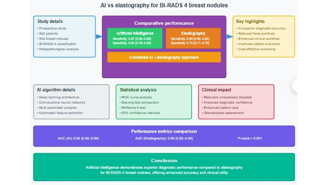
Breast imaging reporting and data system (BI-RADS) category 4 breast lesions represent a heterogeneous category with moderate suspicion of malignancy, which pose significant diagnostic challenges. Both artificial intelligence (AI) and elastography have demonstrated potential adjunctive roles in improving the evaluation of these lesions. Given the increasingly pervasive use of AI in the medical field, a systematic and critical evaluation of its diagnostic efficacy, clinical utility, and practical applications, compared with elastography techniques, is warranted for the assessment of BI-RADS 4 breast nodules. A systematic literature search was conducted across multiple databases from January 2010 to December 2024, and the studies were critically appraised using standardized quality assessment tools (e.g., quality assessment of diagnostic accuracy studies-2). Due to the significant heterogeneity in study populations and methodologies, a narrative synthesis approach with comprehensive critical appraisal was employed. A total of 23 studies met the inclusion criteria for AI assessment (n = 15,847 lesions) and 31 for elastography (n = 12,456 lesions). Critical appraisal revealed significant methodological limitations—67% of the studies had a high risk of bias in patient selection, 45% in index-test conduct, and 56% in flow and timing. Only 25% of the studies were considered high quality. AI systems demonstrated promising diagnostic performance in individual studies (reported area under the curve range: 0.82–0.94), while elastography showed consistent but more modest performance (reported area under the curve range: 0.72–0.87). However, the quality of the evidence was insufficient for a reliable comparative assessment. While both technologies show promise, the existing evidence is limited by significant methodological constraints, precluding reliable comparative conclusions. These gaps highlight the need for high-quality prospective head-to-head comparison studies with standardized protocols and rigorous methodology.
- American College of Radiology. Breast Imaging Reporting and Data System (BI-RADS) Atlas. 5th ed. United States: American College of Radiology; 2013.
- D’Orsi CJ, Sickles EA, Mendelson EB, Morris EA. ACR BI-RADS Atlas: Breast Imaging Reporting and Data System. 5th ed. United States: American College of Radiology; 2013.
- Rao AA, Feneis J, Lalonde C, Ojeda-Fournier H. A Pictorial Review of Changes in the BI-RADS Fifth Edition. Radiographics. 2016;36(3):623-639. doi: 10.1148/rg.2016150178
- Sigrist RMS, Liau J, Kaffas AE, Chammas MC, Willmann JK. Ultrasound Elastography: Review of Techniques and Clinical Applications. Theranostics. 2017;7(5):1303-1329. doi: 10.7150/thno.18650
- Berg WA, Blume JD, Cormack JB, et al. Combined screening with ultrasound and mammography versus mammography alone in women at an elevated risk of breast cancer. JAMA. 2008;299(18):2151-2163. doi: 10.1001/jama.299.18.2151
- Garra BS. Elastography: Current status, future prospects, and making work for you. Ultrasound Q. 2011;27(3): 177-186. doi: 10.1097/RUQ.0b013e31822a2138
- Liberman L, Abramson AF, Squires FB, Glassman JR, Morris EA, Dershaw DD. The breast imaging reporting and data system: Positive predictive value of mammographic features and final assessment categories. AJR Am J Roentgenol. 1998;171(1):35-40. doi: 10.2214/ajr.171.1.9648759
- Rodriguez-Ruiz A, Krupinski E, Mordang JJ, et al. Detection of breast cancer using mammography: The effect of an artificial intelligence support system. Radiology. 2019;290(2):305-314. doi: 10.1148/radiol.2018181371
- Cosgrove D, Piscaglia F, Bamber J, et al. EFSUMB guidelines and recommendations for the clinical use of ultrasound elastography. Part 2: Clinical applications. Ultraschall Med. 2013;34(3):238-253. doi: 10.1055/s-0033-1335375
- Burnside ES, Ochsner JE, Fowler KJ, et al. Use of microcalcification descriptors in BI-RADS 4th edition to stratify risk of malignancy. Radiology. 2007;242(2):388-395. doi: 10.1148/radiol.2422052130
- Brewer NT, Salz T, Lillie SE. Systematic review: The long-term effects of false-positive mammograms. Ann Intern Med. 2007;146(7):502-510. doi: 10.7326/0003-4819-146-7-200704030-00006
- Tosteson AN, Stout NK, Fryback DG, et al. Cost-effectiveness of digital mammography breast cancer screening. Ann Intern Med. 2008;148(1):1-10. doi: 10.7326/0003-4819-148-1-200801010-00002
- Mazurowski MA, Buda M, Saha A, Bashir MR. Deep learning in radiology: An overview of the concepts and a survey of the state of the art with focus on MRI. Magnetic Resonance Imaging. 2018;49(4):939-954. doi: 10.1002/jmri.26534
- Itoh A, Ueno E, Tohno E, et al. Breast disease: Clinical application of US elastography for diagnosis. Radiology. 2006;239(2):341-350. doi: 10.1148/radiol.2391041676
- Barr RG, Nakashima K, Amy D, et al. WFUMB guidelines and recommendations for clinical use of ultrasound elastography: Part 2: Breast. Ultrasound Med Biol. 2015;41(5):1148-1160. doi: 10.1016/j.ultrasmedbio.2015.03.008
- Yoon JH, Kim MJ, Kim EK, et al. Interobserver variability of ultrasound elastography: How does it affect the diagnosis of breast lesions? AJR Am J Roentgenol. 2011;196(3):730-736. doi: 10.2214/AJR.10.4654
- Berg WA, Cosgrove DO, Doré CJ, et al. Shear-wave elastography improves the specificity of breast US: A multicenter, retrospective study of 939 masses. Radiology. 2012;262(2):435-449. doi: 10.1148/radiol.11110640
- Sadigh G, Carlos RC, Neal CH, Dwamena BA. Ultrasonographic differentiation of malignant from benign breast lesions: A meta-analytic comparison of elasticity and BI-RADS scoring. Breast Cancer Res Treat. 2012;133(1):23-35. doi: 10.1007/s10549-011-1857-8
- Evans A, Whelehan P, Thomson K, et al. Invasive breast cancer: Relationship between shear-wave elastographic findings and histologic prognostic factors. Radiology. 2012;263(3):673-677. doi: 10.1148/radiol.12111317
- Han S, Kang HK, Jeong JY, et al. A deep learning framework for supporting the classification of breast lesions in ultrasound images. Phys Med Biol. 2017;62(19):7714-7728. doi: 10.1088/1361-6560/aa82ec
- Fujioka T, Kubota K, Mori M, et al. Distinction between benign and malignant breast masses at breast US using a deep convolutional neural network. Jpn J Radiol. 2019;37(6):466-472. doi: 10.1007/s11604-019-00831-5
- McKinney SM, Sieniek M, Godbole V, et al. International evaluation of an AI system for breast cancer screening. Nature. 2020;577(7788):89-94. doi: 10.1038/s41586-019-1799-6
- Becker AS, Marcon M, Ghafoor S, et al. Deep learning in mammography: Diagnostic accuracy of a multipurpose image analysis software in the detection of breast cancer. Invest Radiol. 2017;52(7):434-440. doi: 10.1097/RLI.0000000000000358
- Geras KJ, Mann RM, Moy L. Artificial intelligence for mammography and digital breast tomosynthesis: Current concepts and future perspectives. Radiology. 2019;293(2):246-259. doi: 10.1148/radiol.2019182627
- Kooi T, Litjens G, van Ginneken B, et al. Large scale deep learning for computer aided detection of mammographic lesions. Med Image Anal. 2017;35:303-312. doi: 10.1016/j.media.2016.07.007
- Huynh BQ, Li H, Giger ML. Digital mammographic tumor classification using transfer learning from deep convolutional neural networks. J Med Imaging (Bellingham). 2016;3(3):034501. doi: 10.1117/1.JMI.3.3.034501
- Hosny A, Parmar C, Quackenbush J, Schwartz LH, Aerts HJWL. Artificial intelligence in radiology. Nat Rev Cancer. 2018;18(8):500-510. doi: 10.1038/s41568-018-0016-5
- Liu X, Faes L, Kale AU, et al. A comparison of deep learning performance against health-care professionals in detecting diseases from medical imaging: A systematic review and meta-analysis. Lancet Digit Health. 2019;1(6):e271-e297. doi: 10.1016/S2589-7500(19)30123-2
- Brady AP, Neri E. Artificial intelligence in radiology-ethical considerations. Diagnostics (Basel). 2020;10(4):231. doi: 10.3390/diagnostics10040231
- Giger ML, Chan HP, Boone J. Anniversary paper: History and status of CAD and quantitative image analysis: The role of medical physics and AAPM. Med Phys. 2008;35(12):5799-5820. doi: 10.1118/1.3013555
- Zhou LQ, Wu XL, Huang SY, et al. Lymph node metastasis prediction from primary breast cancer US images using deep learning. Radiology. 2020;294(1):19-28. doi: 10.1148/radiol.2019190372
- Byra M, Galperin M, Ojeda-Fournier H, et al. Breast mass classification in sonography with transfer learning using a deep convolutional neural network and color conversion. Med Phys. 2019;46(2):746-755. doi: 10.1002/mp.13361
- Wang J, Yang X, Cai H, et al. Discrimination of breast cancer with microcalcifications on mammography by deep learning. Sci Rep. 2016;6:27327. doi: 10.1038/srep27327
- Chartrand G, Cheng PM, Vorontsov E, et al. Deep learning: A primer for radiologists. Radiographics. 2017;37(7): 2113-2131. doi: 10.1148/rg.2017170077
- Erickson BJ, Korfiatis P, Akkus Z, Kline TL. Machine learning for medical imaging. Radiographics. 2017;37(2):505-515. doi: 10.1148/rg.2017160130
- Yassin NIR, Omran S, El Houby EMF, Allam H. Machine learning techniques for breast cancer computer aided diagnosis using different image modalities: A systematic review. Comput Methods Programs Biomed. 2018;156:25-45. doi: 10.1016/j.cmpb.2017.12.012
- Tang A, Tam R, Cadrin-Chênevert A, et al. Canadian Association of Radiologists white paper on artificial intelligence in radiology. Can Assoc Radiol J. 2018;69(2):120-135. doi: 10.1016/j.carj.2018.02.002
- Thrall JH, Li X, Li Q, et al. Artificial intelligence and machine learning in radiology: Opportunities, challenges, pitfalls, and criteria for success. J Am Coll Radiol. 2018;15(3):504-508. doi: 10.1016/j.jacr.2017.12.026
- Zech JR, Badgeley MA, Liu M, Costa AB, Titano JJ, Oermann EK. Variable generalization performance of a deep learning model to detect pneumonia in chest radiographs: A cross-sectional study. PLoS Med. 2018;15(11):e1002683. doi: 10.1371/journal.pmed.1002683
- Antropova N, Huynh BQ, Giger ML. A deep feature fusion methodology for breast cancer diagnosis demonstrated on three imaging modality datasets. Med Phys. 2017;44(10):5162-5171. doi: 10.1002/mp.12453
- Rajpurkar P, Irvin J, Ball RL, et al. Deep learning for chest radiograph diagnosis: A retrospective comparison of the CheXNeXt algorithm to practicing radiologists. PLoS Med. 2018;15(11):e1002686. doi: 10.1371/journal.pmed.1002686
- Zheng X, Yao Z, Huang Y, et al. Deep learning radiomics can predict axillary lymph node status in early-stage breast cancer. Nat Commun. 2020;11(1):1236. doi: 10.1038/s41467-020-15027-z
- Bai HX, Wang R, Xiong Z, et al. Artificial intelligence augmentation of radiologist performance in distinguishing COVID-19 from pneumonia of other origin at chest CT. Radiology. 2020;296(3):E156-E165. doi: 10.1148/radiol.2020201491
- Koh DM, Papanikolaou N, Bick U, et al. Artificial intelligence and machine learning in cancer imaging. Commun Med (Lond). 2022;2:133. doi: 10.1038/s43856-022-00199-0
- Pesapane F, Codari M, Sardanelli F. Artificial intelligence in medical imaging: Threat or opportunity? Radiologists again at the forefront of innovation in medicine. Eur Radiol Exp. 2018;2(1):35. doi: 10.1186/s41747-018-0061-6
- Lehman CD, Wellman RD, Buist DS, et al. Diagnostic accuracy of digital screening mammography with and without computer-aided detection. JAMA Intern Med. 2015;175(11):1828-1837. doi: 10.1001/jamainternmed.2015.5231
- Van der Velden BH, Kuijf HJ, Gilhuijs KG, Viergever MA. Explainable artificial intelligence (XAI) in deep learning-based medical image analysis. Med Image Anal. 2022;79:102470. doi: 10.1016/j.media.2022.102470
- Shen L, Margolies LR, Rothstein JH, Fluder E, McBride R, Sieh W. Deep learning to improve breast cancer detection on screening mammography. Sci Rep. 2019;9(1):12495. doi: 10.1038/s41598-019-48995-4
- Kapoor A, Kapur A, Sidhu BS, Singh J. Use of next-generation shear wave elastography for blue breast cancer evaluation. Int Surg J. 2025;12(8):1288-1295. doi: 10.18203/2349-2902.isj20252278
- Wei Q, Yan YJ, Wu GG, et al. Added value of a new strain elastography technique in conventional ultrasound for the diagnosis of breast masses: A prospective multicenter study. Front Oncol. 2021;11:779612. doi: 10.3389/fonc.2021.779612
- Gennisson JL, Deffieux T, Fink M, Tanter M. Ultrasound elastography: Principles and techniques. Diagnostic and Interventional Imaging. 2013;94(5):487-495. doi: 10.1016/j.diii.2013.01.022