

Hollow spherical mineralized scaffold integrated with a bone marrow mesenchymal stem cell-laden three-dimensional delivery system for regeneration of critical-sized bone defects

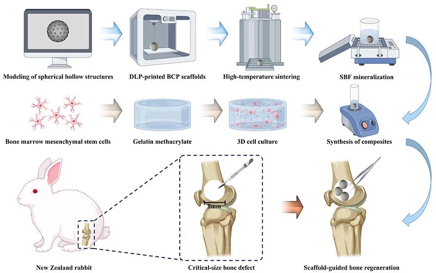
Treating critical-sized bone defects is a significant clinical challenge. Three-dimensional (3D) printing combined with bone tissue engineering (BTE) has emerged as a promising strategy for bone regeneration; however, key limitations persist, including a mismatch between scaffold degradation and osteogenesis, as well as insufficient bioactivity. In this study, we aimed to fabricate a hollow spherical mineralized biphasic calcium phosphate scaffold by 3D printing (photopolymerization via digital light processing) and incorporated within its cavity a 3D delivery system composed of methacryloyl-modified gelatin hydrogel loaded with bone marrow mesenchymal stem cells (BMSCs). The composite scaffold was systematically evaluated using material characterization, in vitro cytocompatibility analysis, and in vivo rabbit bone defect models. Our findings demonstrated that the scaffold exhibited favorable mechanical properties, biocompatibility, and enhanced osteogenic differentiation, migration, and pro-osteogenic gene expression in BMSCs. Notably, the scaffold effectively repaired critical-sized bone defects in rabbit models within 12 weeks. This novel BTE composite scaffold provides a groundbreaking design philosophy and an innovative therapeutic strategy for complex bone defect repair.
- Zhang Y, Lin T, Meng H, et al. 3D gel-printed porous magnesium scaffold coated with dibasic calcium phosphate dihydrate for bone repair in vivo. J Orthop Transl. 2022;33:13–23. doi: 10.1016/j.jot.2021.11.005
- Turnbull G, Clarke J, Picard F, et al. 3D bioactive composite scaffolds for bone tissue engineering. Bioact Mater. 2018;3(3):278-314. doi: 10.1016/j.bioactmat.2017.10.001
- Wang W, Yeung KWK. Bone grafts and biomaterials substitutes for bone defect repair: A review. Bioact Mater. 2017;2(4):224-247. doi: 10.1016/j.bioactmat.2017.05.007
- Zhu G, Zhang T, Chen M, et al. Bone physiological microenvironment and healing mechanism: Basis for future bone-tissue engineering scaffolds. Bioact Mater. 2021;6(11):4110-4140. doi: 10.1016/j.bioactmat.2021.03.043
- Beigi MH, Safaie N, Nasr-Esfahani MH, Kiani A. 3D Titania Nanofiber-Like Webs Induced by Plasma Ionization: A New Direction for Bioreactivity and Osteoinductivity Enhancement of Biomaterials. Sci Rep. 2019;9(1):17999. doi: 10.1038/s41598-019-54533-z
- Qin Y, Wen P, Guo H, et al. Additive manufacturing of biodegradable metals: Current research status and future perspectives. Acta Biomater. 2019;98:3-22. doi: 10.1016/j.actbio.2019.04.046
- Valtanen RS, Yang YP, Gurtner GC, Maloney WJ, Lowenberg DW. Synthetic and bone tissue engineering graft substitutes: What is the future? Injury. 2021;52 Suppl 2:S72-S77. doi: 10.1016/j.injury.2020.07.040
- Yin S, Zhang W, Zhang Z, Jiang X. Recent Advances in Scaffold Design and Material for Vascularized Tissue- Engineered Bone Regeneration. Adv Healthcare Mater. 2019;8(10):e1801433. doi: 10.1002/adhm.201801433
- He F, Ye J. In vitro degradation, biocompatibility, and in vivo osteogenesis of poly(lactic-co-glycolic acid)/calcium phosphate cement scaffold with unidirectional lamellar pore structure. J Biomed Mater Res Part A. 2012;100(12):3239- 3250. doi: 10.1002/jbm.a.34265
- Wang X, Lin M, Kang Y. Engineering Porous β-Tricalcium Phosphate (β-TCP) Scaffolds with Multiple Channels to Promote Cell Migration, Proliferation, and Angiogenesis. ACS Appl. Mater Interfaces. 2019;11(9):9223-9232. doi: 10.1021/acsami.8b22041
- Freytes DO, Kang JW, Marcos-Campos I, Vunjak-Novakovic G. Macrophages modulate the viability and growth of human mesenchymal stem cells. J Cell Biochem. 2013;114(1):220- 229. doi: 10.1002/jcb.24357
- Duda GN, Geissler S, Checa S, Tsitsilonis S, Petersen A, Schmidt-Bleek K. The decisive early phase of bone regeneration. Nat Rev Rheumatol. 2023;19(2):78-95. doi: 10.1038/s41584-022-00887-0
- Liu X, Gao J, Liu J, Zhang L, Li M. Inhibiting the “isolated island” effect in simulated bone defect repair using a hollow structural scaffold design. Front Bioeng Biotechnol. 2024;12:1362913. doi: 10.3389/fbioe.2024.1362913
- Liu X, Gao J, Liu J, et al. Three-Dimensional-Printed Spherical Hollow Structural Scaffolds for Guiding Critical-Sized Bone Regeneration. ACS Biomater Sci Eng. 2024;10(4):2581-2594. doi: 10.1021/acsbiomaterials.3c01956
- Kim HD, Amirthalingam S, Kim SL, Lee SS, Rangasamy J, Hwang NS. Biomimetic Materials and Fabrication Approaches for Bone Tissue Engineering. Adv Healthcare Mater. 2017;6(23). doi: 10.1002/adhm.201700612
- Lowe B, Ottensmeyer MP, Xu C, He Y, Ye Q, Troulis MJ. The Regenerative Applicability of Bioactive Glass and Beta-Tricalcium Phosphate in Bone Tissue Engineering: A Transformation Perspective. J Funct Biomater. 2019;10(1). doi: 10.3390/jfb10010016
- Chen CS. 3D Biomimetic Cultures: The Next Platform for Cell Biology. Trends Cell Biol. 2016;26(11):798-800. doi: 10.1016/j.tcb.2016.08.008
- Payr S, Rosado-Balmayor E, Tiefenboeck T, et al. Direct comparison of 3D and 2D cultivation reveals higher osteogenic capacity of elderly osteoblasts in 3D. J Orthop Surg Res. 2021;16(1):13. doi: 10.1186/s13018-020-02153-z
- Laschke MW, Menger MD. Life is 3D: Boosting Spheroid Function for Tissue Engineering. Trends Biotechnol. 2017;35(2):133-144. doi: 10.1016/j.tibtech.2016.08.004
- Bicer M, Cottrell GS, Widera D. Impact of 3D cell culture on bone regeneration potential of mesenchymal stromal cells. Stem Cell Res Ther. 2021;12(1):31. doi: 10.1186/s13287-020-02094-8
- Wang J, Wang T, Zhang F, et al. Roles of circular RNAs in osteogenic differentiation of bone marrow mesenchymal stem cells (Review). Mol Med Rep. 2022;26(1). doi: 10.3892/mmr.2022.12743
- Hou Y, Lin W, Li Y, et al. De-osteogenic-differentiated mesenchymal stem cells accelerate fracture healing by mir- 92b. J Orthop Transl. 2021;27:25-32. doi: 10.1016/j.jot.2020.10.009
- Ma L, Wang X, Zhou Y, et al. Biomimetic Ti-6Al-4V alloy/gelatin methacrylate hybrid scaffold with enhanced osteogenic and angiogenic capabilities for large bone defect restoration. Bioact Mater. 2021;6(10):3437-3448. doi: 10.1016/j.bioactmat.2021.03.010
- Zhu J, Marchant RE. Design properties of hydrogel tissue-engineering scaffolds. Expert Rev Med Devices. 2011;8(5):607-26. doi: 10.1586/erd.11.27
- Gao C, Sow WT, Wang Y, et al. Hydrogel composite scaffolds with an attenuated immunogenicity component for bone tissue engineering applications. J Mater Chem B. 2021;9(8):2033-2041. doi: 10.1039/d0tb02588g
- Li J, Chen M, Fan X, Zhou H. Recent advances in bioprinting techniques: approaches, applications and future prospects. J Transl Med. 2016;14:271. doi: 10.1186/s12967-016-1028-0
- Li R, Zhou C, Chen J, et al. Synergistic osteogenic and angiogenic effects of KP and QK peptides incorporated with an injectable and self-healing hydrogel for efficient bone regeneration. Bioact Mater. 2022;18:267-283. doi: 10.1016/j.bioactmat.2022.02.011
- Li J, Cui X, Lindberg GCJ, et al. Hybrid fabrication of photo-clickable vascular hydrogels with additive manufactured titanium implants for enhanced osseointegration and vascularized bone formation. Biofabrication. 2022;14(3). doi: 10.1088/1758-5090/ac6051
- Liu X, Gao J, Cui X, et al. Functionalized 3D-Printed PLA Biomimetic Scaffold for Repairing Critical-Size Bone Defects. Bioengineering. 2023;10(9). doi: 10.3390/bioengineering10091019
- Kokubo T, Takadama H. How useful is SBF in predicting in vivo bone bioactivity? Biomaterials. 2006;27(15):2907-2915. doi: 10.1016/j.biomaterials.2006.01.017
- Heise T, Sawyer AY, Hirai T, Schaible S, Sy H, Wickramasekara S. Report on investigation of ISO 10993-12 extraction conditions. Regul Toxicol Pharmacol RTP. 2022;131:105164. doi: 10.1016/j.yrtph.2022.105164
- Zhang Y, Xu Y, Kong H, et al. Microneedle system for tissue engineering and regenerative medicine. Exploration. 2023;3(1):20210170. doi: 10.1002/exp.20210170
- Fan Y, Zheng L, Jin M, Li X, Li ZA, Wang X. Mussel-mimetic polysaccharide-based injectable hydrogels for biomedical applications. BMEMat. 2024;2(4):e12089. doi: 10.1002/bmm2.12089
- Miller MI, Brightman AO, Epstein FH, et al. BME 2.0: Engineering the Future of Medicine. BME Front. 2023;4:0001. doi: 10.34133/bmef.0001
- LeGeros RZ. Properties of osteoconductive biomaterials: calcium phosphates. Clin Orthop Relat Res. 2002;(395):81- 98. doi: 10.1097/00003086-200202000-00009
- Ko CL, Chen WC, Chen JC, et al. Properties of osteoconductive biomaterials: calcium phosphate cement with different ratios of platelet-rich plasma as identifiers. Mater Sci Eng C Mater Biol Appl. 2013;33(6):3537-44. doi: 10.1016/j.msec.2013.04.042
- Lu T, Li G, Zhang L, Yuan X, Wu T, Ye J. Optimizing silicon doping levels for enhanced osteogenic and angiogenic properties of 3D-printed biphasic calcium phosphate scaffolds: An in vitro screening and in vivo validation study. Mater Today Bio. 2024;28:101203. doi: 10.1016/j.mtbio.2024.101203
- Wang Y, Liu Y, Chen S, et al. Enhancing bone regeneration through 3D printed biphasic calcium phosphate scaffolds featuring graded pore sizes. Bioact Mater. 2025;46:21-36. doi: 10.1016/j.bioactmat.2024.11.024
- Touri M, Moztarzadeh F, Osman NAA, Dehghan MM, Mozafari M. 3D-printed biphasic calcium phosphate scaffolds coated with an oxygen generating system for enhancing engineered tissue survival. Mater Sci Eng C Mater Biol Appl. 2018;84:236-242. doi: 10.1016/j.msec.2017.11.037
- Bajpai I, Kim DY, Kyong-Jin J, Song IH, Kim S. Response of human bone marrow-derived MSCs on triphasic Ca-P substrate with various HA/TCP ratio. J Biomed Mater Res Part B Appl Biomater. 2017;105(1):72-80. doi: 10.1002/jbm.b.33538
- Ng AM, Tan KK, Phang MY, et al. Differential osteogenic activity of osteoprogenitor cells on HA and TCP/HA scaffold of tissue engineered bone. J Biomed Mater Res Part A. 2008;85(2):301-12. doi: 10.1002/jbm.a.31324
- Xue J, Qin C, Wu C. 3D printing of cell-delivery scaffolds for tissue regeneration. Regen Biomater. 2023;10:rbad032. doi: 10.1093/rb/rbad032
- Bose S, Sarkar N. Natural Medicinal Compounds in Bone Tissue Engineering. Trends Biotechnol. 2020;38(4):404-417. doi: 10.1016/j.tibtech.2019.11.005
- Lefèvre E, Farlay D, Bala Y, et al. Compositional and mechanical properties of growing cortical bone tissue: a study of the human fibula. Sci Rep. 2019;9(1):17629. doi: 10.1038/s41598-019-54016-1
- Qin Y, Liu A, Guo H, et al. Additive manufacturing of Zn-Mg alloy porous scaffolds with enhanced osseointegration: In vitro and in vivo studies. Acta Biomater. 2022;145:403-415. doi: 10.1016/j.actbio.2022.03.055
- Qu X, Cui W, Yang F, et al. The effect of oxygen plasma pretreatment and incubation in modified simulated body fluids on the formation of bone-like apatite on poly(lactide-co-glycolide) (70/30). Biomaterials. 2007;28(1):9-18. doi: 10.1016/j.biomaterials.2006.08.024
- Feng B, Zhang M, Qin C, et al. 3D printing of conch-like scaffolds for guiding cell migration and directional bone growth. Bioact Mater. 2023;22:127-140. doi: 10.1016/j.bioactmat.2022.09.014
- Futrega K, Mosaad E, Chambers K, Lott WB, Clements J, Doran MR. Bone marrow-derived stem/stromal cells (BMSC) 3D microtissues cultured in BMP-2 supplemented osteogenic induction medium are prone to adipogenesis. Cell Tissue Res. 2018;374(3):541-553. doi: 10.1007/s00441-018-2894-y
- Potapova IA, Gaudette GR, Brink PR, et al. Mesenchymal stem cells support migration, extracellular matrix invasion, proliferation, and survival of endothelial cells in vitro. Stem Cells. 2007;25(7):1761-1768. doi: 10.1634/stemcells.2007-0022
- Yue K, Trujillo-de Santiago G, Alvarez MM, Tamayol A, Annabi N, Khademhosseini A. Synthesis, properties, and biomedical applications of gelatin methacryloyl (GelMA) hydrogels. Biomaterials. 2015;73:254-271. doi: 10.1016/j.biomaterials.2015.08.045
- Gungor-Ozkerim PS, Inci I, Zhang YS, Khademhosseini A, Dokmeci MR. Bioinks for 3D bioprinting: an overview. Biomater Sci. 2018;6(5):915-946. doi: 10.1039/c7bm00765e
- Anada T, Pan CC, Stahl AM, et al. Vascularized Bone- Mimetic Hydrogel Constructs by 3D Bioprinting to Promote Osteogenesis and Angiogenesis. Int J Mol Sci. 2019;20(5):1096. doi: 10.3390/ijms20051096
- Li J, Wang W, Li M, et al. Biomimetic Methacrylated Gelatin Hydrogel Loaded With Bone Marrow Mesenchymal Stem Cells for Bone Tissue Regeneration. Front Bioeng Biotechnol. 2021;9:770049. doi: 10.3389/fbioe.2021.770049
- Guo C, Qi J, Liu J, et al. The Ability of Biodegradable Thermosensitive Hydrogel Composite Calcium-Silicon- Based Bioactive Bone Cement in Promoting Osteogenesis and Repairing Rabbit Distal Femoral Defects. Polymers. 2022;14(18). doi: 10.3390/polym14183852
- Zhang Y, Cui X, Zhao S, et al. Evaluation of injectable strontium-containing borate bioactive glass cement with enhanced osteogenic capacity in a critical-sized rabbit femoral condyle defect model. ACS Appl Mater Interfaces. 2015;7(4):2393-2403. doi: 10.1021/am507008z
- Kelly CN, Wang T, Crowley J, et al. High-strength, porous additively manufactured implants with optimized mechanical osseointegration. Biomaterials. 2021;279:121206. doi: 10.1016/j.biomaterials.2021.121206
- Zhi W, Wang X, Sun D, et al. Optimal regenerative repair of large segmental bone defect in a goat model with osteoinductive calcium phosphate bioceramic implants. Bioact Mater. 2022;11:240-253. doi: 10.1016/j.bioactmat.2021.09.024
- Xu R-J, Ma J-J, Xiao Y, et al. A biphasic calcium phosphate/ acylated methacrylate gelatin composite hydrogel promotes osteogenesis and bone repair. Connect Tissue Res. 2023;64(5):445-456. doi: 10.1080/03008207.2023.2212067
- Lai J, Wang C, Liu J, et al. Low temperature hybrid 3D printing of hierarchically porous bone tissue engineering scaffolds within situ delivery of osteogenic peptide and mesenchymal stem cells. Biofabrication. 2022;14(4). doi: 10.1088/1758-5090/ac84b0
- Kim SS, Sun Park M, Jeon O, Yong Choi C, Kim BS. Poly(lactide-co-glycolide)/hydroxyapatite composite scaffolds for bone tissue engineering. Biomaterials. 2006;27(8):1399-1409. doi: 10.1016/j.biomaterials.2005.08.016
61. Albrektsson T, Johansson C. Osteoinduction, osteoconduction and osseointegration. Eur Spine J. 2001;10(Suppl 2):S96-S101. doi: 10.1007/s005860100282