

Epigenetic regulation role of SUV420H in sexual dimorphism of adipose tissue and obesity

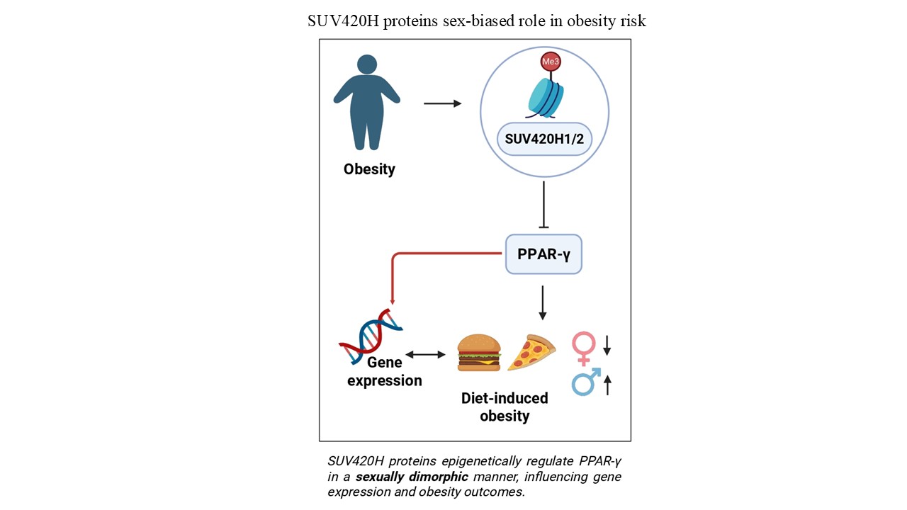
Obesity is a worldwide health crisis linked to numerous harmful medical conditions. It arises from complex interactions between behavioral, environmental, and genetic factors. Interestingly, there is evidence of sexual dimorphism in the function of mitochondrial activity and metabolic flexibility of adipose tissue in obesity. Peroxisome proliferator-activated receptor-gamma (PPAR-γ), a critical factor in the formation of fat cells, glucose processing, and fat production, plays an essential role in the onset of obesity. Recently, it has been implicated in sex-based differences in adipose tissue gene expression and fat distribution. Epigenetic modifications mediate the effects of environmental factors by controlling the activity of genes related to metabolism. SUV420H1 and SUV420H2, histone methyltransferases that catalyze the addition of di- and trimethyl groups to lysine 20 of histone H4, suppress gene expression. Previous research has shown that SUV420H proteins respond to environmental signals by directly repressing PPAR-γ activity. In this study, we demonstrated that SUV420H1 and SUV420H2 are expressed differently between sexes in both human and mouse models. Interestingly, mice with a double knockout of Suv420h1 and Suv420h2 exhibit a sex-specific difference in their resistance to diet-induced obesity. Gene expression analysis of adipose tissue samples from obese humans reveals a significant inverse relationship between SUV420H and PPAR-γ expression in females, while no such correlation is observed in males. These findings highlight a potentially novel role for SUV420H proteins in modulating PPAR-γ activity, metabolic function, and the risk of obesity, with a clear sexual dimorphism in their effects.
- Blüher M. Obesity: Global epidemiology and pathogenesis. Nat Rev Endocrinol. 2019;15(5):288-298. doi: 10.1038/s41574-019-0176-8
- Li J, Ruggiero-Ruff RE, He Y, et al. Sexual dimorphism in obesity is governed by RELMα regulation of adipose macrophages and eosinophils. eLife. 2023;12:e86001. doi: 10.7554/eLife.86001
- Link JC, Reue K. Genetic basis for sex differences in obesity and lipid metabolism. Annu Rev Nutr. 2017;37(1):225-245. doi: 10.1146/annurev-nutr-071816-064827
- Gerdts E, Regitz-Zagrosek V. Sex differences in cardiometabolic disorders. Nat Med. 2019;25(11):1657-1666. doi: 10.1038/s41591-019-0643-8
- Parks BW, Sallam T, Mehrabian M, et al. Genetic architecture of insulin resistance in the mouse. Cell Metab. 2015;21(2):334-347. doi: 10.1016/j.cmet.2015.01.002
- Shungin D, Winkler TW, Croteau-Chonka DC, et al. New genetic loci link adipose and insulin biology to body fat distribution. Nature. 2015;518(7538):187-196. doi: 10.1038/nature14132
- Emdin CA, Khera AV, Natarajan P, et al. Genetic association of waist-to-hip ratio with cardiometabolic traits, type 2 diabetes, and coronary heart disease. JAMA. 2017;317(6):626-634. doi: 10.1001/jama.2016.21042
- Chait A, Wang S, Goodspeed L, et al. Sexually dimorphic relationships among saa3 (serum amyloid a3), inflammation, and cholesterol metabolism modulate atherosclerosis in mice. Arterioscler Thromb Vasc Biol. 2021;41(6):e299-e313. doi: 10.1161/atvbaha.121.316066
- Kurt Z, Barrere-Cain R, LaGuardia J, et al. Tissue-specific pathways and networks underlying sexual dimorphism in non-alcoholic fatty liver disease. Biol Sex Differ. 2018;9(1):46. doi: 10.1186/s13293-018-0205-7
- Norheim F, Hasin-Brumshtein Y, Vergnes L, et al. Gene-by-sex interactions in mitochondrial functions and cardio-metabolic traits. Cell Metab. 2019;29(4):932-949.e4. doi: 10.1016/j.cmet.2018.12.013
- Yang X, Schadt EE, Wang S, et al. Tissue-specific expression and regulation of sexually dimorphic genes in mice. Genome Res. 2006;16(8):995-1004. doi: 10.1101/gr.5217506
- Ahmadian M, Suh JM, Hah N, et al. PPARγ signaling and metabolism: The good, the bad and the future. Nat Med. 2013;19(5):557-566. doi: 10.1038/nm.3159
- Soccio RE, Li Z, Chen ER, et al. Targeting PPARγ in the epigenome rescues genetic metabolic defects in mice. J Clin Invest. 2017;127(4):1451-1462. doi: 10.1172/JCI91211
- Berhouma R, Kouidhi S, Ammar M, Abid H, Ennafaa H, Benammar-Elgaaied A. Correlation of peroxisome proliferator-activated receptor (PPAR-γ) mRNA expression with Pro12Ala polymorphism in obesity. Biochem Genet. 2013;51(3-4):256-263. doi: 10.1007/s10528-012-9560-y
- Moreno-Navarrete JM, Petrov P, Serrano M, et al. Decreased RB1 mRNA, protein, and activity reflect obesity-induced altered adipogenic capacity in human adipose tissue. Diabetes. 2013;62(6):1923-1931. doi: 10.2337/db12-0977
- Hammes TO, Costa CS, Rohden F, et al. Parallel down-regulation of FOXO1, PPARγ and adiponectin mRNA expression in visceral adipose tissue of class III obese individuals. Obes Facts. 2012;5(3):452-459. doi: 10.1159/000339574
- Vidal-Puig AJ, Considine RV, Jimenez-Liñan M, et al. Peroxisome proliferator-activated receptor gene expression in human tissues. Effects of obesity, weight loss, and regulation by insulin and glucocorticoids. J Clin Invest. 1997;99(10):2416-2422. doi: 10.1172/JCI119424
- Sato H, Sugai H, Kurosaki H, et al. The effect of sex hormones on peroxisome proliferator-activated receptor gamma expression and activity in mature adipocytes. Biol Pharm Bull. 2013;36(4):564-573. doi: 10.1248/bpb.b12-00868
- Trang K, Grant SFA. Genetics and epigenetics in the obesity phenotyping scenario. Rev Endocr Metab Disord. 2023;24(5):775-793. doi: 10.1007/s11154-023-09804-6
- Lin X, Li H. Obesity: Epidemiology, pathophysiology, and therapeutics. Front Endocrinol (Lausanne). 2021;12:706978. doi: 10.3389/fendo.2021.706978
- Ling C, Rönn T. Epigenetics in human obesity and type 2 diabetes. Cell Metab. 2019;29(5):1028-1044. doi: 10.1016/j.cmet.2019.03.009
- Schotta G, Lachner M, Sarma K, et al. A silencing pathway to induce H3-K9 and H4-K20 trimethylation at constitutive heterochromatin. Genes Dev. 2004;18(11):1251-1262. doi: 10.1101/gad.300704
- Gabellini D, Pedrotti S. The SUV4-20H histone methyltransferases in health and disease. Int J Mol Sci. 2022;23(9):4736. doi: 10.3390/ijms23094736
- Pedrotti S, Caccia R, Neguembor MV, et al. The Suv420h histone methyltransferases regulate PPAR-γ and energy expenditure in response to environmental stimuli. Sci Adv. 2019;5(4):eaav1472. doi: 10.1126/sciadv.aav1472
- Kahn D, Macias E, Zarini S, et al. Exploring visceral and subcutaneous adipose tissue secretomes in human obesity: Implications for metabolic disease. Endocrinology. 2022;163(11):bqac140. doi: 10.1210/endocr/bqac140
- Chang E, Varghese M, Singer K. Gender and sex differences in adipose tissue. Curr Diab Rep. 2018;18(9):69. doi: 10.1007/s11892-018-1031-3
- Bromberg KD, Mitchell TRH, Upadhyay AK, et al. The SUV4-20 inhibitor A-196 verifies a role for epigenetics in genomic integrity. Nat Chem Biol. 2017;13(3):317-324. doi: 10.1038/nchembio.2282
- Ryu TY, Lee J, Kang Y, et al. Epigenetic regulation of DHRS2 by SUV420H2 inhibits cell apoptosis in renal cell carcinoma. Biochem Biophys Res Commun. 2023;663:41-46. doi: 10.1016/j.bbrc.2023.04.066
- Kautzky-Willer A, Harreiter J, Pacini G. Sex and gender differences in risk, pathophysiology and complications of Type 2 diabetes mellitus. Endocr Rev. 2016;37(3):278-316. doi: 10.1210/er.2015-1137
- Ng M, Fleming T, Robinson M, et al. Global, regional, and national prevalence of overweight and obesity in children and adults during 1980-2013: A systematic analysis for the global burden of disease study 2013. Lancet. 2014;384(9945):766-781. doi: 10.1016/S0140-6736(14)60460-8
- Multhaup ML, Seldin MM, Jaffe AE, et al. Mouse-human experimental epigenetic analysis unmasks dietary targets and genetic liability for diabetic phenotypes. Cell Metab. 2015;21(1):138-149. doi: 10.1016/j.cmet.2014.12.014
- Baca P, Barajas-Olmos F, Mirzaeicheshmeh E, et al. DNA methylation and gene expression analysis in adipose tissue to identify new loci associated with T2D development in obesity. Nutr Diabetes. 2022;12(1):50. doi: 10.1038/s41387-022-00228-w
- Boulet N, Briot A, Galitzky J, Bouloumié A. The sexual dimorphism of human adipose depots. Biomedicines. 2022;10(10):2615. doi: 10.3390/biomedicines10102615
- Chiodi V, Rappa F, Lo Re O, et al. Deficiency of histone variant macroH2A1.1 is associated with sexually dimorphic obesity in mice. Sci Rep. 2023;13:19123. doi: 10.1038/s41598-023-46304-8
- Torres JL, Usategui-Martín R, Hernández-Cosido L, et al. PPAR-γ gene expression in human adipose tissue is associated with weight loss after sleeve gastrectomy. J Gastrointest Surg. 2022;26(2):286-297. doi: 10.1007/s11605-021-05216-6
- Núñez Ruiz A, Cortés-Garcia JD, Cortez-Espinosa N, et al. Diminished levels of regulatory T cell subsets (CD8+Foxp3, CD4+Foxp3 and CD4+CD39+Foxp3) but increased Foxp3 expression in adipose tissue from overweight subjects. Nutrition. 2016;32(9):943-954. doi: 10.1016/j.nut.2016.02.006
- Boughanem H, Cabrera-Mulero A, Millán-Gómez M, et al. Transcriptional analysis of FOXO1, C/EBP-α and PPAR-γ2 Genes and their association with obesity-related insulin resistance. Genes (Basel). 2019;10(9):706. doi: 10.3390/genes10090706
- Tryggestad JB, Teague AM, Sparling DP, Jiang S, Chernausek SD. Macrophage-derived microRNA-155 increases in obesity and influences adipocyte metabolism by targeting peroxisome proliferator-activated receptor gamma. Obesity (Silver Spring). 2019;27(11):1856-1864. doi: 10.1002/oby.22616
- Redonnet A, Bonilla S, Noël-Suberville C, et al. Relationship between peroxisome proliferator-activated receptor gamma and retinoic acid receptor alpha gene expression in obese human adipose tissue. Int J Obes Relat Metab Disord. 2002;26(7):920-927. doi: 10.1038/sj.ijo.0802025
- Lee EK, Lee MJ, Abdelmohsen K, et al. miR-130 suppresses adipogenesis by inhibiting peroxisome proliferator-activated receptor gamma expression. Mol Cell Biol. 2011;31(4):626-638. doi: 10.1128/MCB.00894-10
- Gao H, Fält S, Sandelin A, Gustafsson JÅ, Dahlman-Wright K. Genome-wide identification of estrogen receptor α-binding sites in mouse liver. Mol Endocrinol. 2008;22(1):10-22. doi: 10.1210/me.2007-0121
- Foryst-Ludwig A, Kintscher U. Metabolic impact of estrogen signalling through ERalpha and ERbeta. J Steroid Biochem Mol Biol. 2010;122(1-3):74-81. doi: 10.1016/j.jsbmb.2010.06.012
- Disteche CM. Dosage compensation of the sex chromosomes and autosomes. Semin Cell Dev Biol. 2016;56:9-18. doi: 10.1016/j.semcdb.2016.04.013
- Berletch JB, Yang F, Xu J, Carrel L, Disteche CM. Genes that escape from X inactivation. Hum Genet. 2011;130(2):237-245. doi: 10.1007/s00439-011-1011-z
- Wijchers PJ, Festenstein RJ. Epigenetic regulation of autosomal gene expression by sex chromosomes. Trends Genet. 2011;27(4):132-140. doi: 10.1016/j.tig.2011.01.004
- Arnold AP, Cassis LA, Eghbali M, Reue K, Sandberg K. Sex hormones and sex chromosomes cause sex differences in the development of cardiovascular diseases. Arterioscler Thromb Vasc Biol. 2017;37(5):746-756. doi: 10.1161/atvbaha.116.307301