

Evaluation of inter-fractional tumor target volume changes in ViewRay MRIdian LINAC adaptive radiotherapy using similarity metrics

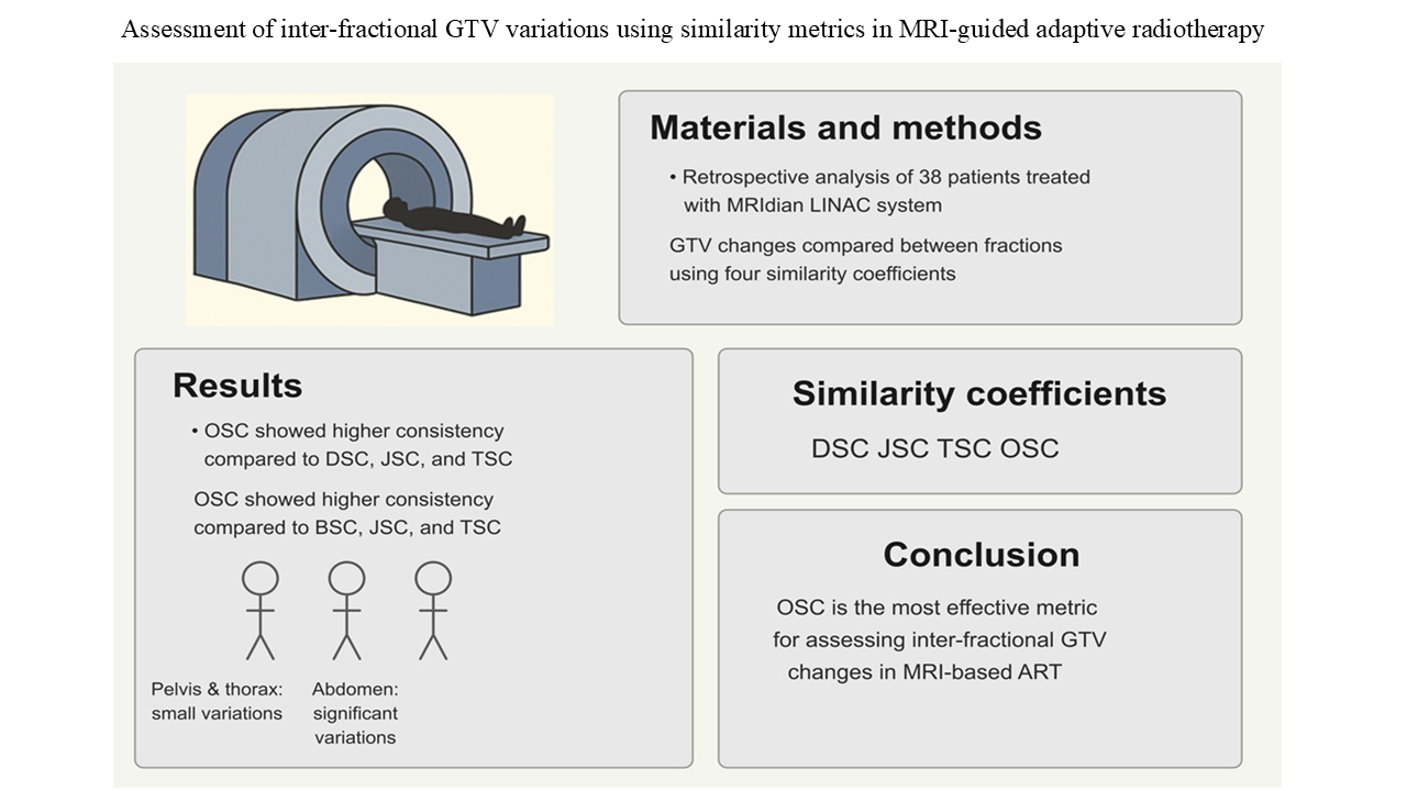
Tumor geometry can change during radiotherapy, and interfractional anatomical variations may compromise target coverage and dose conformity if not properly addressed. Magnetic resonance–guided adaptive radiotherapy (MRgART) enables ongoing visualization of tumor morphology and provides a framework for individualized treatment adaptation. This study quantitatively evaluated interfractional gross tumor volume (GTV) changes in patients treated with a ViewRay MRIdian LINAC system and investigated which similarity metric most reliably characterizes these variations. Retrospective MR images from 37 patients were analyzed and grouped by anatomical region into the pelvis, abdomen, and thorax. Baseline GTV (GTV₀) was compared with GTVs from five consecutive fractions (GTV₁–GTV₅). Geometric agreement was assessed using four similarity metrics: Dice similarity coefficient (DSC), Jaccard similarity coefficient (JSC), Tanimoto similarity coefficient (TSC), and Ochiai similarity coefficient (OSC). Interfractional stability was evaluated using mean similarity values and standard deviations across fractions. The abdominal region exhibited the greatest interfractional variability, with marked volume changes observed particularly in pancreatic cancer patients. Pelvic and thoracic tumors demonstrated relatively greater geometric stability, with lung tumors showing comparatively consistent agreement across fractions. Across all anatomical regions, OSC showed the highest mean similarity values and the lowest variability, indicating greater robustness than the other metrics. Statistical analysis confirmed that OSC performed significantly better than DSC, JSC, and TSC in the pelvis and abdomen (p<0.05), while showing comparable behavior to DSC in the thorax. These findings indicate that OSC is a reliable metric for monitoring interfractional tumor geometry in MR-guided adaptive radiotherapy and may support more precise and efficient adaptive treatment strategies.
- Van Timmeren JE, Chamberlain M, Bogowicz M, et al. MR-guided adaptive radiotherapy for head and neck cancer: Prospective evaluation of migration and anatomical changes of the major salivary glands. Cancers (Basel). 2021;13(21):5404. doi: 10.3390/cancers13215404
- Hirotaki K, Moriya S, Tachibana H, Sakae T. Detection of anatomical changes using two-dimensional x-ray images for head and neck adaptive radiotherapy. Med Phys. 2022;49(5):3288-3297. doi: 10.1002/mp.15587
- Brock KK. Adaptive radiotherapy: Moving into the future. Semin Radiat Oncol. 2019;29(3):181-184. doi: 10.1016/j.semradonc.2019.02.011
- Green OL, Henke LE, Hugo GD. Practical clinical workflows for online and offline adaptive radiation therapy. Semin Radiat Oncol. 2019;29(3):219-227. doi: 10.1016/j.semradonc.2019.02.004
- Sonke JJ, Aznar M, Rasch C. Adaptive radiotherapy for anatomical changes. Semin Radiat Oncol. 2019;29(3):245-257. doi: 10.1016/j.semradonc.2019.02.007
- Zhao JZ, Zheng H, Li LY, Zhang LY, Zhao Y, Jiang N. Predictors for weight loss in head and neck cancer patients undergoing radiotherapy: A systematic review. Cancer Nurs. 2015;38(6):245-257. doi: 10.1097/ncc.0000000000000231
- Ding S, Liu B, Zheng S, et al. An exploratory analysis of MR-guided fractionated stereotactic radiotherapy in patients with brain metastases. Clin Transl Radiat Oncol. 2023;40:100602. doi: 10.1016/j.ctro.2023.100602
- Mannerberg A, Persson E, Jonsson J, et al. Dosimetric effects of adaptive prostate cancer radiotherapy in an MR-linac workflow. Radiat Oncol. 2020;15(1):168. doi: 10.1186/s13014-020-01604-5
- Bhide SA, Davies M, Burke K, et al. Weekly volume and dosimetric changes during chemoradiotherapy with intensity-modulated radiation therapy for head and neck cancer: A prospective observational study. Int J Radiat Oncol Biol Phys. 2010;76(5):1360. doi: 10.1016/j.ijrobp.2009.04.005
- Ramsey CR, Langen KM, Kupelian PA, et al. A technique for adaptive image-guided helical tomotherapy for lung cancer. Int J Radiat Oncol Biol Phys. 2006;64(4):12. doi: 10.1016/j.ijrobp.2005.11.012
- McDonald BA, Zachiu C, Christodouleas J, et al. Dose accumulation for MR-guided adaptive radiotherapy: From practical considerations to state-of-the-art clinical implementation. Front Oncol. 2023;12:1086258. doi: 10.3389/fonc.2022.1086258
- Fischer-Valuck BW, Henke L, Green O, et al. Two-and-a-half-year clinical experience with the world’s first magnetic resonance image guided radiation therapy system. Adv Radiat Oncol. 2017;2(3):485-493. doi: 10.1016/j.adro.2017.05.006
- Kishan AU, Lee P. MRI-guided radiotherapy: Opening our eyes to the future. Integr Cancer Sci Ther. 2016;3(2):420-427. doi: 10.15761/icst.1000181
- Noel CE, Parikh PJ, Spencer CR, et al. Comparison of onboard low-field magnetic resonance imaging versus onboard computed tomography for anatomy visualization in radiotherapy. Acta Oncol (Madr). 2015;54(9):1474-1482. doi: 10.3109/0284186X.2015.1062541
- Pollard JM, Wen Z, Sadagopan R, Wang J, Ibbott GS. The future of image-guided radiotherapy will be MR guided. Br J Radiol. 2017;90(1073):20160667. doi: 10.1259/bjr.20160667
- Klüter S, Katayama S, Spindeldreier CK, et al. First prospective clinical evaluation of feasibility and patient acceptance of magnetic resonance-guided radiotherapy in Germany. Strahlentherapie Onkol. 2020;196(8):691-698. doi: 10.1007/s00066-020-01578-z
- Redler G, Stevens T, Cammin J, et al. Dosimetric feasibility of utilizing the ViewRay magnetic resonance guided linac system for image-guided spine stereotactic body radiation therapy. Cureus. 2019;11:e6364. doi: 10.7759/cureus.6364
- Gani C, Boldrini L, Valentini V. Online MR guided radiotherapy for rectal cancer. New opportunities. Clin Transl Radiat Oncol. 2019;18:66-67. doi: 10.1016/j.ctro.2019.04.005
- Henke L, Kashani R, Robinson C, et al. Phase I trial of stereotactic MR-guided online adaptive radiation therapy (SMART) for the treatment of oligometastatic or unresectable primary malignancies of the abdomen. Radiother Oncol. 2018;126(3):519-526. doi: 10.1016/j.radonc.2017.11.032
- Murgić J, Gregov M, Mrčela I, et al. MRI-guided radiotherapy for prostate cancer: A new paradigm. Acta Clin Croat. 2022;61:65-70. doi: 10.20471/acc.2022.61.S3.9
- Klüter S. Technical design and concept of a 0.35 T MR-Linac. Clin Transl Radiat Oncol. 2019;18:98-101. doi: 10.1016/j.ctro.2019.04.007
- Wen N, Kim J, Doemer A, et al. Evaluation of a magnetic resonance guided linear accelerator for stereotactic radiosurgery treatment. Radiother Oncol. 2018;127(3):460-466. doi: 10.1016/j.radonc.2018.04.034
- Ericsson-Szecsenyi R, Zhang G, Redler G, et al. Robustness assessment of images from a 0.35T scanner of an integrated MRI-linac: Characterization of radiomics features in phantom and patient data. Technol Cancer Res Treat. 2022;21:15330338221099113. doi: 10.1177/15330338221099113
- Han Z, Sudhyadhom A, Hsu S, et al. Comparison of MR‐soft tissue based versus biliary stent based alignment for image guidance in pancreatic SBRT. J Appl Clin Med Phys. 2023;24:e13965. doi: 10.1002/acm2.13965
- Kim JI, Park JM, Choi CH, An HJ, Kim YJ, Kim JH. Retrospective study comparing MR-guided radiation therapy (MRgRT) setup strategies for prostate treatment: Repositioning vs. replanning. Radiat Oncol. 2019;14(1):139. doi: 10.1186/s13014-019-1349-2
- Nousiainen K, Santurio GV, Lundahl N, Cronholm R, Siversson C, Edmund JM. Evaluation of MRI-only based online adaptive radiotherapy of abdominal region on MR-linac. J Appl Clin Med Phys. 2023;24(3):e13838. doi: 10.1002/acm2.13838
- Thorwarth D, Low DA. Technical challenges of real-time adaptive MR-guided radiotherapy. Front Oncol. 2021;11:634507. doi: 10.3389/fonc.2021.634507
- Boldrini L, Romano A, Chiloiro G, et al. Magnetic resonance guided SBRT reirradiation in locally recurrent prostate cancer: A multicentric retrospective analysis. Radiat Oncol. 2023;18(1):84. doi: 10.1186/s13014-023-02271-y
- Park JM, Wu HG, Kim HJ, Choi CH, Kim JI. Comparison of treatment plans between IMRT with MR-linac and VMAT for lung SABR. Radiat Oncol. 2019;14(1):105. doi: 10.1186/s13014-019-1314-0
- Maziero D, Straza MW, Ford JC, et al. MR-guided radiotherapy for brain and spine tumors. Front Oncol. 2021;11:626100. doi: 10.3389/fonc.2021.626100
- Sahin B, Zoto Mustafayev T, Gungor G, et al. First 500 fractions delivered with a magnetic resonance-guided radiotherapy system: Initial experience. Cureus. 2019;11:e6457. doi: 10.7759/cureus.6457
- Anderson P, Dogan N, Ford JC, et al. Repeatability, reproducibility, and the effects of radiotherapy on radiomic features of lowfield MR-LINAC images of the prostate. Front Oncol. 2024;14:1408752. doi: 10.3389/fonc.2024.1408752
- Turkkan G, Bilici N, Sertel H, et al. Clinical utility of a 1.5 T magnetic resonance imaging-guided linear accelerator during conventionally fractionated and hypofractionated prostate cancer radiotherapy. Front Oncol. 2022;12:909402. doi: 10.3389/fonc.2022.909402
- Nierer L, Eze C, Da Silva Mendes V, et al. Dosimetric benefit of MR-guided online adaptive radiotherapy in different tumor entities: Liver, lung, abdominal lymph nodes, pancreas and prostate. Radiat Oncol. 2022;17(1):53. doi: 10.1186/s13014-022-02021-6
- Rachi T, Ariji T, Takahashi S. Development of machine-learning prediction programs for delivering adaptive radiation therapy with tumor geometry and body shape changes in head and neck volumetric modulated arc therapy. Adv Radiat Oncol. 2023;8(4):101172. doi: 10.1016/j.adro.2023.101172
- Dice LR. Measures of the amount of ecologic association between species. Ecology. 1945;26(3):297-302. doi: 10.2307/1932409
- Jaccard P. The distribution of the flora in the alpine zone. New Phytol. 1912;11(2):37-50. doi: 10.1111/j.1469-8137.1912.tb05611.x
- Tanimoto TT. An Elementary Mathematical Theory of Classification and Prediction. In: Proceedings IBM Internal Report; 1958.
- Taha AA, Hanbury A. Metrics for evaluating 3D medical image segmentation: Analysis, selection, and tool. BMC Med Imaging. 2015;15(1):29. doi: 10.1186/s12880-015-0068-x
- Romesburg HC. Cluster Analysis for Researchers. United States: Lulu Press; 1984.