

State-of-the-art review: Commercial artificial intelligence applications in cardiopulmonary imaging—from coronary fractional flow reserve to pulmonary embolism triage

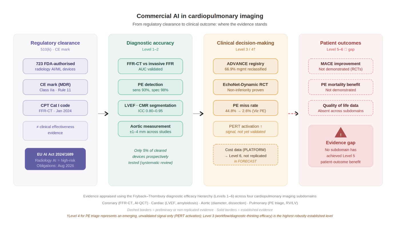
Commercial artificial intelligence (AI) applications in cardiopulmonary imaging have expanded rapidly—723 Food and Drug Administration (FDA)-authorized radiology AI/machine learning (ML) devices had been identified by June 2024, representing 76% of 950 total cleared AI/ML medical devices, yet evidence supporting clinical adoption remains heterogeneous. This state-of-the-art review synthesizes published evidence across four cardiopulmonary subdomains (coronary, cardiac, aortic, and pulmonary artery imaging), applying the Fryback–Thornbury diagnostic efficacy hierarchy as a unified framework for cross-domain comparison, and integrating regulatory, ethical, and equity considerations aligned to the European Union (EU) AI Act (Regulation 2024/1689) and FDA lifecycle management milestones through early 2026. Evidence maturity varies markedly. Computed tomography-derived fractional flow reserve is supported by randomized trials and prospective registry data addressing clinical decision-making (Level 4); preliminary, non-replicated cost data are also available (Level 6), but no trial has demonstrated improvement in major adverse cardiac events (Level 5) as a primary endpoint. Cardiac AI achieves high reproducibility for automated quantification (intraclass correlation coefficient 0.80–0.95) but remains predominantly at Levels 1–2, with a single non-inferiority randomized trial. Aortic AI evidence is almost entirely retrospective (Level 2). Pulmonary embolism triage systems have demonstrated prospective workflow improvements, including reductions in time-to-diagnosis and radiologist miss rates, without demonstrated mortality benefit. Across all domains, only 5% of FDA-cleared devices underwent prospective testing, and only four of 213 (2%) European regulatory-approved radiology AI devices were clearly labeled for pediatric use. Algorithmic bias, explainability limitations, and immature post-market surveillance frameworks represent consistent cross-domain concerns. Realizing patient benefit requires prospective outcome-driven evaluation, representative training datasets, and rigorous post-market monitoring, particularly as EU AI Act high-risk obligations apply from August 2026.
- Stark BA, DeCleene NK, Desai EC, et al. Global, regional, and national burden of cardiovascular diseases and risk factors in 204 countries and territories, 1990-2023. J Am Coll Cardiol. 2025;86(22):2167-2243. doi: 10.1016/j.jacc.2025.08.015
- Wendelboe A, Weitz JI. Global health burden of venous thromboembolism. Arterioscler Thromb Vasc Biol. 2024;44(5):1007-1011. doi: 10.1161/ATVBAHA.124.320151
- Rao SV, O’Donoghue ML, Ruel M, et al. 2025 ACC/AHA/ACEP/NAEMSP/SCAI guideline for the management of patients with acute coronary syndromes: A report of the American college of cardiology/american heart association joint committee on clinical practice guidelines. Circulation. 2025;151(13):e37-e200. doi: 10.1161/CIR.0000000000001309
- Hollenberg SM, Stevenson LW, Ahmad T, et al. 2024 ACC expert consensus decision pathway on clinical assessment, management, and trajectory of patients hospitalized with heart failure focused update. J Am Coll Cardiol. 2024;84(13):1241-1267. doi: 10.1016/j.jacc.2024.06.002
- Bossone E, Eagle KA. Epidemiology and management of aortic disease: Aortic aneurysms and acute aortic syndromes. Nat Rev Cardiol. 2021;18(5):331-348. doi: 10.1038/s41569-020-00472-6
- Writing Committee Members, Creager MA, Barnes GD, et al. 2026 AHA/ACC/ACCP/ACEP/CHEST/SCAI/SHM/SIR/SVM/SVN guideline for the evaluation and management of acute pulmonary embolism in adults: A report of the American college of cardiology/american heart association joint committee on clinical practice guidelines. Circulation. 2026;153(12):e364-e553. doi: 10.1161/CIR.0000000000001415
- Topol EJ. High-performance medicine: The convergence of human and artificial intelligence. Nat Med. 2019;25(1):44-56. doi: 10.1038/s41591-018-0300-7
- Van Leeuwen KG, Schalekamp S, Rutten MJCM, Van Ginneken B, De Rooij M. Artificial intelligence in radiology: 100 commercially available products and their scientific evidence. Eur Radiol. 2021;31(6):3797-3804. doi: 10.1007/s00330-021-07892-z
- Sivakumar R, Lue B, Kundu S. FDA approval of artificial intelligence and machine learning devices in radiology: A systematic review. JAMA Netw Open. 2025;8(11):e2542338. doi: 10.1001/jamanetworkopen.2025.42338
- Aldhafeeri FM. Governing artificial intelligence in radiology: A systematic review of ethical, legal, and regulatory frameworks. Diagnostics. 2025;15(18):2300. doi: 10.3390/diagnostics15182300
- Hill DLG. AI in imaging: The regulatory landscape. Br J Radiol. 2024;97(1155):483-491. doi: 10.1093/bjr/tqae002
- Fryback DG, Thornbury JR. The efficacy of diagnostic imaging. Med Decis Making. 1991;11(2):88-94. doi: 10.1177/0272989X9101100203
- Van Leeuwen KG, Hedderich DM, Harvey H, Schalekamp S. How AI should be used in radiology: Assessing ambiguity and completeness of intended use statements of commercial AI products. Insights Imaging. 2024;15(1):51. doi: 10.1186/s13244-024-01616-9
- Boverhof BJ, Redekop WK, Bos D, et al. Radiology AI deployment and assessment rubric (RADAR) to bring value-based AI into radiological practice. Insights Imaging. 2024;15(1):34. doi: 10.1186/s13244-023-01599-z
- Douglas PS, De Bruyne B, Pontone G, et al. 1-year outcomes of FFRCT-guided care in patients with suspected coronary disease. J Am Coll Cardiol. 2016;68(5):435-445. doi: 10.1016/j.jacc.2016.05.057
- Fairbairn TA, Nieman K, Akasaka T, et al. Real-world clinical utility and impact on clinical decision-making of coronary computed tomography angiography-derived fractional flow reserve: Lessons from the ADVANCE registry. Eur Heart J. 2018;39(41):3701-3711. doi: 10.1093/eurheartj/ehy530
- Patel MR, Nørgaard BL, Fairbairn TA, et al. 1-year impact on medical practice and clinical outcomes of FFRCT. JACC Cardiovasc Imaging. 2020;13(1):97-105. doi: 10.1016/j.jcmg.2019.03.003
- Curzen N, Nicholas Z, Stuart B, et al. Fractional flow reserve derived from computed tomography coronary angiography in the assessment and management of stable chest pain: The FORECAST randomized trial. Eur Heart J. 2021;42(37):3844-3852. doi: 10.1093/eurheartj/ehab444
- Hlatky MA, Wilding S, Stuart B, et al. Randomized comparison of chest pain evaluation with FFRCT or standard care: Factors determining US costs. J Cardiovasc Comput Tomogr. 2023;17(1):52-59. doi: 10.1016/j.jcct.2022.09.005
- Yang J, Shan D, Wang X, et al. On-site computed tomography–derived fractional flow reserve to guide management of patients with stable coronary artery disease: The TARGET randomized trial. Circulation. 2023;147(18):1369-1381. doi: 10.1161/CIRCULATIONAHA.123.063996
- Min JK, Leipsic J, Pencina MJ, et al. Diagnostic accuracy of fractional flow reserve from anatomic CT angiography. JAMA. 2012;308(12):1237-1245. doi: 10.1001/2012.jama.11274
- Xu PP, Li JH, Zhou F, et al. The influence of image quality on diagnostic performance of a machine learning–based fractional flow reserve derived from coronary CT angiography. Eur Radiol. 2020;30(5):2525-2534. doi: 10.1007/s00330-019-06571-4
- Rodriguez-Lozano PF, Waheed A, Evangelou S, et al. CT derived fractional flow reserve: Part 2 – critical appraisal of the literature. J Cardiovasc Comput Tomogr. 2025;19(4):397-408. doi: 10.1016/j.jcct.2025.05.241
- Mittal TK, Hothi SS, Venugopal V, et al. The use and efficacy of FFR-CT. JACC Cardiovasc Imaging. 2023;16(8):1056-1065. doi: 10.1016/j.jcmg.2023.02.005
- Nurmohamed NS, Bom MJ, Jukema RA, et al. AI-guided quantitative plaque staging predicts long-term cardiovascular outcomes in patients at risk for atherosclerotic CVD. JACC Cardiovasc Imaging. 2024;17(3):269-280. doi: 10.1016/j.jcmg.2023.05.020
- Williams MC, Kwiecinski J, Doris M, et al. Low-attenuation noncalcified plaque on coronary computed tomography angiography predicts myocardial infarction: Results from the multicenter SCOT-HEART trial (Scottish computed tomography of the HEART). Circulation. 2020;141(18):1452-1462. doi: 10.1161/CIRCULATIONAHA.119.044720
- Ruijsink B, Puyol-Antón E, Oksuz I, et al. Fully automated, quality-controlled cardiac analysis from CMR. JACC Cardiovasc Imaging. 2020;13(3):684-695. doi: 10.1016/j.jcmg.2019.05.030
- Backhaus SJ, Schuster A, Lange T, et al. Impact of fully automated assessment on interstudy reproducibility of biventricular volumes and function in cardiac magnetic resonance imaging. Sci Rep. 2021;11(1):11648. doi: 10.1038/s41598-021-90702-9
- Chadalavada S, Rauseo E, Salih A, et al. Quality control of cardiac magnetic resonance imaging segmentation, feature tracking, aortic flow, and native T1 analysis using automated batch processing in the UK Biobank study. Eur Heart J Imaging Methods Pract. 2024;2(3):qyae094. doi: 10.1093/ehjimp/qyae094
- Ouyang D, He B, Ghorbani A, et al. Video-based AI for beat-to-beat assessment of cardiac function. Nature. 2020;580(7802):252-256. doi: 10.1038/s41586-020-2145-8
- He B, Kwan AC, Cho JH, et al. Blinded, randomized trial of sonographer versus AI cardiac function assessment. Nature. 2023;616(7957):520-524. doi: 10.1038/s41586-023-05947-3
- Kagiyama N, Abe Y, Kusunose K, et al. Multicenter validation study for automated left ventricular ejection fraction assessment using a handheld ultrasound with artificial intelligence. Sci Rep. 2024;14(1):15359. doi: 10.1038/s41598-024-65557-5
- Sveric KM, Ulbrich S, Dindane Z, et al. Improved assessment of left ventricular ejection fraction using artificial intelligence in echocardiography: A comparative analysis with cardiac magnetic resonance imaging. Int J Cardiol. 2024;394:131383. doi: 10.1016/j.ijcard.2023.131383
- Tromp J, Bauer D, Claggett BL, et al. A formal validation of a deep learning-based automated workflow for the interpretation of the echocardiogram. Nat Commun. 2022;13(1):6776. doi: 10.1038/s41467-022-34245-1
- Goto S, Mahara K, Beussink-Nelson L, et al. Artificial intelligence-enabled fully automated detection of cardiac amyloidosis using electrocardiograms and echocardiograms. Nat Commun. 2021;12(1):2726. doi: 10.1038/s41467-021-22877-8
- Ioannou A, Khouri MG, Kitai T, et al. Diagnosis of cardiac amyloidosis on echocardiography using artificial intelligence. Circ Cardiovasc Imaging. 2026;19(2):e018991. doi: 10.1161/CIRCIMAGING.125.018991
- Backhaus SJ, Aldehayat H, Kowallick JT, et al. Artificial intelligence fully automated myocardial strain quantification for risk stratification following acute myocardial infarction. Sci Rep. 2022;12(1):12220. doi: 10.1038/s41598-022-16228-w
- Pradella M, Weikert T, Sperl JI, et al. Fully automated guideline-compliant diameter measurements of the thoracic aorta on ECG-gated CT angiography using deep learning. Quant Imaging Med Surg. 2021;11(10):4245-4257. doi: 10.21037/qims-21-142
- Adam C, Fabre D, Mougin J, et al. Pre-surgical and post-surgical aortic aneurysm maximum diameter measurement: Full automation by artificial intelligence. Eur J Vasc Endovasc Surg. 2021;62(6):869-877. doi: 10.1016/j.ejvs.2021.07.013
- Sieren MM, Widmann C, Weiss N, et al. Automated segmentation and quantification of the healthy and diseased aorta in CT angiographies using a dedicated deep learning approach. Eur Radiol. 2022;32(1):690-701. doi: 10.1007/s00330-021-08130-2
- Norajitra T, Baumgartner MA, Cusumano LR, et al. Automated AI detection of thoracic aortic dissection on CT imaging. Eur Radiol Exp. 2025;9(1):102. doi: 10.1186/s41747-025-00640-8
- Hata A, Yanagawa M, Yamagata K, et al. Deep learning algorithm for detection of aortic dissection on non-contrast-enhanced CT. Eur Radiol. 2021;31(2):1151-1159. doi: 10.1007/s00330-020-07213-w
- Huang LT, Tsai YS, Liou CF, et al. Automated Stanford classification of aortic dissection using a 2-step hierarchical neural network at computed tomography angiography. Eur Radiol. 2022;32(4):2277-2285. doi: 10.1007/s00330-021-08370-2
- Liu HH, Chang CB, Chen YS, et al. Automated detection and differentiation of Stanford type A and type B aortic dissections in CTA scans using deep learning. Diagnostics. 2024;15(1):12. doi: 10.3390/diagnostics15010012
- Lindquist Liljeqvist M, Bogdanovic M, Siika A, Gasser TC, Hultgren R, Roy J. Geometric and biomechanical modeling aided by machine learning improves the prediction of growth and rupture of small abdominal aortic aneurysms. Sci Rep. 2021;11(1):18040. doi: 10.1038/s41598-021-96512-3
- Soffer S, Klang E, Shimon O, et al. Deep learning for pulmonary embolism detection on computed tomography pulmonary angiogram: A systematic review and meta-analysis. Sci Rep. 2021;11(1):15814. doi: 10.1038/s41598-021-95249-3
- Lanza E, Ammirabile A, Francone M. Meta-analysis of AI-based pulmonary embolism detection: How reliable are deep learning models? Comput Biol Med. 2025;193:110402. doi: 10.1016/j.compbiomed.2025.110402
- DePry EX, Parmar V, Rajendran S, et al. Performance of FDA-approved AI algorithms in detecting acute pulmonary embolism on computed tomographic pulmonary angiography (CTPA): A meta-analysis of real-world retrospective studies. Cureus. 2025;17(6):e94391. doi: 10.7759/cureus.94391
- González G, Jiménez-Carretero D, Rodríguez-López S, et al. Automated axial right ventricle to left ventricle diameter ratio computation in computed tomography pulmonary angiography. PLOS ONE. 2015;10(5):e0127797. doi: 10.1371/journal.pone.0127797
- Foley RW, Glenn-Cox S, Rossdale J, et al. Automated calculation of the right ventricle to left ventricle ratio on CT for the risk stratification of patients with acute pulmonary embolism. Eur Radiol. 2021;31(8):6013-6020. doi: 10.1007/s00330-020-07605-y
- Rothenberg SA, Savage CH, Abou Elkassem A, et al. Prospective evaluation of AI triage of pulmonary emboli on CT pulmonary angiograms. Radiology. 2023;309(1):e230702. doi: 10.1148/radiol.230702
- Topff L, Ranschaert ER, Bartels-Rutten A, et al. Artificial intelligence tool for detection and worklist prioritization reduces time to diagnosis of incidental pulmonary embolism at CT. Radiol Cardiothorac Imaging. 2023;5(2):e220163. doi: 10.1148/ryct.220163
- Momin E, Cook T, Gershon G, Barr J, De Cecco CN, Van Assen M. Systematic review on the impact of deep learning-driven worklist triage on radiology workflow and clinical outcomes. Eur Radiol. 2025;35(11):6879-6893. doi: 10.1007/s00330-025-11674-2
- Khosla A, Singh I, Pollak J, Mojibian H. Impact of an artificial intelligence-guided pulmonary embolism response team (AIPERT) on patient transfers, diagnosis, and management: A healthcare system experience. Clin Pract. 2025;15(11):207. doi: 10.3390/clinpract15110207
- Packhäuser K, Gündel S, Münster N, Syben C, Christlein V, Maier A. Deep learning-based patient re-identification is able to exploit the biometric nature of medical chest X-ray data. Sci Rep. 2022;12(1):14851. doi: 10.1038/s41598-022-19045-3
- De Almeida JG, Alberich LC, Tsakou G, et al. Foundation models for radiology—The position of the AI for health imaging (AI4HI) network. Insights Imaging. 2025;16(1):168. doi: 10.1186/s13244-025-02056-9
- Suleman MU, Mursaleen M, Khalil U, et al. Assessing the generalizability of artificial intelligence in radiology: A systematic review of performance across different clinical settings. Ann Med Surg. 2025;87(12):8803-8811. doi: 10.1097/MS9.0000000000004166
- Tripathi S, Gabriel K, Dheer S, et al. Understanding biases and disparities in radiology AI datasets: A review. J Am Coll Radiol. 2023;20(9):836-841. doi: 10.1016/j.jacr.2023.06.015
- Seyyed-Kalantari L, Zhang H, McDermott MBA, Chen IY, Ghassemi M. Underdiagnosis bias of artificial intelligence algorithms applied to chest radiographs in under-served patient populations. Nat Med. 2021;27(12):2176-2182. doi: 10.1038/s41591-021-01595-0
- Gichoya JW, Banerjee I, Bhimireddy AR, et al. AI recognition of patient race in medical imaging: A modelling study. Lancet Digit Health. 2022;4(6):e406-e414. doi: 10.1016/S2589-7500(22)00063-2
- Banerjee I, Bhattacharjee K, Burns JL, et al. “Shortcuts” causing bias in radiology artificial intelligence: Causes, evaluation, and mitigation. J Am Coll Radiol. 2023;20(9):842-851. doi: 10.1016/j.jacr.2023.06.025
- Kelly BS, Lee J, Antram E, Arthurs O, Shelmerdine SC. Safe for kids? AI medical devices in radiology overlook paediatric suitability. Eur Radiol. 2025;36(3):2083-2090. doi: 10.1007/s00330-025-11975-6
- Shelmerdine SC, Naidoo J, Kelly BS, et al. Artificial intelligence implementation in pediatric radiology for patient safety: A multisociety statement from the ACR, ESPR, SPR, SLARP, AOSPR, SPIN. J Am Coll Radiol. 2026;23(1):89-101. doi: 10.1016/j.jacr.2025.08.019
- Busch F, Kather JN, Johner C, et al. Navigating the European Union Artificial Intelligence Act for healthcare. NPJ Digit Med. 2024;7(1):210. doi: 10.1038/s41746-024-01213-6
- Cuocolo R, Bernardini D, Pinto Dos Santos D, et al. AI medical device post-market surveillance regulations: Consensus recommendations by the European Society of Radiology. Insights Imaging. 2025;16(1):275. doi: 10.1186/s13244-025-02146-8
- Zhang J, Chao H, Dasegowda G, Wang G, Kalra MK, Yan P. Revisiting the trustworthiness of saliency methods in radiology AI. Radiol Artif Intell. 2024;6(1):e220221. doi: 10.1148/ryai.220221
- Reyes M, Meier R, Pereira S, Silva CA, Dahlweid FM, von Tengg-Kobligk H, Summers RM, Wiest R. On the interpretability of artificial intelligence in radiology: Challenges and opportunities. Radiol Artif Intell. 2020;2(3):e190043. doi: 10.1148/ryai.2020190043
- Hlatky MA, De Bruyne B, Pontone G, et al. Quality-of-life and economic outcomes of assessing fractional flow reserve with computed tomography angiography: PLATFORM. J Am Coll Cardiol. 2015;66(21):2315-2323. doi: 10.1016/j.jacc.2015.09.051
- Acosta JN, Falcone GJ, Rajpurkar P, Topol EJ. Multimodal biomedical AI. Nat Med. 2022;28(9):1773-1784. doi: 10.1038/s41591-022-01981-2
- Moor M, Banerjee O, Abad ZSH, et al. Foundation models for generalist medical artificial intelligence. Nature. 2023;616(7956):259-265. doi: 10.1038/s41586-023-05881-4
- Zhang S, Xu Y, Usuyama N, et al. A multimodal biomedical foundation model trained from fifteen million image–text pairs. NEJM AI. 2025;2(1):AIoa2400640. doi: 10.1056/AIoa2400640
- Paschali M, Chen Z, Blankemeier L, et al. Foundation models in radiology: What, how, why, and why not. Radiology. 2025;314(2):e240597. doi: 10.1148/radiol.240597