

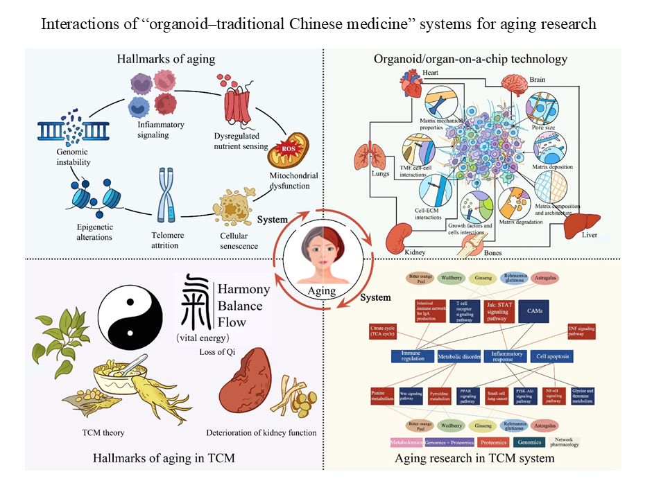
Organoids in aging research: Bridging cutting-edge biotechnology with the wisdom of traditional Chinese medicine

The growth of the global older population has necessitated the development of advanced models to investigate biological processes in aging and to explore viable anti-aging strategies. This review focuses on two objectives: First, to explore organoid technology as a paradigm for observing and intervening in human aging, and second, to examine the possible overlaps and crossroads between organoid-based biomedical paradigms and traditional Chinese medicine (TCM). Recent advancements in this field have increased the physiological relevance of organoids, incorporating both a dynamic microenvironment and multi-organ interactions. Such accomplishments have enhanced the accuracy of models of neurodegeneration, cardiovascular deterioration, metabolic diseases, and age-related conditions. In addition to pharmacological and genetic treatments, organoid models offer new opportunities to study well-known anti-aging techniques, such as those that rely on the TCM system, which aims to balance the body using natural substances with antioxidant and repair properties. Together, organoid-based aging research and mechanistic evaluation of TCM-derived compounds may help identify interventions that support healthy aging.
- Wang XY, Jia QN, Li J, Zheng HY. Organoids as tools for investigating skin aging: Mechanisms, applications, and insights. Biomolecules. 2024;14(11):1436. doi: 10.3390/biom14111436
- Mou X, Zhang A, He TJ, et al. Organoid models for Chinese herbal medicine studies. Acta Materia Med. 2023;2(1):64-71. doi: 10.15212/AMM-2022-0047
- Sun X, Sun F, Zhang Y, Qu J, Zhang W, Liu GH. A narrative review of organoids for investigating organ aging: Opportunities and challenges. J BioX Res. 2023;6(1):3-14. doi: 10.1097/JBR.0000000000000139
- Yang J, Jiang Y, Li M, et al. Organoid, organ-on-a-chip and traditional Chinese medicine. Chin Med. 2025;20(1):22. doi: 10.1186/s13020-025-01071-8
- Zhang Y, Chen H, Huang C. Optimizing health-span: Advances in stem cell medicine and longevity research. Med Rev (2021). 2023;3(4):351-355. doi: 10.1515/mr-2023-0040
- Lu M, Han Y, Zhang Y, et al. Investigating aging‐related endometrial dysfunction using endometrial organoids. Cell Prolif. 2025;58(4):e13780. doi: 10.1111/cpr.13780
- Liu Y, Li Y, Yin Y, Yu L, Ma H. Micro/nanoplastic-driven cardiovascular senescence and multi-target intervention by traditional Chinese medicine. Ageing Res Rev. 2025;111:102841. doi: 10.1016/j.arr.2025.102841
- Xue J, Gao S, You Z, et al. In vitro technology and ADMET research in traditional Chinese medicine. Front Pharmacol. 2025;16:1605330. doi: 10.3389/fphar.2025.1605330
- Cong R, Lu C, Li X, Xu Z, Wang Y, Sun S. Tumor organoids in cancer medicine: From model systems to natural compound screening. Pharm Biol. 2025;63(1):89-109. doi: 10.1080/13880209.2025.2458149
- Torrens-Mas M, Perelló-Reus C, Navas-Enamorado C, et al. Organoids: An emerging tool to study aging signature across human tissues. Modeling aging with patient-derived organoids. Int J Mol Sci. 2021;22(19):10547. doi: 10.3390/ijms221910547
- Hu JL, Todhunter ME, LaBarge MA, Gartner ZJ. Opportunities for organoids as new models of aging. J Cell Biol. 2018;217(1):39-50. doi: 10.1083/jcb.201709054
- Chan K, Shaw D, Simmonds MS, et al. Good practice in reviewing and publishing studies on herbal medicine, with special emphasis on traditional Chinese medicine and Chinese materia medica. J Ethnopharmacol. 2012;140(3):469-475. doi: 10.1016/j.jep.2012.01.038
- World Health Organization. Air Quality Guidelines for Europe. Geneva: World Health Organization; 2020.
- Lancaster, MA, Knoblich JA. Generation of cerebral organoids from human pluripotent stem cells. Nat Protoc. 2014;9(10):2329-2340. doi: 10.1038/nprot.2014.158
- López-Otín C, Blasco MA, Partridge L, Serrano M, Kroemer G. The hallmarks of aging. Cell. 2013;153(6):1194-217. doi: 10.1016/j.cell.2022.11.001
- Shi Y, Inoue H, Wu JC, Yamanaka S. Induced pluripotent stem cell technology: A decade of progress. Nat Rev Drug Discov. 2017;16(2):115-130. doi: 10.1038/nrd.2016.245
- Huch M, Koo BK. Modeling mouse and humandevelopment using organoid cultures. Development. 2015;142(18):3113-3125. doi: 10.1242/dev.118570
- Kim J, Koo BK, Knoblich JA. Human organoids: Model systems for human biology and medicine. Nat Rev Mol Cell Biol. 2020;21(10):571-584. doi: 10.1038/s41580-020-0259-3
- Raja WK, Mungenast AE, Lin YT, et al. Self-organizing 3D human neural tissue derived from induced pluripotent stem cells recapitulate Alzheimer’s disease phenotypes. PLoS One. 2016;11(9):e0161969. doi: 10.1371/journal.pone.0161969
- Seok J, Warren HS, Cuenca AG, et al. Genomic responses in mouse models poorly mimic human inflammatory diseases. Proc Natl Acad Sci. 2013;110(9):3507-3512. doi: 10.1073/pnas.1222878110
- Takasato M, Er PX, Chiu HS, et al. Kidney organoids from human iPS cells contain multiple lineages and model human nephrogenesis. Nature. 2015;526(7574):564-568. doi: 10.1038/nature15695
- Mills RJ, Titmarsh DM, Koenig X, et al. Functional screening in human cardiac organoids reveals a metabolic mechanism for cardiomyocyte cell cycle arrest. Proc Natl Acad Sci. 2017;114(40):E8372-E8381. doi: 10.1073/pnas.1707316114
- Clevers H. Modeling development and disease with organoids. Cell. 2016;165(7):1586-1597. doi: 10.1016/j.cell.2016.05.082
- Bhatia SN, Ingber DE. Microfluidic organs-on-chips. Nat Biotechnol. 2014;32(8):760-772. doi: 10.1038/nbt.2989
- Zhu Y, Tchkonia T, Pirtskhalava T, et al. The Achilles’ heel of senescent cells: From transcriptome to senolytic drugs. Aging Cell. 2015;14(4):644-658. doi: 10.1111/acel.12344
- Tartiere AG, Freije JM, López-Otín C. The hallmarks of aging as a conceptual framework for health and longevity research. Front Aging. 2024;5:1334261. doi: 10.3389/fragi.2024.1334261
- Li Y, Kilian KA. Bridging the gap: From 2D cell culture to 3D microengineered extracellular matrices. Adv Healthc Mater. 2015;4(18):2780-2796. doi: 10.1002/adhm.201500427
- Cacciamali A, Villa R, Dotti S. 3D cell cultures: Evolution of an ancient tool for new applications. Front Physiol. 2022;13:836480. doi: 10.3389/fphys.2022.836480
- Chen YW, Huang SX, De Carvalho ALRT, et al. A three-dimensional model of human lung development and disease from pluripotent stem cells. Nat Cell Biol. 2017;19(5):542-549. doi: 10.1038/ncb3510
- Guerville F, Barreto PDS, Ader I, et al. Revisiting the hallmarks of aging to identify markers of biological age. J Prevent Alzheimers Dis. 2020;7(1):56-64. doi: 10.14283/jpad.2019.50
- Di Micco R, Krizhanovsky V, Baker D, D’Adda di Fagagna F. Cellular senescence in ageing: From mechanisms to therapeutic opportunities. Nat Rev Mol Cell Biol. 2021;22(2):75-95. doi: 10.1038/s41580-020-00314-w
- Rossiello F, Jurk D, Passos JF, D’Adda di Fagagna F. Telomere dysfunction in ageing and age-related diseases. Nat Cell Biol. 2022;24(2):135-147. doi: 10.1038/s41556-022-00842-x
- Jun Y, Albarran E, Wilson DL, Ding J, Kool EAO. Fluorescence imaging of mitochondrial DNA base excision repair reveals dynamics of oxidative stress responses. Angew Chem Int Ed Engl. 2022;61(6):e202111829. doi: 10.1002/anie.202111829
- Vijg J, Montagna C. Genome instability and aging: Cause or effect? Transl Med Aging. 2017;1:5-11. doi: 10.1016/j.tma.2017.09.003
- Yu P, Liu B, Dong C, Chang Y. Induced pluripotent stem cells-based regenerative therapies in treating human aging-related functional decline and diseases. Cells. 2025;14(8):619. doi: 10.3390/cells14080619
- Lee S, Hong CI. Organoids as model systems to investigate circadian clock-related diseases and treatments. Front Genet. 2022;13:874288. doi: 10.3389/fgene.2022.874288
- Wang L, Li M, Yu B, et al. Recapitulating lipid accumulation and related metabolic dysregulation in human liver-derived organoids. J Mol Med. 2022;100(3):471-484. doi: 10.1007/s00109-021-02176-x
- Baker DJ, Childs BG, Durik M, et al. Naturally occurring p16Ink4a-positive cells shorten healthy lifespan. Nature. 2016;530(7589):184-189. doi: 10.1038/nature16932
- Yousefzadeh MJ, Zhu Y, McGowan SJ, et al. Fisetin is a senotherapeutic that extends health and lifespan. EBioMedicine. 2018;36:18-28. doi: 10.1016/j.ebiom.2018.09.015
- Gorgoulis V, Adams PD, Alimonti A, et al. Cellular senescence: Defining a path forward. Cell. 2019;179(4):813-827. doi: 10.1016/j.cell.2019.10.005
- Campisi J, Kapahi P, Lithgow GJ, Melov S, Newman JC, Verdin E. From discoveries in ageing research to therapeutics for healthy ageing. Nature. 2019;571(7764):183-192. doi: 10.1038/s41586-019-1365-2
- López-Otín C, Blasco MA, Partridge L, Serrano M, Kroemer G. Hallmarks of aging: An expanding universe. Cell. 2023;186(2):243-278. doi: 10.1016/j.cell.2022.11.001
- Artegiani B, Lyubimova A, Muraro M, van EJH, van Oudenaarden A, Clevers H. A single-cell RNA sequencing study reveals cellular and molecular dynamics of the hippocampal neurogenic niche. Cell Rep. 2017;21(11):3271-3284. doi: 10.1016/j.celrep.2017.11.050
- Horvath S, Raj K. DNA methylation-based biomarkers and the epigenetic clock theory of ageing. Nat Rev Genet. 2018;19(6):371-384. doi: 10.1038/s41576-018-0004-3
- Fajardo VM, Feng I, Chen BY, et al. GLUT1 overexpression enhances glucose metabolism and promotes neonatal heart regeneration. Sci Rep. 2021;11(1):8669. doi: 10.1038/s41598-021-88159-x
- Hu JL, Todhunter ME, LaBarge MA, Gartner ZJ. Opportunities for organoids as new models of aging. J Cell Biol. 2018;217:39-50. doi: 10.1083/jcb.201709054
- Bian Z, Zhang R, Zhang X, et al. Extraction, structure and bioactivities of polysaccharides from Rehmannia glutinosa: A review. J Ethnopharmacol. 2023;305:116132. doi: 10.1016/j.jep.2022.116132
- De Jaeger, C, Kruiskamp, S, Voronska, E, et al. A natural astragalus-based nutritional supplement lengthens telomeres in a middle-aged population: A randomized, double-blind, placebo-controlled study. Nutrients. 2024;16(17):2963. doi: 10.3390/nu16172963
- Li X, Zheng K, Chen H, Li W. Ginsenoside Re regulates oxidative stress through the PI3K/Akt/Nrf2 signaling pathway in mice with scopolamine-induced memory impairments. Curr Issues Mol Biol. 2024;46:11359-11374. doi: 10.3390/cimb46100677
- Cheng X, Su X, Chen X, et al. Biological ingredient analysis of traditional Chinese medicine preparation based on high-throughput sequencing: The story for Liuwei Dihuang Wan. Sci Rep. 2014;4(1):5147. doi: 10.1038/srep05147
- OuYang Y, Chen B, Yi J, et al. Study on the molecular mechanisms of Liuwei Dihuang decoction against aging-related cognitive impairment based on network pharmacology and experimental verification. Heliyon. 2024;10(11):e32526. doi: 10.1016/j.heliyon.2024.e32526
- Smits LM, Reinhardt L, Reinhardt P, et al. Modeling Parkinson’s disease in midbrain-like organoids. NPJ Parkinsons Dis. 2019;5(1):5. doi: 10.1038/s41531-019-0078-4
- Lu Z, Yuan Y, Han Q, Wang Y, Liang Q. Lab-on-a-chip: An advanced technology for the modernization of traditional Chinese medicine. Chin Med. 2024;19(1):80. doi: 10.1186/s13020-024-00956-4
- Wu L, Ai Y, Xie R, Xiong J, Wang Y, Liang Q. Organoids/organs-on-a-chip: New frontiers of intestinal pathophysiological models. Lab Chip. 2023;23(5):1192-1212. doi: 10.1039/d2lc00804a
- Qin W, He Y, Xiao J, et al. A successive laminar flow extraction for plant medicine preparation by microfluidic chip. Microfluidics Nanofluidics. 2019;23(4):61. doi: 10.1007/s10404-019-2228-8
- Shen Y, Chen B, Zuilhof H, van Beek TA. Microfluidic chip-based induced phase separation extraction as a fast and efficient miniaturized sample preparation method. Molecules. 2021;26(1):38. doi: 10.3390/molecules26010038
- Tetala KK, Swarts JW, Chen B, Janssen AE, van Beek TA. A three-phase microfluidic chip for rapid sample clean-up of alkaloids from plant extracts. Lab Chip. 2009;9(14):2085-2092. doi: 10.1039/b822106e
- Mu X, Liang Q, Hu P, Ren K, Wang Y, Luo G. Selectively modified microfluidic chip for solvent extraction of Radix Salvia miltiorrhiza using three-phase laminar flow to provide double liquid-liquid interface area. Microfluidics Nanofluidics. 2010;9(2):365-373. doi: 10.1007/s10404-009-0554-y
- Sun Y, Li Y, Zeng J, Lu Q, Li PC. Microchip electrophoretic separation and fluorescence detection of chelerythrine and sanguinarine in medicinal plants. Talanta. 2015;142:90-96. doi: 10.1016/j.talanta.2015.04.008
- Li OL, Tong YL, Chen ZG, Liu C, Zhao S, Mo JY. A glass/ PDMS hybrid microfluidic chip embedded with integrated electrodes for contactless conductivity detection. Chromatographia. 2008;68(11):1039-1044. doi: 10.1365/s10337-008-0808-y
- Wei Y, Liu C, Zhang Y, et al. All-Fiber SPR microfluidic chip for arctigenin detection. IEEE Sensors J. 2023;23(12):12838-12844. doi: 10.1109/JSEN.2023.3269032
- Li ZH, Ai N, Yu LX, Qian ZZ, Cheng YY. A multiple biomarker assay for quality assessment of botanical drugs using a versatile microfluidic chip. Sci Rep. 2017;7(1):12243. doi: 10.1038/s41598-017-12453-w
- Guo G, Wu X, Liu D, et al. A self-regulated microfluidic device with thermal bubble micropumps. Micromachines (Basel). 2022;13(10):1620. doi: 10.3390/mi13101620
- Fan JX, Bao YR, Meng XS, Wang S, Li TJ. Study on relationship between efficacy against lung cancer and different parts of Schizonepeta tenuifolia based on microfluidic chip technology. Zhongguo Zhong Yao Za Zhi. 2017;42(9):1717-1721. doi: 10.19540/j.cnki.cjcmm.20170224.004
- Fan JX, Wang S, Meng XS, Bao YR, Li TJ. Study of cancer cell apoptosis induced by Schizonepeta tenuifolia with microfluidic chip technology. Yao Xue Xue Bao. 2017;52(1):126-131. doi: 10.16438/j.0513-4870.2016-0466
- Han Q, Bing W, Di Y, et al. Kinsenoside screening with a microfluidic chip attenuates gouty arthritis through inactivating NF-κB signaling in macrophages and protecting endothelial cells. Cell Death Dis. 2016;7(9):e2350. doi: 10.1038/cddis.2016.255
- Gao Y, Peng H, Li L, et al. Screening of high-efficiency and low-toxicity antitumor active components in Macleaya cordata seeds based on the competitive effect of drugs on double targets by a new laminar flow chip. Analyst. 2021;146(15):4934-4944. doi: 10.1039/d1an00754h
- Niu Y, Bai J, Kamm RD, Wang Y, Wang C. Validating antimetastatic effects of natural products in an engineered microfluidic platform mimicking tumor microenvironment. Mol Pharm. 2014;11(7):2022-2029. doi: 10.1021/mp500054h
- Kwok HC, Lau PM, Wu SY, et al. Allergy testing and drug screening on an ITO-coated lab-on-a-disc. Micromachines (Basel). 2016;7(3):38. doi: 10.3390/mi7030038
- Liu Y, Wang M, Liu R, Qiu F. Label-free microfluidic device reveals single cell phagocytic activity and screens plant medicine rapidly. Lab Chip. 2023;23(3):553-559. doi: 10.1039/d2lc01021f
- Guo S, Lin X, Wang Y, Gong X. Fabrication of paper-based enzyme immobilized microarray by 3D-printing technique for screening alpha-glucosidase inhibitors in mulberry leaves and lotus leaves. Chin Med. 2019;14:13. doi: 10.1186/s13020-019-0236-y
- Li N, Men W, Zheng Y, Wang H, Meng X. Oroxin B induces apoptosis by down-regulating MicroRNA-221 resulting in the inactivation of the PTEN/PI3K/AKT pathway in liver cancer. Molecules. 2019;24(23):4384. doi: 10.3390/molecules24234384
- Li H, van den Driesche S, Bunge F, Yang B, Vellekoop MJ. Optimization of on-chip bacterial culture conditions using the Box-Behnken design response surface methodology for faster drug susceptibility screening. Talanta. 2019;194:627-633. doi: 10.1016/j.talanta.2018.10.048
- Shi YW, Cai Y, He XL, Hong ZY, Chai YF. Construction of a blood-brain barrier microfluidic chip model and evaluation of the permeability of active components in traditional Chinese medicine. Journal article. Acta Pharm Sin. 2022;57(3):802-808. doi: 10.16438/j.0513-4870.2021-1811
- Li Z, Li J, Sun M, et al. Analysis of metabolites and metabolism-mediated biological activity assessment of ginsenosides on microfluidic co-culture system. Front Pharmacol. 2023;14:1046722. doi: 10.3389/fphar.2023.1046722
- Zhang Y, Chen S, Fan F, et al. Neurotoxicity mechanism of aconitine in HT22 cells studied by microfluidic chip-mass spectrometry. J Pharm Anal. 2023;13(1):88-98. doi: 10.1016/j.jpha.2022.11.007
- Fan F, Xu N, Sun Y, et al. Uncovering the metabolic mechanism of salidroside alleviating microglial hypoxia inflammation based on microfluidic chip-mass spectrometry. J Proteome Res. 2022;21(4):921-929. doi: 10.1021/acs.jproteome.1c00647
- Yang, Z, Qin, W, Chen, D, et al. In vitro study of emodin-induced nephrotoxicity in human renal glomerular endothelial cells on a microfluidic chip. Biocell. 2023;47(1):125-131. doi: 10.32604/biocell.2023.022937
- Han CH, Ma JY, Zou W, et al. 3D microfluidic system for evaluating inhibitory effect of Chinese herbal medicine Oldenlandia diffusa on human malignant glioma invasion combined with network pharmacology analysis. Chin J Integr Med. 2023;29(1):52-60. doi: 10.1007/s11655-021-3726-1
- Wang H, Li T, Bao Y, Wang S, Meng X. A multifunctional integrated simultaneously online screening microfluidic biochip for the examination of “efficacy-toxicity” and compatibility of medicine. Chin Chem Lett. 2019;30(2):403-405. doi: 10.1016/j.cclet.2018.08.016
- Shi Y, He X, Wang H, et al. Construction of a novel blood brain barrier-glioma microfluidic chip model: Applications in the evaluation of permeability and anti-glioma activity of traditional Chinese medicine components. Talanta. 2023;253:123971. doi: 10.1016/j.talanta.2022.123971
- Xu T, Wu Z, Yao H, et al. Evaluation of aconitine cardiotoxicity with a heart-on-a-particle prepared by a microfluidic device. Chem Commun (Camb). 2024;60(37):4898-4901. doi: 10.1039/d4cc00396a
- Xu M, Bradley EW, Weivoda MM, et al. Transplanted senescent cells induce an osteoarthritis-like condition in mice. J Gerontol A Biol Sci Med Sci. 2017;72(6):780-785. doi: 10.1093/gerona/glw154
- Kulkarni AS, Gubbi S, Barzilai N. Benefits of metformin in attenuating the hallmarks of aging. Cell Metab. 2020;32(1):15-30. doi: 10.1016/j.cmet.2020.04.001
- Wilkinson JE, Burmeister L, Brooks SV, et al. Rapamycin slows aging in mice. Aging Cell. 2012;11(4):675-682. doi: 10.1111/j.1474-9726.2012.00832.x
- Yoshino J, Baur JA, Imai SI. NAD(+) intermediates: The biology and therapeutic potential of NMN and NR. Cell Metab. 2018;27(3):513-528. doi: 10.1016/j.cmet.2017.11.002
- Kennedy BK, Berger SL, Brunet A, et al. Geroscience: Linking aging to chronic disease. Cell. 2014;159(4):709-713. doi: 10.1016/j.cell.2014.10.039
- McCauley ME, Baloh RH. Inflammation in ALS/FTD pathogenesis. Acta Neuropathol. 2019;137(5):715-730. doi: 10.1007/s00401-018-1933-9
- Wyss-Coray T. Ageing, neurodegeneration and brain rejuvenation. Nature. 2016;539(7628):180-186. doi: 10.1038/nature20411
- Hou Y, Dan X, Babbar M, et al. Ageing as a risk factor for neurodegenerative disease. Nat Rev Neurol. 2019;15(10):565-581. doi: 10.1038/s41582-019-0244-7
- Katsimpardi L, Litterman NK, Schein PA, et al. Vascular and neurogenic rejuvenation of the aging mouse brain by young systemic factors. Science. 2014;344(6184):630-634. doi: 10.1126/science.1251141
- Xu M, Pirtskhalava T, Farr JN, et al. Senolytics improve physical function and increase lifespan in old age. Nat Med. 2018;24(8):1246-1256. doi: 10.1038/s41591-018-0092-9
- Baar MP, Brandt RM, Putavet DA, et al. Targeted apoptosis of senescent cells restores tissue homeostasis in response to chemotoxicity and aging. Cell. 2017;169(1):132-147.e16. doi: 10.1016/j.cell.2017.02.031
- Song P, An J, Zou MH. Immune clearance of senescent cells to combat ageing and chronic diseases. Cells. 2020;9(3):671. doi: 10.3390/cells9030671
- Childs BG, Baker DJ, Kirkland JL, Campisi J, van Deursen JM. Senescence and apoptosis: Dueling or complementary cell fates? EMBO Rep. 2014;15(11):1139-1153. doi: 10.15252/embr.201439245
- Tchkonia T, Kirkland JL. Aging, cell senescence, and chronic disease: Emerging therapeutic strategies. JAMA. 2018;320(13):1319-1320. doi: 10.1001/jama.2018.12440
- Khosla S, Farr JN, Tchkonia T, Kirkland JL. The role of cellular senescence in ageing and endocrine disease. Nat Rev Endocrinol. 2020;16(5):263-275. doi: 10.1038/s41574-020-0335-y
- Omori S, Wang TW, Johmura Y, et al. Generation of a p16 reporter mouse and its use to characterize and target p16high cells in vivo. Cell Metab. 2020;32(5):814-828.e6. doi: 10.1016/j.cmet.2020.09.006
- Zhu S, Ke X, Li Y, et al. The application of microfluidics in traditional Chinese medicine research. Biosensors (Basel). 2025;15(12):770. doi: 10.3390/bios15120770
- Li Y, Lin Z, Wang Y, et al. Unraveling the mystery of efficacy in Chinese medicine formula: New approaches and technologies for research on pharmacodynamic substances. Arab J Chem. 2022;15(11):104302. doi: 10.1016/j.arabjc.2022.104302
- Li T, Yang Y, Yang F, et al. Organoids and organoids-on-chip in traditional chinese medicine research: Applications, advantages, and future prospects. Cell Biol Int. 2025;49(10):1233-1244. doi: 10.1002/cbin.70067
- Ren YB, Huang JH, Cai WJ, Shen ZY. Shen-Jing as a Chinese medicine concept might be a counterpart of stem cells in regenerative medicine. Chin J Integr Med. 2019;15:64-70. doi: 10.1007/s11655-015-2136-z
- Ceccotti E, Semnani A, Bussolati B, Bruno S. Human kidney organoids for modeling the development of different diseases. Curr Top Dev Biol. 2025;163:364-393. doi: 10.1016/bs.ctdb.2024.12.001