
A hybrid 3D-printed/electrospun biodegradable drug-eluting scaffold for heterotopic ossification-related local drug delivery

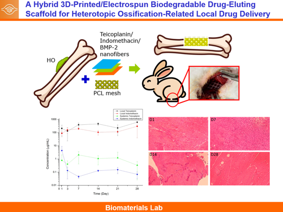
Heterotopic ossification (HO) is a clinically challenging complication after trauma or orthopedic surgery. This study evaluated a hybrid biodegradable scaffold for localized peri-osseous delivery of agents relevant to HO-risk and bone-healing environments. Polycaprolactone (PCL) mesh scaffolds were fabricated using solvent-cast additive manufacturing as flexible macro-scale barriers, while poly(lactic-co-glycolic acid) (PLGA) nanofibers incorporating indomethacin, teicoplanin, and bone morphogenetic protein-2 (BMP-2) were prepared using electrospinning and coaxial electrospinning. Scaffold morphology, wettability, mechanical behavior, Fourier-transform infrared spectroscopy and differential scanning calorimetry profiles, in vitro release, rabbit local/systemic release, and peri-implant histology were evaluated. The PCL mesh showed an ultimate tensile strength of 26.2 ± 2.6 MPa and a maximum strain of 337%. After 3 days in phosphate-buffered saline, the assembled PCL mesh/PLGA nanofiber scaffold retained comparable tensile properties, with an ultimate tensile strength of 24.8 ± 2.0 MPa and maximum strain of 334 ± 6%, indicating preserved flexibility under hydrated conditions. Drug-loaded PLGA nanofibers showed reduced tensile strength compared with pristine PLGA fibers, indicating that drug incorporation affected nanofiber handling and durability. In vitro testing demonstrated initial burst release of indomethacin and teicoplanin followed by sustained release, whereas BMP-2 release persisted for more than 30 days. In healthy rabbits, local teicoplanin and indomethacin levels were sustained for 28 days with substantially lower systemic levels. Histology demonstrated an early peri-implant inflammatory response that decreased over time. As no validated HO model or ectopic bone quantification was used, the findings support scaffold feasibility and localized delivery, not proven HO prevention. Further disease-model efficacy, biological activity, dose optimization, degradation, and safety studies are required before clinical translation.
- Ranganathan K, Loder S, Agarwal S, et al. Heterotopic ossification: basic-science principles and clinical correlates. J Bone Joint Surg Am. 2015;97(13):1101-1111. doi: 10.2106/JBJS.N.01056
- Meyers C, Lisiecki J, Miller S, et al. Heterotopic ossification: a comprehensive review. JBMR Plus. 2019;3(4):e10172. doi: 10.1002/jbm4.10172
- Ramirez DM, Ramirez MR, Reginato AM, Medici D. Molecular and cellular mechanisms of heterotopic ossification. Histol Histopathol. 2014;29(10):1281-1285. doi: 10.14670/HH-29.1281
- Bei M, Cao Q, Zhao C, Wu X. Heterotopic ossification: current developments and emerging potential therapies. Chin Med J (Engl). 2025;138(4):389-404. doi: 10.1097/CM9.0000000000003244
- Felix-Ilemhenbhio F, Pickering GAE, Kiss-Toth E, Wilkinson JM. Pathophysiology and emerging molecular therapeutic targets in heterotopic ossification. Int J Mol Sci. 2022;23(13):6983. doi: 10.3390/ijms23136983
- Zhou L, Gee SM, Hansen JA, Posner MA. Heterotopic ossification after arthroscopic procedures: a scoping review of the literature. Orthop J Sports Med. 2022;10(1):23259671211060040. doi: 10.1177/23259671211060040
- Kontokostopoulos AP, Gkiatas I, Vasileiadis GI, et al. Heterotopic ossification around the elbow revisited. Life (Basel). 2023;13(12):2358. doi: 10.3390/life13122358
- Pakos EE, Ioannidis JPA. Radiotherapy vs nonsteroidal anti-inflammatory drugs for the prevention of heterotopic ossification after major hip procedures: a meta-analysis of randomized trials. Int J Radiat Oncol Biol Phys. 2004;60(3):888-895. doi: 10.1016/j.ijrobp.2003.11.015
- Geller JS, Allegra PR, Seldon CS, et al. Prophylactic radiotherapy for prevention of heterotopic ossification after periacetabular fractures: a review of efficacy and associated conditions. J Surg Orthop Adv. 2022;31(2):113-118. PMID:35820098
- Georhakopoulos I, Kouloulias V, Kougiountzopoulou A, et al. Radiation therapy for the prevention of heterotopic ossification: efficacy and toxicity of single-fraction radiotherapy. Orthop Rev (Pavia). 2020;12(2):8577. doi: 10.4081/or.2020.8577
- Zhao P, Gu H, Mi H, Rao C, Fu J, Turng LS. Fabrication of scaffolds in tissue engineering: a review. Front Mech Eng. 2018;13(1):107-119. doi: 10.1007/s11465-018-0496-8
- Almouemen N, Kelly HM, O’Leary C. Tissue engineering: understanding the role of biomaterials and biophysical forces on cell functionality through computational and structural biotechnology analytical methods. Comput Struct Biotechnol J. 2019;17:591-598. doi: 10.1016/j.csbj.2019.04.008
- Ganesan O, Kiwanuka H, Hamaguchi R, Orgill DP. A review of regenerative medicine and tissue engineering with a focus on wound healing and anti-aging. Front Surg. 2025;12:1504563. doi: 10.3389/fsurg.2025.1504563
- Chen JM, Lee D, Yang JW, Lin SH, Lin YT, Liu SJ. Solution extrusion additive manufacturing of biodegradable polycaprolactone. Appl Sci. 2020;10(9):3189. doi: 10.3390/app10093189
- Sayet G, Sinegre M, Reguiga MB. Development of a Fourier transform infrared spectroscopy coupled to UV-Visible analysis technique for aminosides and glycopeptides quantitation in antibiotic locks. Ann Pharm Fr. 2014;72(1):41-50. doi: 10.1016/j.pharma.2013.10.002
- Amiri N, Ajami S, Shahroodi A, et al. Teicoplanin-loaded chitosan-PEO nanofibers for local antibiotic delivery and wound healing. Int J Biol Macromol. 2020;162:645-656. doi: 10.1016/j.ijbiomac.2020.06.195
- Liu M, Liu J, Wang Q, et al. Quantitative analysis of amorphous form in indomethacin binary polymorphic mixtures using infrared spectroscopy analytical techniques combined with chemometrics methods. Arab J Chem. 2023;16(8):104885. doi: 10.1016/j.arabjc.2023.104885
- Otsuka Y, Kuwashima W, Tanaka Y, Yamaki Y, Shimada Y, Goto S. Effects of heat treatment on indomethacin-cimetidine mixture: investigation of drug-drug interaction using singular value decomposition in FTIR spectroscopy. J Pharm Sci. 2021;110(3):1142-1147. doi: 10.1016/j.xphs.2020.09.049
- Flieger J, Orzel A, Kowalska-Kepczynska A, et al. Teicoplanin-modified HPLC column as a source of experimental parameters for prediction of the anticonvulsant activity of 1,2,4-triazole-3-thiones by the regression models. Materials (Basel). 2020;13(11):2650. doi: 10.3390/ma13112650
- Ornik J, Heidrich L, Schesny R, Castro-Camus E, Keck CM, Koch M. Non-destructive crystallinity assessment of indomethacin in tablets made from smartFilms using terahertz time-domain spectroscopy. Sci Rep. 2022;12(1):6099. doi: 10.1038/s41598-022-10041-1
- Liu CY, Lee WS, Fung CP, et al. Comparative study of teicoplanin vs vancomycin for the treatment of methicillin-resistant Staphylococcus aureus bacteraemia. Clin Drug Investig. 1996;12(2):80-87. doi: 10.2165/00044011-199612020-00003
- Song R, Murphy M, Li C, Ting K, Soo C, Zheng Z. Current development of biodegradable polymeric materials for biomedical applications. Drug Des Devel Ther. 2018;12:3117- 3145. doi: 10.2147/DDDT.S165440
- Kalirajan C, Dukle A, Nathanael AJ, Oh TH, Manivasagam G. A critical review on polymeric biomaterials for biomedical applications. Polymers (Basel). 2021;13(17):3015. doi: 10.3390/polym13173015
- Woodruff MA, Hutmacher DW. The return of a forgotten polymer-polycaprolactone in the 21st century. Prog Polym Sci. 2010;35(10):1217-1256. doi: 10.1016/j.progpolymsci.2010.04.002
- Backes EH, Harb SV, Beatrice CAG, et al. Polycaprolactone usage in additive manufacturing strategies for tissue engineering applications: a review. J Biomed Mater Res B Appl Biomater. 2022;110(6):1479-1503. doi: 10.1002/jbm.b.34997
- Yang J, Zeng H, Luo Y, et al. Recent applications of PLGA in drug delivery systems. Polymers (Basel). 2024;16(18):2606. doi: 10.3390/polym16182606
- Marquina S, Ozgul M, Robertson-Brown K, Kenney MC. A review on PLGA particles as a sustained drug-delivery system and its effect on the retina. Exp Eye Res. 2023;235:109626. doi: 10.1016/j.exer.2023.109626
- Ngo TD, Kashani A, Imbalzano G, Nguyen KTQ, Hui D. Additive manufacturing (3D printing): a review of materials, methods, applications and challenges. Compos B Eng. 2018;143:172-196. doi: 10.1016/j.compositesb.2018.02.012
- Rasiya G, Shukla A, Saran K. Additive manufacturing-a review. Mater Today Proc. 2021;47:6896-6901. doi: 10.1016/j.matpr.2021.05.181
- Bozkurt Y, Karayel E. 3D printing technology; methods, biomedical applications, future opportunities and trends. J Mater Res Technol. 2021;14:1430-1450. doi: 10.1016/j.jmrt.2021.07.050
- Xue J, Wu T, Dai Y, Xia Y. Electrospinning and electrospun nanofibers: methods, materials, and applications. Chem Rev. 2019;119(8):5298-5415. doi: 10.1021/acs.chemrev.8b00593
- Zhao J, Chen L, Ma A, et al. Recent advances in coaxial electrospun nanofibers for wound healing. Mater Today Bio. 2024;29:101309. doi: 10.1016/j.mtbio.2024.101309
- Ali SA, Doherty PJ, Williams DF. Mechanisms of polymer degradation in implantable devices. 2. Poly(DL-lactic acid). J Biomed Mater Res. 1993;27(11):1409-1418. doi: 10.1002/jbm.820271108
- Kamaly N, Yameen B, Wu J, Farokhzad OC. Degradable controlled-release polymers and polymeric nanoparticles: mechanisms of controlling drug release. Chem Rev. 2016;116(4):2602-2663. doi: 10.1021/acs.chemrev.5b00346
- Benhabbour SR, Sheardown H, Adronov A. Cell adhesion and proliferation on hydrophilic dendritically modified surfaces. Biomaterials. 2008;29(31):4177-4186. doi: 10.1016/j.biomaterials.2008.07.016
- Torres E, Valles-Lluch A, Fombuena V, Napiwocki B, Turng LS. Influence of the hydrophobic-hydrophilic nature of biomedical polymers and nanocomposites on in vitro biological development. Macromol Mater Eng. 2017;302(12):1700259. doi: 10.1002/mame.201700259