

Three-dimensional bioprinted personalized repair scaffolds based on CT/MRI fusion imaging after hepatocellular carcinoma resection

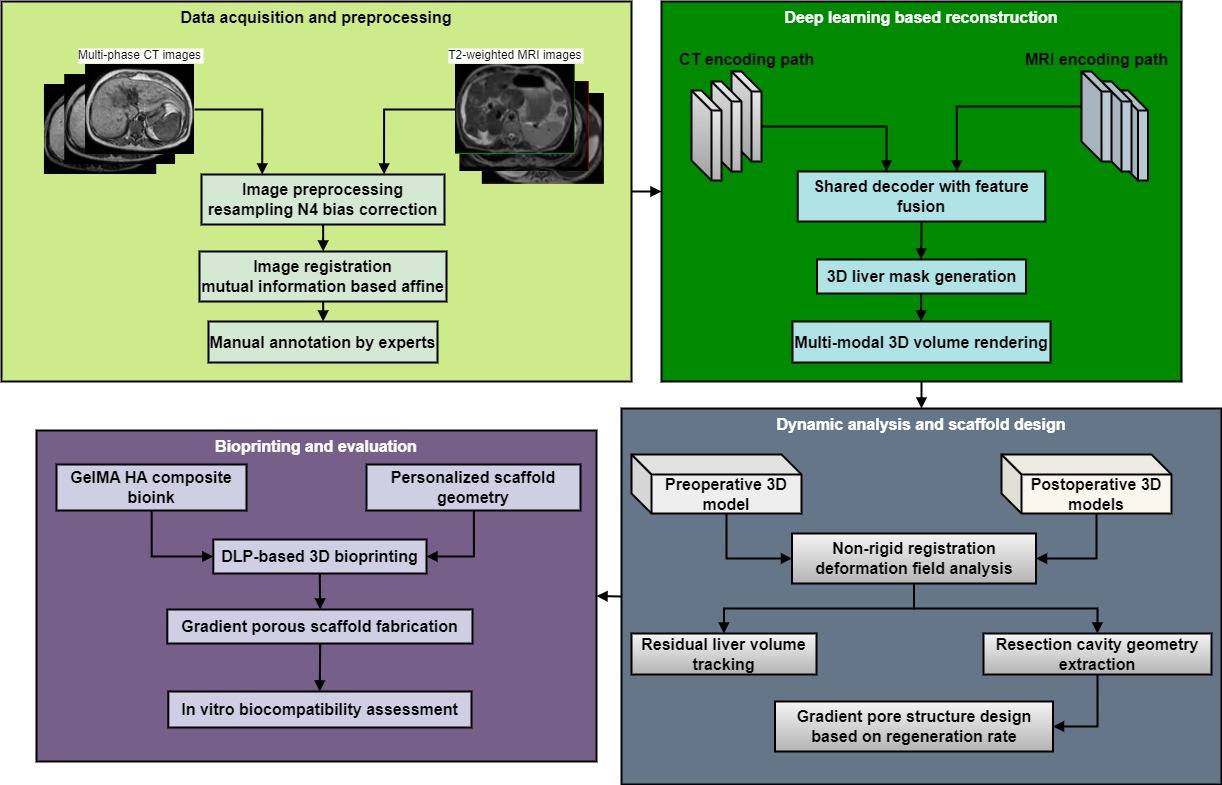
Accurate assessment of residual liver regeneration following hepatocellular carcinoma resection remains a major challenge due to the dynamic nature of the regenerative process and limitations in conventional image-based evaluation. This study develops a three-dimensional (3D) bioprinted personalized repair scaffold based on multimodal fusion of computed tomography (CT) and magnetic resonance imaging (MRI). Multi-temporal CT/MRI images of patients with hepatocellular carcinoma were acquired, registered, and preprocessed using standardization. A dual-channel deep learning network was employed to achieve high-precision liver segmentation and multimodal fusion 3D reconstruction. Using the deformation field from non-rigid registration, the dynamic changes in residual liver volume after surgery were accurately tracked, and the geometry of the resected cavity was extracted to serve as the basis for scaffold design. Subsequently, the internal pore gradient was optimized by combining regeneration rate data. Using a gelatin methacryloyl (GelMA)/hyaluronic acid (HA) composite bio-ink, a repair scaffold with a personalized shape and functional internal structure was manufactured via digital light processing photopolymerization 3D printing. Experiments showed that, in terms of structural fidelity, the GelMA/HA composite scaffold exhibited excellent performance in overall shape fidelity (94.82%) and key structural wall thickness deviation (12.3 μm). Regarding in vitro cell compatibility, the relative cell proliferation rate achieved 0.85 ± 0.04 under low-serum conditions. In vitro experiments have shown that the scaffold has good cell compatibility and structural stability, providing a theoretical and technical approach for precise repair after liver cancer surgery.
- Primavesi F, Maglione M, Cipriani F, et al. E-AHPBA–ESSO– ESSR Innsbruck consensus guidelines for preoperative liver function assessment before hepatectomy. Br J Surg. 2023;110(10):1331-1347. doi: 10.1093/bjs/znad233
- Kimura J, Takagi K, Fuji T, et al. Risk Factors and Strategies for Failure to Rescue Following Hepatectomy: A Review. J Hepato Biliary Pancreat Sci. 2025;32(11):801-809. doi: 10.1002/jhbp.70014
- Birgin E, Abdelhadi S, Seyfried S, et al. Robotic or laparoscopic repeat hepatectomy after open hepatectomy: a cohort study. Surg Endosc. 2024;38(3):1296-1305. doi: 10.1007/s00464-023-10645-2
- Raptis DA, Elsheikh Y, Alnemary Y, et al. Robotic living donor hepatectomy is associated with superior outcomes for both the donor and the recipient compared with laparoscopic or open-A single-center prospective registry study of 3448 cases. Am J Transplant. 2024;24(11):2080-2091. doi: 10.1016/j.ajt.2024.04.020
- Broering DC, Raptis DA, Elsheikh Y. Pioneering fully robotic donor hepatectomy and robotic recipient liver graft implantation–a new horizon in liver transplantation. Int J Surg. 2024;110(3):1333-1336. doi: 10.1097/JS9.0000000000001031
- Wong P, Vien P, Kessler J, et al. Augmenting the future liver remnant prior to major hepatectomy: a review of options on the menu. Ann Surg Oncol. 2025;32(8):5694-5709. doi: 10.1245/s10434-025-17607-z
- Mao B, Zhu S, Li D, et al. Comparison of safety and effectiveness between robotic and laparoscopic major hepatectomy: a systematic review and meta-analysis. Int J Surg. 2023;109(12):4333-4346. doi: 10.1097/JS9.0000000000000750
- Berardi G, Cucchetti A, Colasanti M, et al. Prehabilitation with exercise and nutrition to reduce morbidity of major hepatectomy in patients with sarcopenia: the PREHEP randomized clinical trial. JAMA surgery, 2025, 160(10): 1068-1075. doi: 10.1001/jamasurg.2025.3102
- Hong SK, Kim J Y, Lee J, et al. Pure laparoscopic donor hepatectomy: experience of 556 cases at Seoul National University Hospital. Am J Transplant. 2024;24(2):222-238. doi: 10.1016/j.ajt.2023.06.007
- Zhu XD, Huang C, Shen YH, et al. Hepatectomy after conversion therapy using tyrosine kinase inhibitors plus anti-PD-1 antibody therapy for patients with unresectable hepatocellular carcinoma. Ann Surg Oncol. 2023;30(5):2782- 2790. doi: 10.1245/s10434-022-12530-z
- Cheah YL, Yang HY, Simon CJ, et al. The learning curve for robotic living donor right hepatectomy: Analysis of outcomes in 2 specialized centers. Liver Transplant. 2025;31(2):190-200. doi: 10.1097/LVT.0000000000000480
- Sun M, Li M, Hu M, et al. Fully bioactive nanodrugs: stem cell-derived exosomes engineered with biomacromolecules to treat CCl4-and extreme hepatectomy-induced acute liver failure. ACS Nano. 2024;18(50):33907-33921. doi: 10.1021/acsnano.4c07408
- Yang DL, Peng N, Nong JL, et al. Survival benefit of hepatectomy after complete or partial response to conversion therapy in unresectable hepatocellular carcinoma (GUIDANCE003): a multicenter study. Liver Cancer. 2025;14(6):687-703. doi: 10.1159/000546052
- Cusumano C, Kansoun A, Tougoue FK, et al. Incidence and outcomes of post-hepatectomy diaphragmatic hernia: a systematic review. HPB. 2023;25(12):1466-1474. doi: 10.1016/j.hpb.2023.08.008
- Liu Y, Wang Q, Du B, et al. A meta-analysis of the three-dimensional reconstruction visualization technology for hepatectomy. Asian J Surg. 2023;46(2):669-676. doi: 10.1016/j.asjsur.2022.07.006
- Sambommatsu Y, Kumaran V, Imai D, et al. Early outcomes of robotic vs open living donor right hepatectomy in a US Center. Surg Endosc. 2025;39(3):1643-1652. doi: 10.1007/s00464-024-11469-4
- Zhang XP, Zhang TC, Wu FF, et al. Patterns and outcomes of early and late recurrence after hepatectomy for hepatocellular carcinoma with microvascular invasion: a multicenter study in China. Hepatol Int. 2025;19(4):903-914. doi: 10.1007/s12072-025-10802-w
- Zhang Y, Li L, Dong L, et al. Hydrogel-based strategies for liver tissue engineering. Chem Bio Eng. 2024;1(11):887-915. doi: 10.1021/cbe.4c00079
- Du Y, Bai Y, Lang S, et al. Gelatin sponges with a uniform interoperable pore structure and biodegradability for liver injury hemostasis and tissue regeneration. Biomacromolecules. 2023;24(11):5313-5327. doi: 10.1021/acs.biomac.3c00803
- Farasati Far B, Isfahani AA, Nasiriyan E, et al. An updated review on advances in hydrogel-based nanoparticles for liver cancer treatment. Livers. 2023;3(2):161-189. doi: 10.3390/livers3020012
- Mukherjee P, Guha S, Ghosh A, et al. Porous Organic Polymer-Based Nanocomposites for Hypoxia Relieving and Enhanced Chemotherapy in Hepatocellular Carcinoma. ACS Appl Bio Mater. 2024;7(9):6138-6151. doi: 10.1021/acsabm.4c00723
- Caruso D, De Santis D, Del Gaudio A, et al. Low-dose liver CT: image quality and diagnostic accuracy of deep learning image reconstruction algorithm. Eur Radiol. 2024;34(4):2384-2393. doi: 10.1007/s00330-023-10171-8
- Wu T, Yang D, Wee A, et al. Identification of MRI features associated with injury type, severity, and prognosis in drug-induced liver injury. Eur Radiol. 2023;33(1):666-677. doi: 10.1007/s00330-022-09041-6
- Jeong B, Heo S, Lee SS, et al. Predicting post-hepatectomy liver failure in patients with hepatocellular carcinoma: nomograms based on deep learning analysis of gadoxetic acid-enhanced MRI. Eur Radiol. 2025;35(5):2769-2782. doi: 10.1007/s00330-024-11173-w
- Görgec B, Hansen IS, Kemmerich G, et al. MRI in addition to CT in patients scheduled for local therapy of colorectal liver metastases (CAMINO): an international, multicentre, prospective, diagnostic accuracy trial. Lancet Oncol. 2024;25(1):137-146. doi: 10.1016/S1470-2045(23)00572-7
- Cha H, Choi JY, Park YN, et al. Comparison of imaging findings of macrotrabecular-massive hepatocellular carcinoma using CT and gadoxetic acid–enhanced MRI. Eur Radiol. 2023;33(2):1364-1377. doi: 10.1007/s00330-022-09105-7
- Pei C, Wu F, Yang M, et al. Multi-source domain adaptation for medical image segmentation. IEEE Trans Med Imaging 2023;43(4):1640-1651. doi: 10.1109/TMI.2023.3346285
- Odisio BC, Albuquerque J, Lin YM, et al. Software-based versus visual assessment of the minimal ablative margin in patients with liver tumours undergoing percutaneous thermal ablation (COVER-ALL): a randomised phase 2 trial. Lancet Gastroenterol Hepatol. 2025;10(5):442-451. doi: 10.1016/S2468-1253(25)00024-X
- Wu Y, Su H, Li M, et al. Digital light processing‐based multi‐material bioprinting: Processes, applications, and perspectives. J Biomed Mater Res Part A. 2023;111(4):527- 542. doi: 10.1002/jbm.a.37473
- Dhand AP, Davidson MD, Burdick JA. Lithography-based 3D printing of hydrogels. Nat Rev Bioeng. 2025;3(2):108- 125. doi: 10.1038/s44222-024-00251-9
- Gadre M, Vasanthan KS. Engineering a GelMA–dECM-based 3D bioprinted liver fibrosis model: methotrexate-induced functional and molecular validation. RSC Adv. 2025;15(44):37012-37026. doi: 10.1039/D5RA05955K
- Shinn J, Park S, Lee S, et al. Antioxidative hyaluronic acid–bilirubin nanomedicine targeting activated hepatic stellate cells for anti-hepatic-fibrosis therapy. ACS Nano. 2024;18(6):4704-4716. doi: 10.1021/acsnano.3c06107
- Xiao L, Ye M, Fan Y, et al. Dual-cross-linked methylacrylated collagen-DPPA bioinks for precision dlp bioprinting and accelerated skin wound healing. Biomacromolecules. 2025;26(7):4308-4321. doi: 10.1021/acs.biomac.5c00305
- Kim MH, Lin CC. Poly (ethylene glycol)–norbornene as a photoclick bioink for digital light processing 3D bioprinting. ACS Appl Mater Interfaces. 2023;5(2):2737-2746. doi: 10.1021/acsami.2c20098
- Kim D H, Byun J Y, Kim D, et al. Geometric evaluation of biomimetic 3D printed rat femur. J Hard Tissue Biol. 2023;32(2):133-138. doi: 10.2485/jhtb.32.133
- Wang K, Wang J, Jiang P. High-dose-rate three-dimensional image-guided adaptive brachytherapy (3D IGABT) for locally advanced cervical cancer (LACC): A narrative review on imaging modality and clinical evidence. Curr Oncol. 2023;31(1):50-65. doi: 10.3390/curroncol31010004
- Ding H, Chen L, Tao Q, et al. DCU-Net: a dual-channel U-shaped network for image splicing forgery detection. Neural Comput Appl. 2023;35(7):5015-5031. doi: 10.1007/s00521-021-06329-4
- Li M, Zhang W, Hu B, et al. Automatic assessment of depression and anxiety through encoding pupil-wave from HCI in VR scenes. ACM Trans Multimed Comput Commun Appl. 2023;20(2):1-22. doi: 10.1145/3513263