

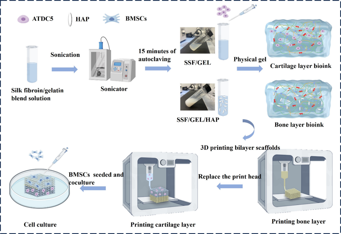
Self-healing silk fibroin–gelatin–based 3D-printed bilayer scaffolds with stable interface and bidirectional lineage control for osteochondral regeneration

The osteochondral interface—characterized by a steep gradient in both composition and mechanical properties—remains one of the most challenging anatomical sites to regenerate. Reconstructing this spatially complex heterogeneity continues to confound conventional osteochondral grafts. Although multilayer scaffolds are widely adopted, interfacial delamination frequently compromises repair outcomes. Here, we report a self-healing, physically crosslinked silk fibroin–based 3D-printing ink that incorporates gelatin and nano-hydroxyapatite for the fabrication of bilayer scaffolds with robust interfacial bonding. By tuning ultrasonication time of silk fibroin and gelatin content, the ink exhibits exceptional printability and cytocompatibility, enabling >90% post-printing cell viability. Leveraging a dual-nozzle alternating-print strategy, we generated bilayer constructs that display a stable interface and layerspecific mechanical heterogeneity. Both upper- and lower-layer inks possess good self-healing capacity, eliminating delamination and yielding a monolithic scaffold. Functional analyses revealed significant upregulation of the chondrogenic marker type II collagen in the upper layer and the osteogenic marker RUNX2 in the lower layer, achieving bidirectional lineage instruction required for osteochondral regeneration. This silk/gelatin-based, physically crosslinked, integrative bilayer scaffold offers a promising therapeutic platform for osteochondral defect repair.
- Long H, Liu Q, Yin H, et al. Prevalence trends of site specific osteoarthritis from 1990 to 2019: findings from the Global Burden of Disease Study 2019. Arthritis Rheumatol. 2019;74(7):1172-1183. doi: 10.1002/art.42089
- Yang M, Zhang ZC, Yuan FZ, et al. An immunomodulatory polypeptide hydrogel for osteochondral defect repair. Bioact Mater. 2023;19:678-689. doi: 10.1016/j.bioactmat.2022.05.008
- Wu Z, Yao H, Sun H, et al. Enhanced hyaline cartilage formation and continuous osteochondral regeneration via 3D-Printed heterogeneous hydrogel with multi-crosslinking inks. Mater Today Bio. 2024;26:101080. doi: 10.1016/j.mtbio.2024.101080
- Li M, Yin H, Yan Z, et al. The immune microenvironment in cartilage injury and repair. Acta Biomater. 2022;140:23-42. doi: 10.1016/j.actbio.2021.12.006
- Singh YP, Bandyopadhyay A, Mandal BB. 3D bioprinting using cross-linker-free silk–gelatin bioink for cartilage tissue engineering. ACS Appl Mater Interfaces. 2019;11(37):33684-33696. doi: 10.1021/acsami.9b11644
- Li C, Zhang W, Nie Y, et al. Integrated and bifunctional bilayer 3D printing scaffold for osteochondral defect repair. Adv Funct Mater. 2023;33(20):2214158. doi: 10.1002/adfm.202214158
- Lowen JM, Wheeler EE, Shimamoto NK, et al. Functionalized annealed microgels for spatial control of osteogenic and chondrogenic differentiation. Adv Funct Mater. 2024;34(30):2311017. doi: 10.1002/adfm.202311017
- Lee J, Lee S, Huh SJ, Kang BJ, Shin H. Directed regeneration of osteochondral tissue by hierarchical assembly of spatially organized composite spheroids. Adv Sci. 2022;9(3):2103525. doi: 10.1002/advs.202103525
- Wang H, Zhang J, Bai H, et al. 3D printed cell-free bilayer porous scaffold based on alginate with biomimetic microenvironment for osteochondral defect repair. Biomater Adv. 2025;167:214092. doi: 10.1016/j.bioadv.2024.214092
- Levingstone TJ, Matsiko A, Dickson GR, O’Brien FJ, Gleeson JP. A biomimetic multi-layered collagen-based scaffold for osteochondral repair. Acta Biomater. 2014;10(5):1996-2004. doi: 10.1016/j.actbio.2014.01.005
- Shimomura K, Moriguchi Y, Murawski CD, Yoshikawa H, Nakamura N. Osteochondral tissue engineering with biphasic scaffold: current strategies and techniques. Tissue Eng Part B Rev. 2014;20(5):468-476. doi: 10.1089/ten.teb.2013.0543
- Barui S, Ghosh D, Laurencin CT. Osteochondral regenerative engineering: challenges, state-of-the-art and translational perspectives. Regen Biomater. 2023;10:rbac109. doi: 10.1093/rb/rbac109
- Du J, Zhu Z, Liu J, et al. 3D-printed gradient scaffolds for osteochondral defects: Current status and perspectives. Int J Bioprint. 2023;9(4):724. doi: 10.18063/ijb.724
- Lai Y, Fan J, Li P, et al. Recent advances in 3D bioprinting for cartilage and osteochondral regeneration. Int J Bioprint. 2025;11(3):154-184. doi: 10.36922/IJB025120098
- Huey DJ, Hu JC, Athanasiou KA. Unlike bone, cartilage regeneration remains elusive. Science. 2012;338(6109):917-921. doi: 10.1126/science.1222454
- Niu X, Li N, Du Z, Li X. Integrated gradient tissue engineered osteochondral scaffolds: Challenges, current efforts and future perspectives. Bioact Mater. 2023;20:574-597. doi: 10.1016/j.bioactmat.2022.06.011
- Li C, Guo C, Fitzpatrick V, et al. Design of biodegradable, implantable devices towards clinical translation. Nat Rev Mater. 2020;5(1):61-81. doi: 10.1038/s41578-019-0150-z
- Sarr MM, Inoue H, Kosaka T. Study on the improvement of interfacial strength between glass fiber and matrix resin by grafting cellulose nanofibers. Compos Sci Technol. 2021;211:108853. doi: 10.1016/j.compscitech.2021.108853
- Sharifi S, Islam MM, Sharifi H, et al. Tuning gelatin-based hydrogel towards bioadhesive ocular tissue engineering applications. Bioact Mater. 2021;6(11):3947-3961. doi: 10.1016/j.bioactmat.2021.03.042
- Zhang X, Liu Y, Zuo Q, et al. 3D bioprinting of biomimetic bilayered scaffold consisting of decellularized extracellular matrix and silk fibroin for osteochondral repair. Int J Bioprint. 2021;7(4):401. doi: 10.18063/ijb.v7i4.401
- Chawla S, Midha S, Sharma A, Ghosh S. Silk-based bioinks for 3D bioprinting. Adv Healthc Mater. 2018;7(8):1701204. doi: 10.1002/adhm.201701204
- Kim SH, Yeon YK, Lee JM, et al. Precisely printable and biocompatible silk fibroin bioink for digital light processing 3D printing. Nat Commun. 2018;9(1):1620. doi: 10.1038/s41467-018-03759-y
- Kokol V, Pottathara YB, Mihelčič M, Perše LS. Rheological properties of gelatine hydrogels affected by flow and horizontally-induced cooling rates during 3D cryo-printing. Colloids Surf A Physicochem Eng Asp. 2021;616:126356. doi: 10.1016/j.colsurfa.2021.126356
- Hou P, Yang X, Liu Z, et al. Advancing knee cartilage repair with 3D printed GelMA/SF/Haps composite hydrogels for enhanced chondrocyte regeneration. J Mater Sci. 2024;59(11):4636-4648. doi: 10.1007/s10853-024-09508-5
- Zheng Z, Wu J, Liu M, et al. 3D bioprinting of self-standing silk-based bioink. Adv Healthc Mater. 2018;7(6):1701026. doi: 10.1002/adhm.201701026
- Zou S, Fan S, Oliveira AL, et al. 3D printed gelatin scaffold with improved shape fidelity and cytocompatibility by using Antheraea pernyi silk fibroin nanofibers. Adv Fiber Mater. 2022;4(4):758-773. doi: 10.1007/s42765-022-00135-w
- Chakraborty J, Fernandez-Perez J, van Kampen KA, et al. Development of a biomimetic arch-like 3D bioprinted construct for cartilage regeneration using gelatin methacryloyl and silk fibroin-gelatin bioinks. Biofabrication. 2023;15(3):035009. doi: 10.1088/1758-5090/acc68f
- Lee H, Shin D, Shin S, Hyun J. Effect of gelatin on dimensional stability of silk fibroin hydrogel structures fabricated by digital light processing 3D printing. J Ind Eng Chem. 2020;89:119-127. doi: 10.1016/j.jiec.2020.03.034
- Chu S, Maples MM, Bryant SJ. Cell encapsulation spatially alters crosslink density of poly(ethylene glycol) hydrogels formed from free-radical polymerizations. Acta Biomater. 2020;109:37-50. doi: 10.1016/j.actbio.2020.03.033
- Yuan X, Li G, Huang L, et al. Hydroxypropyl chitin oxidized chondroitin sulfate double-network hydrogel assists microfracture technique to enhance cartilage regeneration. Mater Des. 2024;238:112656. doi: 10.1016/j.matdes.2024.112656
- Yao D, Li M, Wang T, et al. Viscoelastic silk fibroin hydrogels with tunable strength. ACS Biomater Sci Eng. 2021;7(2):636-647. doi: 10.1021/acsbiomaterials.0c01348
- Ding X, Gao J, Yu X, et al. 3D-printed porous scaffolds of hydrogels modified with TGF-β1 binding peptides to promote in vivo cartilage regeneration and animal gait restoration. ACS Appl Mater Interfaces. 2022;14(14):15982-
- doi: 10.1021/acsami.2c00761
- Song P, Li M, Zhang B, et al. DLP fabricating of precision GelMA/HAp porous composite scaffold for bone tissue engineering application. Compos B Eng. 2022;244:110163. doi: 10.1016/j.compositesb.2022.110163
- Wang X, Kluge JA, Leisk GG, Kaplan DL. Sonication-induced gelation of silk fibroin for cell encapsulation. Biomaterials. 2008;29(8):1054-1064. doi: 10.1016/j.biomaterials.2007.11.003
- Zhong J, Liu X, Wei D, et al. Effect of incubation temperature on the self-assembly of regenerated silk fibroin: A study using AFM. Int J Biol Macromol. 2015;76:195-202. doi: 10.1016/j.ijbiomac.2015.02.045
- Lu Q, Zhu H, Zhang C, et al. Silk self-assembly mechanisms and control from thermodynamics to kinetics. Biomacromolecules. 2012;13(3):826-832. doi: 10.1021/bm201731e
- Mosleh Y, de Zeeuw W, Nijemeisland M, et al. The structure– property correlations in dry gelatin adhesive films. Adv Eng Mater. 2021;23(1):2000716. doi: 10.1002/adem.202000716
- Sun X, Liang H, Wang H, et al. Silk fibroin/polyvinyl alcohol composite film loaded with antibacterial AgNP/ polydopamine-modified montmorillonite; characterization and antibacterial properties. Int J Biol Macromol. 2023;251:126368. doi: 10.1016/j.ijbiomac.2023.126368
- Park S, Edwards S, Hou S, et al. A multi-interpenetrating network (IPN) hydrogel with gelatin and silk fibroin. Biomater Sci. 2019;7(4):1276-1280. doi: 10.1039/c8bm01532e
- Zhanbassynova A, Mukasheva F, Abilev M, et al. Impact of Hydroxyapatite on Gelatin/Oxidized Alginate 3D-Printed Cryogel Scaffolds. Gels. 2024;10(6):406. doi: 10.3390/gels10060406
- Kazemi M, Mirzadeh M, Esmaeili H, et al. Evaluation of the morphological effects of hydroxyapatite nanoparticles on the rheological properties and printability of hydroxyapatite/polycaprolactone nanocomposite inks and final scaffold features. 3D Print Addit Manuf. 2024;11(1):132-142. doi: 10.1089/3dp.2021.0292
- Luo M, Chen M, Bai J, et al. A bionic composite hydrogel with dual regulatory functions for the osteochondral repair. Colloids Surf B Biointerfaces. 2022;219:112821. doi: 10.1016/j.colsurfb.2022.112821
- Choi DJ, Kho Y, Park SJ, et al. Effect of cross-linking on the dimensional stability and biocompatibility of a tailored 3D-bioprinted gelatin scaffold. Int J Biol Macromol. 2019;135:659-667. doi: 10.1016/j.ijbiomac.2019.05.207
- Ren YJ, Zhou ZY, Liu BF, Xu QY, Cui FZ. Preparation and characterization of fibroin/hyaluronic acid composite scaffold. Int J Biol Macromol. 2009;44(4):372-378. doi: 10.1016/j.ijbiomac.2009.02.004
- Hoque M, Alam M, Wang S, et al. Interaction chemistry of functional groups for natural biopolymer-based hydrogel design. Mater Sci Eng R Rep. 2023;156:100758. doi: 10.1016/j.mser.2023.100758
- Guo W, Gao X, Ding X, et al. Self-adhesive and self-healing hydrogel dressings based on quaternary ammonium chitosan and host-guest interacted silk fibroin. Colloids Surf A Physicochem Eng Asp. 2024;684:133145. doi: 10.1016/j.colsurfa.2024.133145
- Yu R, Yang Y, He J, Li M, Guo B. Novel supramolecular self healing silk fibroin-based hydrogel via host–guest interaction as wound dressing to enhance wound healing. Chem Eng J. 2021;417:128278. doi: 10.1016/j.cej.2020.128278
- Koh RH, Kim J, Kim JU, et al. Bioceramic-mediated chondrocyte hypertrophy promotes calcified cartilage formation for rabbit osteochondral defect repair. Bioact Mater. 2024;40:306-317. doi: 10.1016/j.bioactmat.2024.06.018
- Pacheco MO, Aikman EL, Bagnis HK, et al. Degumming Time Governs Self-Assembled Silk Fibroin Hydrogel Properties through Molecular Weight and Amino Acid Composition. Biomacromolecules. 2025;26(8):5069-5085. doi: 10.1021/acs.biomac.5c00506
- Lee H, Gu L, Mooney DJ, Levenston ME, Chaudhuri O. Mechanical confinement regulates cartilage matrix formation by chondrocytes. Nat Mater. 2017;16(12):1243-1251. doi: 10.1038/nmat4993
- Song J, Chen C, Zhu S, et al. Processing bulk natural wood into a high-performance structural material. Nature. 2018;554(7691):224-228. doi: 10.1038/nature25476