

Optimization of 3D bioprinting of mouse preosteoblasts using nanofibrillated cellulose hydrogels

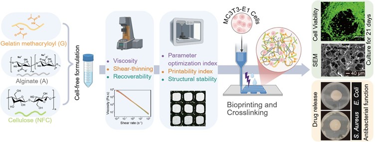
Development and optimization of advanced bioink formulations for living tissue-engineered scaffolds remain a challenging task. Herein, a nanofibrillated cellulose (NFC)-composited gelatin methacryloyl (G)/alginate (A) formulation (G/A/NFC100) was prepared for 3D bioprinting of mouse preosteoblasts MC3T3-E1, whereby the G/A formulations with a mixed NFC/microfibrillated cellulose and without NFC were included for comparison. The rheological properties of G/A formulation were enhanced by the addition of NFC, as evidenced by a decreased viscosity index characterizing shear thinning behavior from 0.52 (G/A) to 0.19 (G/A/NFC100). To construct 3D scaffolds with excellent shape fidelity while minimizing shear damage to cells during extrusion, the bioprinting conditions of the formulations were optimized based on the parameter optimization index. The G/A/NFC100 scaffold printed at a printing speed of 2 mm/s and a dispensing pressure of 30 kPa from a 27-gauge nozzle displayed a high shape fidelity (printability index of 0.883). The mechanical stability of the crosslinked 20-layered G/A/NFC100 structures were demonstrated by three consecutive press-relax cycles. The successful bioprinting of mouse preosteoblasts using the G/A/NFC100 formulation translated into an increased cell viability (above 97.64%) up to 21 days post-bioprinting. These results emphasize the exceptional potential of NFC-composited G/A formulation for bioprinting of bone tissue analogues for biomedical applications. In addition, the long-term controlled release of ampicillin (67.42% after 72 h) by G/A/NFC100 scaffolds demonstrates the feasibility of utilizing porous cellulose fibers as drug-delivery carriers to enable multifunctionality in bone tissue repair.
1 Fang Y, Guo Y, Liu T, et al. Advances in 3D bioprinting. Chin J Mech Eng Addit Manuf Front. 2022;1(1): 100011. doi: 10.1016/j.cjmeam.2022.100011
2 Zhang B, Gao L, Ma L, Luo Y, Yang H, Cui Z. 3D bioprinting: a novel avenue for manufacturing tissues and organs. Engineering. 2019;5(4):777-794. doi: 10.1016/j.eng.2019.03.009
3 Sharma C, Raza MA, Purohit SD, et al. Cellulose-based 3D printing bio-inks for biomedical applications: a review. Int J Biol Macromol. 2025;305:141174. doi: 10.1016/j.ijbiomac.2025.141174
4 Mo Q, Huang L, Sheng Y, et al. Crosslinking strategy and promotion role of cellulose as a composite hydrogel component for three-dimensional printing – a review. Food Hydrocoll. 2024;154:110079. doi: 10.1016/j.foodhyd.2024.110079
5 You P, Sun H, Chen H, et al. Composite bioink incorporating cell-laden liver decellularized extracellular matrix for bioprinting of scaffolds for bone tissue engineering. Biomater Adv. 2024;165:214017. doi: 10.1016/j.bioadv.2024.214017
6 Ouiyangkul P, Hirun N, Suknuntha K, Tantishaiyakul V. Development and characterization of 3D bioprintable and mechanically reinforced hydrogel based on gellan gum/ methylcellulose/cellulose nanocrystals. Polym Adv Technol. 2024;35(1):e6206. doi: 10.1002/pat.6206
7 Suamte L, Tirkey A, Barman J, Jayasekhar Babu P. Various manufacturing methods and ideal properties of scaffolds for tissue engineering applications. Smart Mater Manuf. 2023;1:100011. doi: 10.1016/j.smmf.2022.100011
8 Mohammed A, Jiménez A, Bidare P, et al. Review on engineering of bone scaffolds using conventional and additive manufacturing technologies. 3D Print Addit Manuf. 2023;11(4):1418-1440. doi: 10.1089/3dp.2022.0360
9 Li N, Guo R, Zhang ZJ. Bioink formulations for bone tissue regeneration. Front Bioeng Biotechnol. 2021;9. doi: 10.3389/fbioe.2021.630488
10 Yang J, Wang J, Yang Y, et al. 3D-printed bioactive scaffolds: an emerging strategy for the regeneration of infectious bone defects. Int J Bioprint. 2024;11(2):79-138. doi: 10.36922/ijb.4986
11 Zhao T, Liu Y, Wu Y, Zhao M, Zhao Y. Controllable and biocompatible 3D bioprinting technology for microorganisms: fundamental, environmental applications and challenges. Biotechnol Adv. 2023;69:108243. doi: 10.1016/j.biotechadv.2023.108243
12 Chen A, Wang W, Mao Z, et al. Multimaterial 3D and 4D bioprinting of heterogenous constructs for tissue engineering. Adv Mater. 2024;36(34):2307686. doi: 10.1002/adma.202307686
13 Liu S, Yu J-M, Gan Y-C, et al. Biomimetic natural biomaterials for tissue engineering and regenerative medicine: new biosynthesis methods, recent advances, and emerging applications. Mil Med Res. 2023;10(1):16. doi: 10.1186/s40779-023-00448-w
14 Tarsitano M, Ming CLC, Idais D, et al. Sericin improves alginate-gelatin hydrogels’ mechanical properties, porosity, durability, and viability of fibroblasts in cardiac spheroids. Int J Bioprint. 2024;11(1):327-346. doi: 10.36922/ijb.5678
15 Jahani A, Nourbakhsh MS, Ebrahimzadeh MH, Mohammadi M, Yari D, Moradi A. Biomolecules- loading of 3D-printed alginate-based scaffolds for cartilage tissue engineering applications: a review on current status and future prospective. Arch Bone Jt Surg. 2024;12(2):92-101. doi: 10.22038/abjs.2023.73275.3396
16 Liu F, Jiang J, Zhe M, Yu P, Xing F, Xiang Z. Alginate-based 3D bioprinting strategies for structure–function integrated tissue regeneration. J Mater Chem B. 2025;13(40):12765-12811. doi: 10.1039/D5TB01489A
17 de Souza JR, Rahimnejad M, Mendes Soares IP, et al. 3D printing β-TCP-laden GelMA/alginate interpenetrating-polymer-network biomaterial inks for bone tissue engineering. Bioprinting. 2025;49:e00413. doi: 10.1016/j.bprint.2025.e00413
18 Aldana AA, Valente F, Dilley R, Doyle B. Development of 3D bioprinted GelMA-alginate hydrogels with tunable mechanical properties. Bioprinting. 2021;21:e00105. doi: 10.1016/j.bprint.2020.e00105
19 Seddiqi H, Oliaei E, Honarkar H, et al. Cellulose and its derivatives: towards biomedical applications. Cellulose. 2021;28(4):1893-1931. doi: 10.1007/s10570-020-03674-w
20 Tabatabaei Hosseini BS, Meadows K, Gabriel V, Hu J, Kim K. Biofabrication of cellulose-based hydrogels for advanced wound healing: a special emphasis on 3D bioprinting. Macromol Biosci. 2024;24(5):2300376. doi: 10.1002/mabi.202300376
21 Singh P, Baniasadi H, Gupta S, et al. 3D-printed cellulose nanocrystals and gelatin scaffolds with bioactive cues for regenerative medicine: advancing biomedical applications. Int J Biol Macromol. 2024;278:134402. doi: 10.1016/j.ijbiomac.2024.134402
22 Jiao H, Shi Y, Sun J, et al. Sawdust-derived cellulose nanofibrils with high biosafety for potential bioprinting. Ind Crops Prod. 2024;209:118025. doi: 10.1016/j.indcrop.2024.118025
23 Carvalho JPF, Lameirinhas NS, Teixeira MC, et al. All-cellulose hydrogel-based bioinks for the versatile 3D bioprinting of different cell lines. Biomacromolecules. 2025;26(3):1761-1770. doi: 10.1021/acs.biomac.4c01546
24 Lameirinhas NS, Carvalho JPF, Teixeira MC, et al. Nanocomposite hydrogel-based bioinks composed of a fucose-rich polysaccharide and nanocellulose fibers for 3D-bioprinting applications. Bioprinting. 2025;45: e00382. doi: 10.1016/j.bprint.2024.e00382
25 Li N, Qi S, Buccoli L, et al. Multiscale mechanical properties and enhancement mechanism of cellulose-composited hydrogels. Carbohydr Polym. 2025;357:123421. doi: 10.1016/j.carbpol.2025.123421
26 Li N, Bassett DC, Zhang ZJ. Microfibrillated cellulose (MFC)-composite formulations for 3D bioprinting with excellent printability, mechanical strength, and biological functionality. Chem Eng J. 2025:169037. doi: 10.1016/j.cej.2025.169037
27 Gatenholm P, Martinez H, Karabulut E, et al. Development of nanocellulose-based bioinks for 3D bioprinting of soft tissue. In: Ovsianikov A, Yoo J, Mironov V, eds. 3D Printing and Biofabrication. New York, NY: Springer International Publishing; 2016:1-23. doi: 10.1007/978-3-319-45444-3_14
28 Webb B, Doyle BJ. Parameter optimization for 3D bioprinting of hydrogels. Bioprinting. 2017;8:8-12. doi: 10.1016/j.bprint.2017.09.001
29 Lewicki J, Bergman J, Kerins C, Hermanson O. Optimization of 3D bioprinting of human neuroblastoma cells using sodium alginate hydrogel. Bioprinting. 2019;16:e00053. doi: 10.1016/j.bprint.2019.e00053
30 Ouyang L, Yao R, Zhao Y, Sun W. Effect of bioink properties on printability and cell viability for 3D bioplotting of embryonic stem cells. Biofabrication. 2016;8(3):035020. doi: 10.1088/1758-5090/8/3/035020