

Fibrous bioinks for bioprinting anisotropic micro-and nanoscale scaffolds: A novel strategy for in vitro skeletal muscle engineering

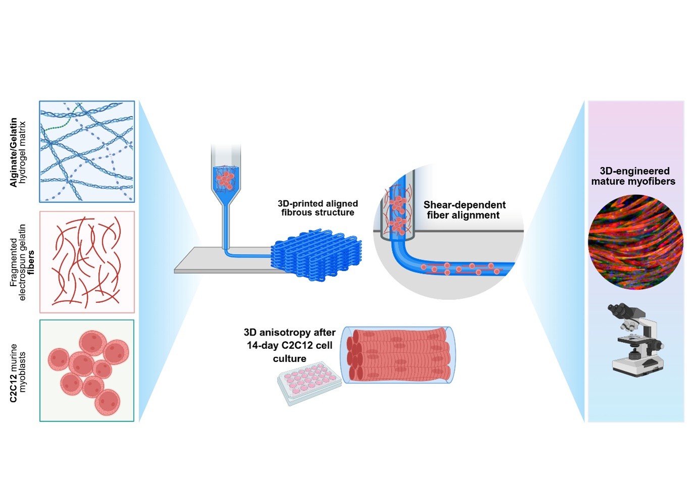
Replicating skeletal muscle architecture remains challenging in 3D bioprinting, as conventional bioinks lack multiscale directional cues. Herein, we propose a next-generation fibrous bioink composed of fragmented electrospun gelatin fibers (f-GFs), uniformly embedded in an alginate/gelatin hydrogel matrix (f-ALG/Gel). Upon microextrusion bioprinting, shear-induced f-GF alignment enabled the fabrication of microfilament-based scaffolds with intrinsic anisotropy. The resulting constructs exhibited high shape fidelity, favorable viscoelastic properties, and physiologically relevant stiffness (Young’s modulus: 16.1 ± 1.7 kPa). In vitro studies using C2C12 murine myoblasts demonstrated that the embedded f-GFs provided strong topographical guidance, enhancing cell alignment and myogenesis. After 14 days of culture, the f-ALG/Gel scaffolds supported a 2.5-fold increase in myotube fusion index and length, alongside reduced angular dispersion. These effects were achieved without the need for biochemical induction with a differentiation medium, underscoring the key role of structural cues at the micro- and nanoscale in C2C12 differentiation and maturation. In conclusion, this work proposes a scalable, cell-compatible strategy to recapitulate the hierarchical organization of skeletal muscle tissue within 3D-printed constructs. The platform holds broad potential for applications in regenerative medicine, skeletal muscle tissue modeling, and the engineering of cultured meat.
- Hulett NA, Scalzo RL, Reusch JEB. Glucose uptake by skeletal muscle within the contexts of type 2 diabetes and exercise: An integrated approach. Nutrients. 2022;14(3):647. doi: 10.3390/nu14030647
- Kim J, Kim IU, Lee ZF, Sim G, Jeon JS. Strategic approaches in generation of robust microphysiological 3D musculoskeletal tissue system. Adv Funct Mater. 2024;34(52). doi: 10.1002/adfm.202410872
- García-Lizarribar A, Villasante A, Lopez-Martin JA, et al. 3D bioprinted functional skeletal muscle models have potential applications for studies of muscle wasting in cancer cachexia. Biomater Adv. 2023;150:213426. doi: 10.1016/j.bioadv.2023.213426
- Zhuang P, An J, Chua CK, Tan LP. Bioprinting of 3D in vitro skeletal muscle models: A review. Mater Des. 2020;193:108794. doi: 10.1016/j.matdes.2020.108794
- Volpi M, Paradiso A, Costantini M, Świȩszkowski W. Hydrogel-based fiber biofabrication techniques for skeletal muscle tissue engineering. ACS Biomater Sci Eng. 2022;8(2):379-405. doi: 10.1021/acsbiomaterials.1c01145
- Lou H, Lu H, Zhang S, et al. Highly aligned myotubes formation of piscine satellite cells in 3D fibrin hydrogels of cultured meat. Int J Biol Macromol. 2024;282:136879. doi: 10.1016/j.ijbiomac.2024.136879
- Li X, Sim D, Wang Y, et al. Fiber-based biomaterial scaffolds for cell support towards the production of cultivated meat. Acta Biomater. 2025;191:292-307. doi: 10.1016/j.actbio.2024.11.006
- Sabetkish S, Currie P, Meagher L. Recent trends in 3D bioprinting technology for skeletal muscle regeneration. Acta Biomater. 2024;181:46-66. doi: 10.1016/j.actbio.2024.04.038
- Schwab A, Hélary C, Richards RG, Alini M, Eglin D, D’Este M. Tissue mimetic hyaluronan bioink containing collagen fibers with controlled orientation modulating cell migration and alignment. Mater Today Bio. 2020;7:100058. doi: 10.1016/j.mtbio.2020.100058
- Mehmood H, Kasher PR, Barrett-Jolley R, Walmsley GL. Aligning with the 3Rs: Alternative models for research into muscle development and inherited myopathies. BMC Vet Res. 2024;20(1):477. doi: 10.1186/s12917-024-04309-z
- Kang MS, Lee SH, Park WJ, Lee JE, Kim B, Han DW. Advanced techniques for skeletal muscle tissue engineering and regeneration. Bioengineering. 2020;7(3):99. doi: 10.3390/bioengineering7030099
- Trovato F, Imbesi R, Conway N, Castrogiovanni P. Morphological and functional aspects of human skeletal muscle. J Funct Morphol Kinesiol. 2016;1(3):289-302. doi: 10.3390/jfmk1030289
- Nikkhah M, Edalat F, Manoucheri S, Khademhosseini A. Engineering microscale topographies to control the cell– substrate interface. Biomaterials. 2012;33(21):5230-5246. doi: 10.1016/j.biomaterials.2012.03.079
- Wang L, Wu Y, Guo B, Ma PX. Nanofiber yarn/hydrogel core–shell scaffolds mimicking native skeletal muscle tissue for guiding 3D myoblast alignment, elongation, and differentiation. ACS Nano. 2015;9(9):9167-9179. doi: 10.1021/acsnano.5b03644
- Mueller C, Trujillo‐Miranda M, Maier M, Heath DE, O’Connor AJ, Salehi S. Effects of external stimulators on engineered skeletal muscle tissue maturation. Adv Mater Interfaces. 2021;8(1). doi: 10.1002/admi.202001167
- Ostrovidov S, Hosseini V, Ahadian S, et al. Skeletal muscle tissue engineering: Methods to form skeletal myotubes and their applications. Tissue Eng Part B Rev. 2014;20(5):403-436. doi: 10.1089/ten.teb.2013.0534
- Kim H, Jang J, Park J, et al. Shear-induced alignment of collagen fibrils using 3D cell printing for corneal stroma tissue engineering. Biofabrication. 2019;11(3):035017. doi: 10.1088/1758-5090/ab1a8b
- Moncal KK, Ozbolat V, Datta P, Heo DN, Ozbolat IT. Thermally-controlled extrusion-based bioprinting of collagen. J Mater Sci Mater Med. 2019;30(5):55. doi: 10.1007/s10856-019-6258-2
- Prendergast ME, Davidson MD, Burdick JA. A biofabrication method to align cells within bioprinted photocrosslinkable and cell-degradable hydrogel constructs via embedded fibers. Biofabrication. 2021;13(4). doi: 10.1088/1758-5090/AC25CC
- Chaudhuri O, Cooper-White J, Janmey PA, Mooney DJ, Shenoy VB. Effects of extracellular matrix viscoelasticity on cellular behaviour. Nature. 2020;584(7822):535-546. doi: 10.1038/s41586-020-2612-2
- Lloyd EM, Hepburn MS, Li J, et al. Multimodal three-dimensional characterization of murine skeletal muscle micro-scale elasticity, structure, and composition: Impact of dysferlinopathy, duchenne muscular dystrophy, and age on three hind-limb muscles. J Mech Behav Biomed Mater. 2024;160:106751. doi: 10.1016/j.jmbbm.2024.106751
- Choi S, Lee KY, Kim SL, et al. Fibre-infused gel scaffolds guide cardiomyocyte alignment in 3d-printed ventricles. Nat Mater. 2023;22(8):1039-1046. doi: 10.1038/s41563-023-01611-3
- Kamaraj M, Rezayof O, Barer A, et al. Development of silk microfiber-reinforced bioink for muscle tissue engineering and in situ printing by a handheld 3d printer. Biomater Adv. 2025;166:214057. doi: 10.1016/j.bioadv.2024.214057
- Li T, Hou J, Wang L, et al. Bioprinted anisotropic scaffolds with fast stress relaxation bioink for engineering 3D skeletal muscle and repairing volumetric muscle loss. Acta Biomater. 2023;156:21-36. doi: 10.1016/j.actbio.2022.08.037
- Stola GP, Paoletti C, Nicoletti L, et al. Internally-crosslinked alginate dialdehyde/alginate/gelatin-based hydrogels as bioinks for prospective cardiac tissue engineering applications. Int J Bioprint. 2024;10(6):544-566. doi: 10.36922/ijb.4014
- Tonda-Turo C, Gentile P, Saracino S, et al. Comparative analysis of gelatin scaffolds crosslinked by genipin and silane coupling agent. Int J Biol Macromol. 2011;49(4):700-706. doi: 10.1016/j.ijbiomac.2011.07.002
- Tonda-Turo C, Cipriani E, Gnavi S, et al. Crosslinked gelatin nanofibres: Preparation, characterisation and in vitro studies using glial-like cells. Mater Sci Eng: C. 2013;33(5):2723-2735. doi: 10.1016/j.msec.2013.02.039
- Giuntoli G, Muzio G, Actis C, et al. In-vitro characterization of a hernia mesh featuring a nanostructured coating. Front Bioeng Biotechnol. 2021;8. doi: 10.3389/fbioe.2020.589223
- John JV., McCarthy A, Wang H, et al. Engineering biomimetic nanofiber microspheres with tailored size, predesigned structure, and desired composition via gas bubble–mediated coaxial electrospray. Small. 2020;16(19). doi: 10.1002/smll.201907393
- Carmagnola I, Chiono V, Ruocco G, et al. PLGA membranes functionalized with gelatin through biomimetic mussel-inspired strategy. Nanomaterials. 2020;10(11):2184. doi: 10.3390/nano10112184
- Salgado-Delgado AM, González-Mondragón EG, Hernández-Pérez R, Salgado-Delgado R, Santana-Camilo JA, Olarte-Paredes A. Obtention and characterization of GO/Epoxy and GO-GPTMS/epoxy nanocompounds with different oxidation degrees and ultrasound methods. C (Basel). 2023;9(1):28. doi: 10.3390/c9010028
- Sitthiracha M, Kilmartin PA, Edmonds NR. Novel organic– inorganic hybrid materials based on epoxy-functionalized silanes. J Solgel Sci Technol. 2015;76(3):542-551. doi: 10.1007/s10971-015-3804-3
- Wang X, Wang M, Xu Y, Yin J, Hu J. A 3D-printable gelatin/alginate/ε-poly-l-lysine hydrogel scaffold to enable porcine muscle stem cells expansion and differentiation for cultured meat development. Int J Biol Macromol. 2024; 271:131980. doi: 10.1016/j.ijbiomac.2024.131980
- Yeong WY, Yu H, Lim KP, et al. Multiscale topological guidance for cell alignment via direct laser writing on biodegradable polymer. Tissue Eng Part C Methods. 2010;16(5):1011-1021. doi: 10.1089/ten.tec.2009.0604
- Guven S, Chen P, Inci F, Tasoglu S, Erkmen B, Demirci U. Multiscale assembly for tissue engineering and regenerative medicine. Trends Biotechnol. 2015;33(5):269-279. doi: 10.1016/j.tibtech.2015.02.003
- Kerativitayanan P, Carrow JK, Gaharwar AK. Nanomaterials for engineering stem cell responses. Adv Healthc Mater. 2015;4(11):1600-1627. doi: 10.1002/adhm.201500272
- Rizzi R, Bearzi C, Mauretti A, Bernardini S, Cannata S, Gargioli C. Tissue engineering for skeletal muscle regeneration. Muscles Ligaments Tendons J. 2012;2(3):230-234.
- Griffith LG, Swartz MA. Capturing complex 3D tissue physiology in vitro. Nat Rev Mol Cell Biol. 2006;7(3):211-224. doi: 10.1038/NRM1858
- Dutta SD, Ganguly K, Jeong MS, et al. Bioengineered lab-grown meat-like constructs through 3D bioprinting of antioxidative protein hydrolysates. ACS Appl Mater Interfaces. 2022;14(30):34513-34526. doi: 10.1021/acsami.2c10620
- Yi H, Forsythe S, He Y, et al. Tissue-specific extracellular matrix promotes myogenic differentiation of human muscle progenitor cells on gelatin and heparin conjugated alginate hydrogels. Acta Biomater. 2017;62:222-233. doi: 10.1016/j.actbio.2017.08.022
- Rao KM, Kim HJ, Won S, Choi SM, Han SS. Effect of grape seed extract on gelatin-based edible 3D-hydrogels for cultured meat application. Gels. 2023;9(1):65. doi: 10.3390/gels9010065
- Valliant EM, Romer F, Wang D, et al. Bioactivity in silica/ poly(γ-glutamic acid) sol–gel hybrids through calcium chelation. Acta Biomater. 2013;9(8):7662-7671. doi: 10.1016/j.actbio.2013.04.037
- Li X, Wang X, Yao D, et al. Effects of aligned and random fibers with different diameter on cell behaviors. Colloids Surf B Biointerfaces. 2018;171:461-467. doi: 10.1016/j.colsurfb.2018.07.045
- Tian F, Hosseinkhani H, Hosseinkhani M, et al. Quantitative analysis of cell adhesion on aligned micro- and nanofibers. J Biomed Mater Res A. 2008;84(2):291-299. doi: 10.1002/jbm.a.31304
- Martino F, Perestrelo AR, Vinarský V, Pagliari S, Forte G. Cellular mechanotransduction: From tension to function. Front Physiol. 2018;9. doi: 10.3389/fphys.2018.00824
- Davidenko N, Schuster CF, Bax D V., et al. Evaluation of cell binding to collagen and gelatin: A study of the effect of 2D and 3D architecture and surface chemistry. J Mater Sci Mater Med. 2016;27(10):148. doi: 10.1007/s10856-016-5763-9
- Abdin M, Salama MA, Riaz A, Akhtar HMS, Elsanat SY. Enhanced the entrapment and controlled release of syzygium cumini seeds polyphenols by modifying the surface and internal organization of alginate‐based microcapsules. J Food Process Preserv. 2021;45(1). doi: 10.1111/jfpp.15100
- Levato R, Jungst T, Scheuring RG, Blunk T, Groll J, Malda J. From shape to function: The next step in bioprinting. Adv Mater. 2020;32(12). doi: 10.1002/adma.201906423
- Tirella A, Orsini A, Vozzi G, Ahluwalia A. A phase diagram for microfabrication of geometrically controlled hydrogel scaffolds. Biofabrication. 2009;1(4):045002. doi: 10.1088/1758-5082/1/4/045002
- Nam SY, Park SH. ECM based bioink for tissue mimetic 3d bioprinting. Adv Exp Med Biol. 2018; 1064:335-353. doi: 10.1007/978-981-13-0445-3_20
- Malda J, Visser J, Melchels FP, et al. 25th anniversary article: Engineering hydrogels for biofabrication. Adv Mater. 2013;25(36):5011-5028. doi: 10.1002/adma.201302042
- Sakai S, Yoshii A, Sakurai S, Horii K, Nagasuna O. Silk fibroin nanofibers: A promising ink additive for extrusion three-dimensional bioprinting. Mater Today Bio. 2020;8:100078. doi: 10.1016/j.mtbio.2020.100078
- Jessop ZM, Al-Sabah A, Gao N, et al. Printability of pulp derived crystal, fibril and blend nanocellulose-alginate bioinks for extrusion 3D bioprinting. Biofabrication. 2019;11(4):045006. doi: 10.1088/1758-5090/ab0631
- Markstedt K, Mantas A, Tournier I, Martínez Ávila H, Hägg D, Gatenholm P. 3D bioprinting human chondrocytes with nanocellulose–alginate bioink for cartilage tissue engineering applications. Biomacromolecules. 2015;16(5):1489-1496. doi: 10.1021/acs.biomac.5b00188
- Seiti M, Mazzoldi EL, Pandini S, et al. FRESH 3D bioprinting of alginate – cellulose – gelatin constructs for soft tissue biofabrication. Procedia CIRP. 2024;125:42-47. doi: 10.1016/j.procir.2024.08.008
- Distler T, McDonald K, Heid S, Karakaya E, Detsch R, Boccaccini AR. Ionically and enzymatically dual cross-linked oxidized alginate gelatin hydrogels with tunable stiffness and degradation behavior for tissue engineering. ACS Biomater Sci Eng. 2020;6(7):3899-3914. doi: 10.1021/acsbiomaterials.0c00677
- Sonaye SY, Ertugral EG, Kothapalli CR, Sikder P. Extrusion 3D (bio)printing of alginate-gelatin-based composite scaffolds for skeletal muscle tissue engineering. Materials. 2022;15(22):7945. doi: 10.3390/ma15227945
- Sonnleitner D, Schrüfer S, Berglund L, Schubert DW, Lang G. Correlating rheology and printing performance of fiber-reinforced bioinks to assess predictive modelling for biofabrication. J Mater Res. 2021;36(19):3821-3832. doi: 10.1557/s43578-021-00276-5
- Hausmann MK, Rühs PA, Siqueira G, et al. Dynamics of cellulose nanocrystal alignment during 3D printing. ACS Nano. 2018;12(7):6926-6937. doi: 10.1021/acsnano.8b02366
- Chaturvedi V, Dye DE, Kinnear BF, van Kuppevelt TH, Grounds MD, Coombe DR. Interactions between skeletal muscle myoblasts and their extracellular matrix revealed by a serum free culture system. PLoS One. 2015;10(6):e0127675. doi: 10.1371/journal.pone.0127675
- Denes LT, Riley LA, Mijares JR, et al. Culturing C2C12 myotubes on micromolded gelatin hydrogels accelerates myotube maturation. Skelet Muscle. 2019;9(1):17. doi: 10.1186/s13395-019-0203-4
- Jensen JH, Cakal SD, Li J, et al. Large-scale spontaneous self-organization and maturation of skeletal muscle tissues on ultra-compliant gelatin hydrogel substrates. Sci Rep. 2020;10(1):13305. doi: 10.1038/s41598-020-69936-6
- Spedicati M, Zoso A, Mortati L, Chiono V, Marcello E, Carmagnola I. Three-dimensional microfibrous scaffold with aligned topography produced via a combination of melt-extrusion additive manufacturing and porogen leaching for in vitro skeletal muscle modeling. Bioengineering. 2024;11(4):332. doi: 10.3390/bioengineering11040332
- Engler AJ, Sen S, Sweeney HL, Discher DE. Matrix elasticity directs stem cell lineage specification. Cell. 2006;126(4):677-689.doi: 10.1016/j.cell.2006.06.044
- Cui W, Huang Y, Chen L, et al. Tiny yet tough: Maximizing the toughness of fiber-reinforced soft composites in the absence of a fiber-fracture mechanism. Matter. 2021;4(11):3646-3661. doi: 10.1016/j.matt.2021.08.013
- Heher P, Maleiner B, Prüller J, et al. A novel bioreactor for the generation of highly aligned 3D skeletal muscle-like constructs through orientation of fibrin via application of static strain. Acta Biomater. 2015;24:251-265. doi: 10.1016/j.actbio.2015.06.033
- Schwab A, Levato R, D’Este M, Piluso S, Eglin D, Malda J. Printability and shape fidelity of bioinks in 3D bioprinting. Chem Rev Am Chem Soc. 2020;120(19):11028-11055. doi: 10.1021/acs.chemrev.0c00084
- Heck T, Faccio G, Richter M, Thöny-Meyer L. Enzyme-catalyzed protein crosslinking. Appl Microbiol Biotechnol. 2013;97(2):461-475. doi: 10.1007/s00253-012-4569-z
- Kashiwagi T, Yokoyama K ichi, Ishikawa K, et al. Crystal Structure Of Microbial Transglutaminase From Streptoverticillium mobaraense. J Biol Chem. 2002;277(46):44252-44260. doi: 10.1074/jbc.M203933200
- Kitzmann M, Carnac G, Vandromme M, Primig M, Lamb NJC, Fernandez A. The muscle regulatory factors MyoD and Myf-5 undergo distinct cell cycle–specific expression in muscle cells. J Cell Biol. 1998;142(6):1447-1459. doi: 10.1083/jcb.142.6.1447
- Yang HS, Lee B, Tsui JH, et al. Electroconductive nanopatterned substrates for enhanced myogenic differentiation and maturation. Adv Healthc Mater. 2016;5(1):137-145. doi: 10.1002/adhm.201500003
- Niknezhad SV, Mehrali M, Khorasgani FR, et al. Enhancing volumetric muscle loss (VML) recovery in a rat model using super durable hydrogels derived from bacteria. Bioact Mater. 2024;38:540-558. doi: 10.1016/j.bioactmat.2024.04.006
- Kim K, Jin S, Shin M. Effect of 3D‐printable anisotropic fibrous hydrogels on fabricating artificial skeletal muscle constructs. Adv Ther (Weinh). 2024;7(1). doi: 10.1002/adtp.202300170
- Lee S, Kim W, Kim G. Efficient myogenic activities achieved through blade-casting-assisted bioprinting of aligned myoblasts laden in collagen bioink. Biomacromolecules. 2023;24(11):5219-5229. doi: 10.1021/acs.biomac.3c00749
- Li Q, Yu S, Wang Y, et al. Programmable embedded bioprinting for one-step manufacturing of arterial models with customized contractile and metabolic functions. Trends Biotechnol. 2025;43(4):918-945. doi: 10.1016/j.tibtech.2024.11.019