

Suspended 3D printing of polycaprolactone/ hydroxyapatite composites for the fabrication of complex bone scaffolds

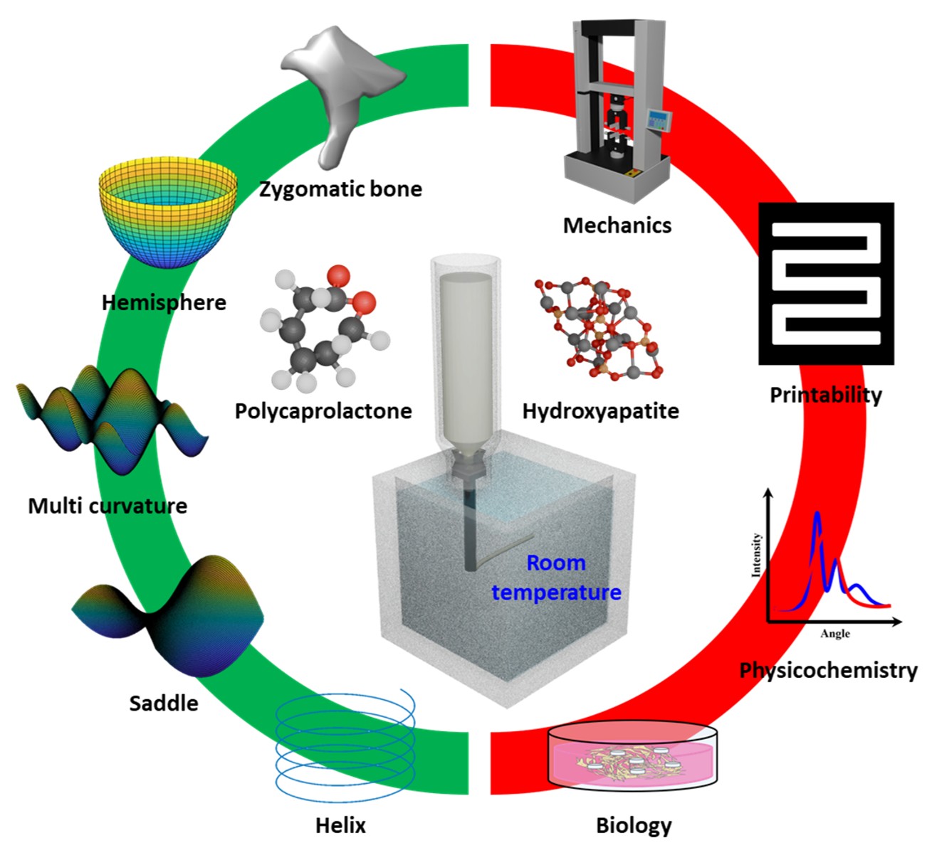
Extrusion-based three-dimensional (3D) printing has been rapidly advancing as a key technique for fabricating tissue-engineering scaffolds. However, printing complex structures with appropriate mechanical strength and biocompatibility remains a challenge. Suspended 3D printing is an emerging fabrication strategy that enables the generation of tissues or organs within a support medium that provides a stable printing environment without the need for additional support structures. This study presents a novel strategy for fabricating intricate scaffolds using suspended 3D printing of bioinks incorporating dissolved polycaprolactone (dPCL) and hydroxyapatite (HA). The optimized dPCL/HA bioink demonstrated up to an 85% reduction in print errors compared to conventional methods, significantly enhancing 3D printability. Moreover, mechanical assessments revealed a compressive Young’s modulus approximately 50 MPa higher in dPCL/HA scaffolds compared to dPCL scaffolds. Furthermore, dPCL/HA scaffolds outperformed both PCL and dPCL scaffolds in cell proliferation tests. Complex 3D shapes, including helices, saddles, multi-curvature structures, hollow hemispheres, and zygomatic bones, were successfully fabricated, demonstrating the ability to mimic natural and intricate anatomical structures of the human body. These approaches pave the way for 3D printing patient-specific and structurally robust bone constructs with enhanced mechanical and biological properties.
- Deng X, Yu C, Zhang X, et al. A chitosan-coated PCL/ nano-hydroxyapatite aerogel integrated with a nanofiber membrane for providing antibacterial activity and guiding bone regeneration. Nanoscale. 2024;16(20):9861-9874. doi: 10.1039/d4nr00563e
- Song T, Zhou J, Shi M, et al. Osteon-mimetic 3D nanofibrous scaffold enhances stem cell proliferation and osteogenic differentiation for bone regeneration. Biomater Sci. 2022;10(4):1090-1103. doi: 10.1039/d1bm01489g
- Romanazzo S, Hudson AR, Shiwarski DJ, et al. Synthetic bone‐like structures through omnidirectional ceramic bioprinting in cell suspensions. Adv Funct Mater. 2021;31(13):2008216. doi: 10.1002/adfm.202008216
- Kim MH, Chalisserry EP, Mondal S, Oh J, Nam SY. Silicon-substituted hydroxyapatite reinforced 3D printed gelatin membrane for guided bone regeneration. Mater Lett. 2021;304:130670. doi: 10.1016/j.matlet.2021.130670
- Lee H, Yoo JM, Ponnusamy NK, Nam SY. 3D-printed hydroxyapatite/gelatin bone scaffolds reinforced with graphene oxide: optimized fabrication and mechanical characterization. Ceram Int. 2022;48(7):10155-10163. doi: 10.1016/j.ceramint.2021.12.227
- Montalbano G, Molino G, Fiorilli S, Vitale-Brovarone C. Synthesis and incorporation of rod-like nano-hydroxyapatite into type I collagen matrix: a hybrid formulation for 3D printing of bone scaffolds. J Eur Ceram Soc. 2020;40(11):3689-3697. doi: 10.1016/j.jeurceramsoc.2020.02.018
- Shirzad M, Kang J, Kim G, Bodaghi M, Nam SY. Bioinspired 3D‐printed auxetic structures with enhanced fatigue behavior. Adv Eng Mater. 2024;26(20):2302036. doi: 10.1002/adem.202302036
- Hu K, Jin S, Wang CC. Support slimming for single material based additive manufacturing. Comput Aided Des. 2015;65:1-10. doi: 10.1016/j.cad.2015.03.001
- Jiang J, Xu X, Stringer J. Support structures for additive manufacturing: a review. J Manuf Mater Process. 2018;2(4):64. doi: 10.3390/jmmp2040064
- Alioglu MA, Yilmaz YO, Singh YP, et al. Nested biofabrication: matryoshka-inspired intra-embedded bioprinting. Small Methods. 2023;8(8):e2301325. doi: 10.1002/smtd.202301325
- Song KH, Highley CB, Rouff A, Burdick JA. Complex 3D‐printed microchannels within cell‐degradable hydrogels. Adv Funct Mater. 2018;28(31). doi: 10.1002/adfm.201801331
- Bhattacharjee T, Zehnder SM, Rowe KG, et al. Writing in the granular gel medium. Sci Adv. 2015;1(8):e1500655. doi: 10.1126/sciadv.1500655
- Wu W, DeConinck A, Lewis JA. Omnidirectional printing of 3D microvascular networks. Adv Mater. 2011;23(24):H178-H183. doi: 10.1002/adma.201004625
- Jin Y, Compaan A, Chai W, Huang Y. Functional nanoclay suspension for printing-then-solidification of liquid materials. ACS Appl Mater Interfaces. 2017;9(23):20057-20066. doi: 10.1021/acsami.7b02398
- Lee A, Hudson A, Shiwarski D, et al. 3D bioprinting of collagen to rebuild components of the human heart. Science. 2019;365(6452):482-487. doi: 10.1126/science.aav9051
- Gao C, Sow WT, Wang Y, et al. Hydrogel composite scaffolds with an attenuated immunogenicity component for bone tissue engineering applications. J Mater Chem B. 2021;9(8):2033-2041. doi: 10.1039/d0tb02588g
- Liu L, Yang B, Wang LQ, et al. Biomimetic bone tissue engineering hydrogel scaffolds constructed using ordered CNTs and HA induce the proliferation and differentiation of BMSCs. J Mater Chem B. 2020;8(3):558-567. doi: 10.1039/c9tb01804b
- Mondal S, Dey A, Pal U. Low temperature wet-chemical synthesis of spherical hydroxyapatite nanoparticles and their in situ cytotoxicity study. Adv Nano Res. 2016;4(4): 295-307. doi: 10.12989/anr.2016.4.4.295
- Shi Y, Wang L, Sun L, et al. Melt electrospinning writing PCL scaffolds after alkaline modification with outstanding cytocompatibility and osteoinduction. Int J Bioprint. 2023;9(6):1071. doi: 10.36922/ijb.1071
- Biscaia S, Branquinho MV, Alvites RD, et al. 3D printed poly(ε-caprolactone)/hydroxyapatite scaffolds for bone tissue engineering: a comparative study on a composite preparation by melt blending or solvent casting techniques and the influence of bioceramic content on scaffold properties. Int J Mol Sci. 2022;23(4):2318. doi: 10.3390/ijms23042318
- Rezaei A, Mohammadi MR. Development of hydroxyapatite nanorods-polycaprolactone composites and scaffolds derived from a novel in-situ sol-gel process. Tissue Eng Regen Med. 2012;9(6):295-303. doi: 10.1007/s13770-012-0002-z
- Wang Y, Liu L, Guo S. Characterization of biodegradable and cytocompatible nano-hydroxyapatite/polycaprolactone porous scaffolds in degradation in vitro. Polym Degrad Stabil. 2010;95(2):207-213. doi: 10.1016/j.polymdegradstab.2009.11.023
- Cestari F, Petretta M, Yang Y, Motta A, Grigolo B, Sglavo VM. 3D printing of PCL/nano-hydroxyapatite scaffolds derived from biogenic sources for bone tissue engineering. Sustain Mater Technol. 2021;29:e00318. doi: 10.1016/j.susmat.2021.e00318
- Zeng X, Meng Z, Qiu Z, He J, Fan J, Li D. Melt-based embedded printing for freeform fabrication of overhanging and flexible polycaprolactone scaffolds. Virt Phys Prototyp. 2023;18(1). doi: 10.1080/17452759.2023.2209778
- Jakus AE, Rutz AL, Jordan SW, et al. Hyperelastic “bone”: a highly versatile, growth factor-free, osteoregenerative, scalable, and surgically friendly biomaterial. Sci Transl Med. 2016;8(358):358ra127. doi: 10.1126/scitranslmed.aaf7704
- Moghadam MZ, Hassanajili S, Esmaeilzadeh F, Ayatollahi M, Ahmadi M. Formation of porous HPCL/LPCL/HA scaffolds with supercritical CO2 gas foaming method. J Mech Behav Biomed Mater. 2017;69:115-127. doi: 10.1016/j.jmbbm.2016.12.014
- Lee H-U, Jeong Y-S, Jeong S-Y, et al. Role of reactive gas in atmospheric plasma for cell attachment and proliferation on biocompatible poly ε-caprolactone film. Appl Surf Sci. 2008;254(18):5700-5705. doi: 10.1016/j.apsusc.2008.03.049
- Liu Z, Liu X, Ramakrishna S. Surface engineering of biomaterials in orthopedic and dental implants: strategies to improve osteointegration, bacteriostatic and bactericidal activities. Biotechnol J. 2021;16(7):2000116. doi: 10.1002/biot.20200011
- Wang L, Wang C, Zhou L, et al. Fabrication of a novel three-dimensional porous PCL/PLA tissue engineering scaffold with high connectivity for endothelial cell migration. Eur Polym J. 2021;161:110834. doi: 10.1016/j.eurpolymj.2021.110834
- Ma J, Lin L, Zuo Y, et al. Modification of 3D printed PCL scaffolds by PVAc and HA to enhance cytocompatibility and osteogenesis. RSC Adv. 2019;9(10):5338-5346. doi: 10.1039/c8ra06652c
- Oh D, Shirzad M, Kim MC, Chung E-J, Nam SY. Rheology-informed hierarchical machine learning model for the prediction of printing resolution in extrusion-based bioprinting. Int J Bioprint. 2023;9(6):1280. doi: 10.36922/ijb.1280
- Kim MH, Lee YW, Jung WK, Oh J, Nam SY. Enhanced rheological behaviors of alginate hydrogels with carrageenan for extrusion-based bioprinting. J Mech Behav Biomed Mater. 2019;98:187-194. doi: 10.1016/j.jmbbm.2019.06.014
- Huang B, Bártolo PJ. Rheological characterization of polymer/ceramic blends for 3D printing of bone scaffolds. Polym Test. 2018;68:365-378. doi: 10.1016/j.polymertesting.2018.04.033
- He M, Zhang F, Li C, et al. Mechanical properties and oral restoration applications of 3D printed aliphatic polyester-calcium composite materials. Alex Eng J. 2024;88:245-252. doi: 10.1016/j.aej.2024.01.042
- Motloung MP, Mofokeng TG, Ray SS. Viscoelastic, thermal, and mechanical properties of melt-processed poly (epsilon-caprolactone) (PCL)/hydroxyapatite (HAP) composites. Materials (Basel). 2021;15(1):104. doi: 10.3390/ma15010104
- Daskalakis E, Hassan MH, Omar AM, Cooper G, Weightman A, Bartolo P. Rheological behaviour of different composite materials for additive manufacturing of 3D bone scaffolds. J Mater Res Technol. 2023;24:3670-3682. doi: 10.1016/j.jmrt.2023.03.231
- Waters R, Alam P, Pacelli S, Chakravarti AR, Ahmed RP, Paul A. Stem cell-inspired secretome-rich injectable hydrogel to repair injured cardiac tissue. Acta Biomater. 2018;69:95-106. doi: 10.1016/j.actbio.2017.12.025
- Guo C, Wu J, Zeng Y, Li H. Construction of 3D bioprinting of HAP/collagen scaffold in gelation bath for bone tissue engineering. Regen Biomater. 2023;10:rbad067. doi: 10.1093/rb/rbad067
- Milazzo M, Fitzpatrick V, Owens CE, et al. 3D printability of silk/hydroxyapatite composites for microprosthetic applications. ACS Biomater Sci Eng. 2023;9(3): 1285-1295. doi: 10.1021/acsbiomaterials.2c01357
- Hinton TJ, Jallerat Q, Palchesko RN, et al. Three-dimensional printing of complex biological structures by freeform reversible embedding of suspended hydrogels. Sci Adv. 2015;1(9):e1500758. doi: 10.1126/sciadv.1500758
- Wu Q, Zhu F, Wu Z, et al. Suspension printing of liquid metal in yield-stress fluid for resilient 3D constructs with electromagnetic functions. NPJ Flex Electron. 2022;6(1):50. doi: 10.1038/s41528-022-00184-6
- Enea S, Moon SK. Guidelines for 3D printed springs using material extrusion. Rapid Prototyp J. 2022;28(3):409-427. doi: 10.1108/RPJ-04-2020-0078
- Okutani C, Yokota T, Miyazako H, Someya T. 3D printed spring‐type electronics with liquid metals for highly stretchable conductors and inductive strain/pressure sensors. Adv Mater Technol. 2022;7(7):2101657. doi: 10.1002/admt.202101657
- Rodriguez-Padilla C, Cuan-Urquizo E, Roman-Flores A, Gordillo JL, Vázquez-Hurtado C. Algorithm for the conformal 3D printing on non-planar tessellated surfaces: applicability in patterns and lattices. Appl Sci. 2021;11(16):7509. doi: 10.3390/app11167509
- Abdullah AM, Dunn ML, Yu K. Robotic 3D printing of continuous fiber reinforced thermoset composites. Adv Mater Technol. 2024;9(24):2400839. doi: 10.1002/admt.202400839
- Murali A, Parameswaran R. Extrusion 3D printing advances for craniomaxillofacial bone tissue engineering. Polym Plast Technol Mater. 2024;63(7):889-912. doi: 10.1080/25740881.2024.2307351
- Tao O, Kort-Mascort J, Lin Y, et al. The applications of 3D printing for craniofacial tissue engineering. Micromachines. 2019;10(7):480. doi: 10.3390/mi10070480
- Meglioli M, Naveau A, Macaluso GM, Catros S. 3D printed bone models in oral and cranio-maxillofacial surgery: a systematic review. 3D Print Med. 2020;6(1):30. doi: 10.1186/s41205-020-00082-5.
- Nyberg E, O’Sullivan A, Grayson W. scafSLICR: a MATLAB-based slicing algorithm to enable 3D-printing of tissue engineering scaffolds with heterogeneous porous microarchitecture. PLoS One. 2019;14(11):e0225007. doi: 10.1371/journal.pone.0225007
- Moiduddin K, Mian SH, Umer U, Alkhalefah H, Ahmed F, Hashmi FH. Design, analysis, and 3D printing of a patient-specific polyetheretherketone implant for the reconstruction of zygomatic deformities. Polymers. 2023;15(4):886. doi: 10.3390/polym15040886
- Van Belleghem S, Torres Jr L, Santoro M, et al. Hybrid 3D printing of synthetic and cell‐laden bioinks for shape retaining soft tissue grafts. Adv Funct Mater. 2020;30(3):1907145. doi: 10.1002/adfm.201907145
- Shiwarski DJ, Hudson AR, Tashman JW, et al. 3D bioprinting of collagen-based high-resolution internally perfusable scaffolds for engineering fully biologic tissue systems. Sci Adv. 2025;11(17):eadu5905. doi: 10.1126/sciadv.adu5905
- Guerrero-de-Mier A, Espinosa M, Domínguez M. Bricking: a new slicing method to reduce warping. Proc Eng. 2015;132:126-131. doi: 10.1016/j.proeng.2015.12.488
- Moeini S, Mohammadi MR, Simchi A. In-situ solvothermal processing of polycaprolactone/hydroxyapat ite nanocomposites with enhanced mechanical and biological performance for bone tissue engineering. Bioact Mater. 2017;2(3):146-155. doi: 10.1016/j.bioactmat.2017.04.004
- Shirzad M, Bodaghi M, Oh D, Yi M, Nam SY. Design and optimization of bioinspired auxetic structure for biomedical applications. Eur J Mech A Solids. 2024;103:105139. doi: 10.1016/j.euromechsol.2023.105139
- Almela T, Brook IM, Khoshroo K, et al. Simulation of cortico-cancellous bone structure by 3D printing of bilayer calcium phosphate-based scaffolds. Bioprinting. 2017;6:1-7. doi: 10.1016/j.bprint.2017.04.001
- Huang B, Caetano G, Vyas C, Blaker JJ, Diver C, Bartolo P. Polymer-ceramic composite scaffolds: the effect of hydroxyapatite and beta-tri-calcium phosphate. Materials (Basel). 2018;11(1):129. doi: 10.3390/ma11010129