

Low-concentration GOQD-functionalized Ti6Al4V scaffolds enhance osteogenesis and angiogenesis for vascularized bone regeneration

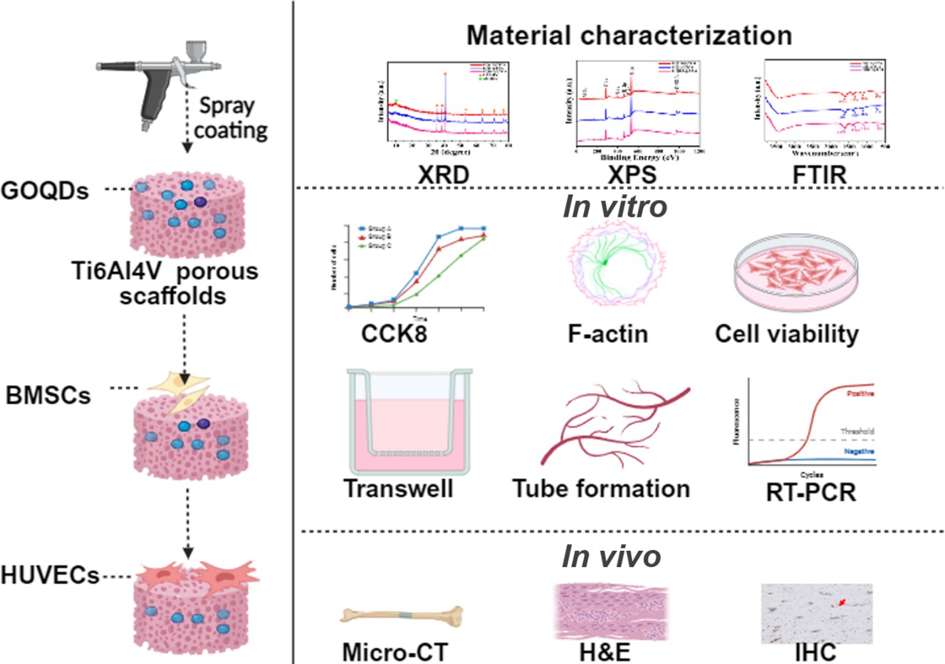
Graphene oxide quantum dots (GOQDs) possess excellent biocompatibility and have demonstrated potential to enhance osteogenesis and angiogenesis. The objective of this work was to construct Ti6Al4V porous scaffolds modified with different GOQD concentrations and investigate their influence on osteogenesis and angiogenesis. Porous Ti6Al4V scaffolds were coated with GOQDs at concentrations of 0.1, 1, and 10 μg/mL. The proliferation and adhesion of bone marrow mesenchymal stem cells (BMSCs) and human umbilical vein endothelial cells (HUVECs) on these scaffolds were evaluated using CCK-8 assay, immunofluorescence staining, and real time-polymerase chain reaction (RT-PCR). In vivo bone regeneration and angiogenesis were assessed through micro-computed tomography imaging and tissue section staining analysis. The results demonstrated successful deposition of GOQDs and the presence of characteristic functional groups. In vitro assays demonstrated that scaffolds coated with 0.1 μg/mL GOQDs significantly promoted the osteogenic/ angiogenic differentiation of BMSCs and HUVECs. In vivo experiments revealed that the 0.1 μg/mL GOQDs-coated scaffold (GQ@TC4) significantly enhanced bone formation and vascularization after 12 weeks. These findings suggest that Ti6Al4V biomimetic porous scaffolds functionalized with an optimal concentration (0.1 μg/ mL) of GOQDs can effectively promote both osteogenesis and angiogenesis, offering a promising strategy for bone defect repair.
- Ding Q, Cui J, Shen H, et al. Advances of nanomaterial applications in oral and maxillofacial tissue regeneration and disease treatment. Wiley Interdiscip Rev Nanomed Nanobiotechnol. 2020;13(2):e1669. doi: 10.1002/wnan.1669
- Liang Y, Li M, Yang Y, Qiao L, Xu H, Guo B. pH/glucose dual responsive metformin release hydrogel dressings with adhesion and self-healing via dual-dynamic bonding for athletic diabetic foot wound healing. ACS Nano. 2022;16(2):3194-3207. doi: 10.1021/acsnano.1c11040
- Li B, Shu R, Dai W, et al. Bioheterojunction-engineered polyetheretherketone implants with diabetic infectious micromilieu twin-engine powered disinfection for boosted osteogenicity. Small. 2022;18(45):e2203619. doi: 10.1002/smll.202203619
- Jang HJ, Kang MS, Jang J, et al. Harnessing 3D printed highly porous Ti-6Al-4V scaffolds coated with graphene oxide to promote osteogenesis. Biomater Sci. 2024;12(21): 5491-5503. doi: 10.1039/d4bm00970c
- Xiao F, Ye JH, Huang CX, et al. Gradient gyroid Ti6Al4V scaffolds with TiO(2) surface modification: promising approach for large bone defect repair. Biomater Adv. 2024;161:213899. doi: 10.1016/j.bioadv.2024.213899
- Zarei M, Hasanzadeh Azar M, Sayedain SS, et al. Material extrusion additive manufacturing of poly(lactic acid)/ Ti6Al4V@calcium phosphate core-shell nanocomposite scaffolds for bone tissue applications. Int J Biol Macromol. 024;255:128040. doi: 10.1016/j.ijbiomac.2023.128040
- Zhang X, Guan S, Qiu J, et al. Atomic layer deposition of tantalum oxide films on 3D-printed Ti6al4v scaffolds with enhanced osteogenic property for orthopedic implants. ACS Biomater Sci Eng. 2023;9(7):4197-4207. doi: 10.1021/acsbiomaterials.3c00217
- Shah NJ, Hyder MN, Moskowitz JS, et al. Surface-mediated bone tissue morphogenesis from tunable nanolayered implant coatings. Sci Transl Med. 2013;5(191):191ra83. doi: 10.1126/scitranslmed.3005576
- Shum JM, Gadomski BC, Tredinnick SJ, et al. Enhanced bone formation in locally-optimised, low-stiffness additive manufactured titanium implants: an in silico and in vivo tibial advancement study. Acta Biomater. 2023;156:202-213. doi: 10.1016/j.actbio.2022.04.006
- Abaricia JO, Farzad N, Heath TJ, Simmons J, Morandini L, Olivares-Navarrete R. Control of innate immune response by biomaterial surface topography, energy, and stiffness. Acta Biomater. 2021;133:58-73. doi: 10.1016/j.actbio.2021.04.021
- Ching HA, Choudhury D, Nine MJ, Abu Osman NA. Effects of surface coating on reducing friction and wear of orthopaedic implants. Sci Technol Adv Mater. 2014;15(1):014402. doi: 10.1088/1468-6996/15/1/014402
- Mediero A, Frenkel SR, Wilder T, He W, Mazumder A, Cronstein BN. Adenosine A2A receptor activation prevents wear particle-induced osteolysis. Sci Transl Med. 2012;4(135):135ra65. doi: 10.1126/scitranslmed.3003393
- Dinoro J, Maher M, Talebian S, et al. Sulfated polysaccharide-based scaffolds for orthopaedic tissue engineering. Biomaterials. 2019;214:119214. doi: 10.1016/j.biomaterials.2019.05.025
- Kapat K, Srivas PK, Rameshbabu AP, et al. Influence of porosity and pore-size distribution in Ti(6)Al(4) V foam on physicomechanical properties, osteogenesis, and quantitative validation of bone ingrowth by micro-computed tomography. ACS Appl Mater Interfaces. 2017;9(45):39235-39248. doi: 10.1021/acsami.7b13960
- Wang C, Xu D, Li S, et al. Effect of pore size on the physicochemical properties and osteogenesis of ti6al4v porous scaffolds with bionic structure. ACS Omega. 2020;5(44):28684-28692. doi: 10.1021/acsomega.0c03824
- Wang C, Xu D, Lin L, et al. Large-pore-size Ti6Al4V scaffolds with different pore structures for vascularized bone regeneration. Mater Sci Eng C Mater Biol Appl. 1;131:112499. doi: 10.1016/j.msec.2021.112499
- Wang C, Wu J, Liu L, et al. Improving osteoinduction and osteogenesis of ti6al4v alloy porous scaffold by regulating the pore structure. Front Chem. 2023;11:1190630. doi: 10.3389/fchem.2023.1190630
- Lee H, Lee MK, Han G, et al. Customizable design of multiple-biomolecule delivery platform for enhanced osteogenic responses via ‘tailored assembly system’. Bio Des Manuf. 2022;5(3):451-464. doi: 10.1007/s42242-022-00190-7
- Lee MK, Lee H, Park C, et al. Accelerated biodegradation of iron-based implants via tantalum-implanted surface nanostructures. Bioact Mater. 2022;9:239-250. doi: 10.1016/j.bioactmat.2021.07.003
- Deshmukh S, Chand A, Ghorpade R. Bio-mechanical analysis of porous Ti-6Al-4V scaffold: a comprehensive review on unit cell structures in orthopaedic application. Biomed Phys Eng Express. 2024;10(6):062003. doi: 10.1088/2057-1976/ad8202
- Unsworth T. Proceedings of the Institution of Mechanical Engineers Part H. Proc Inst Mech Eng H. 2008;222(7):i. doi: 10.1177/095441190822200701
- Awonusi BO, Li H, Yin Z, Zhao J, Yang K, Li J. Surface modification of Zn-Cu Alloy with heparin nanoparticles for urinary implant applications. ACS Appl Bio Mater. 2024;7(3):1748-1762. doi: 10.1021/acsabm.3c01177
- Ehlert M, Radtke A, Bartmanski M, Piszczek P. Evaluation of the cathodic electrodeposition effectiveness of the hydroxyapatite layer used in surface modification of Ti6Al4V-based biomaterials. Materials (Basel). 2022;15(19):6925. doi: 10.3390/ma15196925
- Teixeira-Santos R, Belo S, Vieira R, Mergulhao FJM, Gomes LC. Graphene-based composites for biomedical applications: surface modification for enhanced antimicrobial activity and biocompatibility. Biomolecules. 2023;13(11):1571. doi: 10.3390/biom13111571
- Kang MS, Jeong SJ, Lee SH, et al. Reduced graphene oxide coating enhances osteogenic differentiation of human mesenchymal stem cells on Ti surfaces. Biomater Res. 2021;25(1):4. doi: 10.1186/s40824-021-00205-x
- Li P, Di Stasio F, Eda G, et al. Luminescent properties of a water-soluble conjugated polymer incorporating graphene-oxide quantum dots. Chemphyschem. 2015;16(6):1258-1262. doi: 10.1002/cphc.201402744
- Liu G, Zhang K, Ma K, Care A, Hutchinson MR, Goldys EM. Graphene quantum dot-based “switch-on” nanosensors for intracellular cytokine monitoring. Nanoscale. 2017;9(15):4934-4943. doi: 10.1039/c6nr09381g
- Yang X, Zhao Q, Chen Y, et al. Effects of graphene oxide and graphene oxide quantum dots on the osteogenic differentiation of stem cells from human exfoliated deciduous teeth. Artif Cells Nanomed Biotechnol. 2019;47(1):822-832. doi: 10.1080/21691401.2019.1576706
- Lin L, Zheng Y, Wang C, Li P, Xu D, Zhao W. Concentration-dependent cellular uptake of graphene oxide quantum dots promotes the odontoblastic differentiation of dental pulp cells via the AMPK/mTOR pathway. ACS Omega. 2023;8(6):5393-5405. doi: 10.1021/acsomega.2c06508
- Liao TT, Deng QY, Wu BJ, et al. Dose-dependent cytotoxicity evaluation of graphite nanoparticles for diamond-like carbon film application on artificial joints. Biomed Mater. 2017;12(1):015018. doi: 10.1088/1748-605X/aa52ca
- Qiu J, Li D, Mou X, et al. Effects of graphene quantum dots on the self-renewal and differentiation of mesenchymal stem cells. Adv Healthc Mater. 2016;5(6):702-710. doi: 10.1002/adhm.201500770
- Xu D, Wang C, Wu J, et al. Effects of low-concentration graphene oxide quantum dots on improving the proliferation and differentiation ability of bone marrow mesenchymal stem cells through the Wnt/beta-catenin signaling pathway. ACS Omega. 2022;7(16):13546-13556. doi: 10.1021/acsomega.1c06892
- Xie H, Cao T, Rodriguez-Lozano FJ, Luong-Van EK, Rosa V. Graphene for the development of the next-generation of biocomposites for dental and medical applications. Dental Mater. 2017;33(7):765-774. doi: 10.1016/j.dental.2017.04.008
- Pruna A, Cembrero J, Pullini D, Mocioiu AM, Busquets- Mataix D. Effect of reduced graphene oxide on photocatalytic properties of electrodeposited ZnO. Appl Phys A Mater. 2017;123(12):792. doi: 10.1007/s00339-017-1424-1
- Yan X, An N, Zhang Z, et al. Graphene oxide quantum dots-preactivated dental pulp stem cells/GelMA facilitates mitophagy-regulated bone regeneration. Int J Nanomed. 2024;19:10107-10128. doi: 10.2147/IJN.S480979
- An N, Yan X, Qiu Q, et al. Human periodontal ligament stem cell sheets activated by graphene oxide quantum dots repair periodontal bone defects by promoting mitochondrial dynamics dependent osteogenic differentiation. J Nanobiotechnol. 2024;22(1):133. doi: 10.1186/s12951-024-02422-7
- Jia ZJ, Shi YY, Xiong P, et al. From solution to biointerface: graphene self-assemblies of varying lateral sizes and surface properties for biofilm control and osteodifferentiation. ACS Appl Mater Inter. 2016;8(27):17151-17165. doi: 10.1021/acsami.6b05198
- Kang ES, Song I, Kim DS, et al. Size-dependent effects of graphene oxide on the osteogenesis of human adipose-derived mesenchymal stem cells. Colloid Surface B. 2018;169:20-29. doi: 10.1016/j.colsurfb.2018.04.053
- Li XJ, Lin KL, Wang ZL. Enhanced growth and osteogenic differentiation of MC3T3-E1 cells on Ti6Al4V alloys modified with reduced graphene oxide. Rsc Adv. 2017;7(24):14430-14437. doi: 10.1039/c6ra25832h
- Li D, Dai D, Wang J, Zhang C. Honeycomb bionic graphene oxide quantum dot/layered double hydroxide composite nanocoating promotes osteoporotic bone regeneration via activating mitophagy. Small. 2024;20(50):e2403907. doi: 10.1002/smll.202403907
- Ha HD, Jang MH, Liu F, Cho YH, Seo TS. Upconversion photoluminescent metal ion sensors via two photon absorption in graphene oxide quantum dots. Carbon. 2015;81:367-375. doi: 10.1016/j.carbon.2014.09.069
- Qiu JJ, Geng H, Wang DH, et al. Layer-number dependent antibacterial and osteogenic behaviors of graphene oxide electrophoretic deposited on titanium. ACS Appl Mater Inter. 2017;9(14):12253-12263. doi: 10.1021/acsami.7b00314
- Chen X, Sun Z, Peng X, et al. Graphene oxide/black phosphorus functionalized collagen scaffolds with enhanced near-infrared controlled in situ biomineralization for promoting infectious bone defect repair through PI3K/Akt pathway. ACS Appl Mater Interfaces. 2024;16(38):50369-50388. doi: 10.1021/acsami.4c10284
- Kiroshka VV, Petrova VA, Chernyakov DD, et al. Influence of chitosan-chitin nanofiber composites on cytoskeleton structure and the proliferation of rat bone marrow stromal cells. J Mater Sci Mater M. 2017;28(1):21. doi: 10.1007/s10856-016-5822-2
- Xue DT, Chen EM, Zhong HM, et al. Immunomodulatory properties of graphene oxide for osteogenesis and angiogenesis. Int J Nanomed. 2018;13:5799-5810. doi: 10.2147/Ijn.S170305
- Zhou J, Zhao L. Multifunction Sr, Co and F co-doped microporous coating on titanium of antibacterial, angiogenic and osteogenic activities. Sci Rep. 2016;6:29069. doi: 10.1038/srep29069
- Jiang X, Chen X, Li Q, et al. Synergistic effects of polydopamine-coated reduced graphene oxide on osteogenesis and anti-inflammation in periodontitis. J Mater Sci Mater Med. 2025;36(1):51. doi: 10.1007/s10856-025-06905-3
- Ma R, Tang SC, Tan HL, et al. Preparation, characterization, and in vitro osteoblast functions of a nano-hydroxyapatite/ polyetheretherketone biocomposite as orthopedic implant material. Int J Nanomed. 2014;9:3949-3961. doi: 10.2147/Ijn.S67358
- Sankar D, Shalumon KT, Chennazhi KP, Menon D, Jayakumar R. Surface plasma treatment of poly(caprolactone) micro, nano, and multiscale fibrous scaffolds for enhanced osteoconductivity. Tissue Eng Pt A. 2014;20(11-12):1689-1702. doi: 10.1089/ten.tea.2013.0569
- Park JB, Ahn JY, Yang WS, et al. Stacked graphene with nanoscale wrinkles supports osteogenic differentiation of human adipose-derived stromal cells. 2D Mater. 2021;8(2):025034. doi: 10.1088/2053-1583/abe105
- Lamalice L, Le Boeuf F, Huot J. Endothelial cell migration during angiogenesis. Circ Res. 2007;100(6):782-794. doi: 10.1161/01.RES.0000259593.07661.1e
- Wang Q, Chen MC, Schafer NP, et al. Assemblies of calcium/calmodulin-dependent kinase II with actin and their dynamic regulation by calmodulin in dendritic spines. Proc Natl Acad Sci U S A. 2019;116(38): 18937-18942. doi: 10.1073/pnas.1911452116
- Cota Teixeira S, Silva Lopes D, Santos da Silva M, et al. Pentachloropseudilin impairs angiogenesis by disrupting the actin cytoskeleton, integrin trafficking and the cell cycle. Chembiochem. 2019;20(18):2390-2401. doi: 10.1002/cbic.201900203
- Yadunandanan Nair N, Samuel V, Ramesh L, Marib A, David DT, Sundararaman A. Actin cytoskeleton in angiogenesis. Biol Open. 2022;11(12):bio058899. doi: 10.1242/bio.058899
- Nakayama M, Nakayama A, van Lessen M, et al. Spatial regulation of VEGF receptor endocytosis in angiogenesis. Nat Cell Biol. 2013;15(3):249-260. doi: 10.1038/ncb2679
- Soker S, Machado M, Atala A. Systems for therapeutic angiogenesis in tissue engineering. World J Urol. 2000;18(1):10-18. doi: 10.1007/Pl00007070
- Liu WC, Chen SH, Zheng LZ, Qin L. Angiogenesis assays for the evaluation of angiogenic properties of orthopaedic biomaterials-a general review. Adv Healthc Mater. 2017;6(5):1600434. doi: 10.1002/adhm.201600434