

Neural cell responses to spinal implant biomaterials in a 3D-bioprinted cell culture model

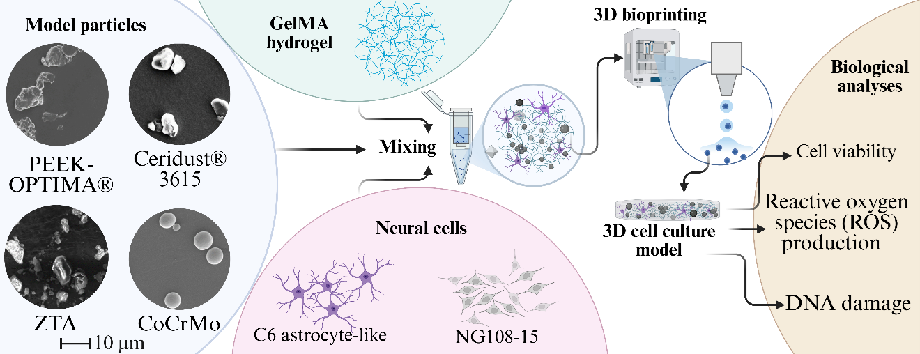
Spinal implants are vital for treating spinal disorders, yet wear particle-induced complications threaten their long-term success. Despite this, the direct effects of implant-derived particles on neural cells remain largely unexplored, especially given the limitations of conventional 2D culture models to capture such complex interactions. The current study introduces a novel in vitro platform consisting of a 3D-bioprinted gelatin methacryloyl (GelMA) hydrogel embedded with neural cells (C6 astrocyte-like and NG108-15 neurons) and spinal implant biomaterial particles, designed to model the spinal cord microenvironment with enhanced physiological relevance. As the first of its kind, this cell-particle-laden system supports the evaluation of neural cell responses to spinal biomaterial particles, including polymers, PEEK-OPTIMA™ and polyethylene Ceridust® 3615, zirconia-toughened alumina (ZTA) ceramic, and CoCrMo metal alloy. The bioprinted platform demonstrated excellent compatibility with various neural cell types and particle compositions, enabling a wide range of biological assays. Cell viability within the 3D model was comparable to traditional 2D cultures, affirming its ability to sustain cell survival while offering improved biomimicry. Biological assays assessing cell viability, reactive oxygen species (ROS) production, and DNA damage provided critical insights into material-specific and time-dependent cellular responses. While no significant cytotoxic effects were observed in short-term cultures, distinct variations in ROS production, and viability emerged based on biomaterial type and exposure duration. Overall, this versatile 3D-bioprinted system presents a robust, scalable tool for mechanistic and toxicological studies of spinal implant wear particles under physiologically relevant conditions.
- Tavakoli J, Diwan AD, Tipper JL. Advanced strategies for the regeneration of lumbar disc annulus fibrosus. Int J Mol Sci. 2020;21(14):4889. doi: 10.3390/ijms21144889
- Wen DJ, Tavakoli J, Tipper JL. Lumbar total disc replacements for degenerative disc disease: a systematic review of outcomes with a minimum of 5 years follow-up. Global Spine J. 2024;14(6):1827-1837. doi: 10.1177/21925682241228756
- Zeegers W, Bohnen L, Laaper M, Verhaegen M. Artificial disc replacement with the modular type SB Charite III: 2-year results in 50 prospectively studied patients. Eur Spine J. 1999;8:210-217. doi: 10.1007/s005860050160
- Vicars R, Hyde PJ, Brown TD, et al. The effect of anterior– posterior shear load on the wear of ProDisc-L TDR. Eur Spine J. 2010;19(8):1356-1362. doi: 10.1007/s00586-010-1396-8
- Vicars R, Prokopovich P, Brown TD, et al. The effect of anterior-posterior shear on the wear of CHARITÉ total disc replacement. Spine (Phila Pa 1976). 2012;37(9):E528-E534. doi: 10.1097/BRS.0b013e31823cbd6e
- Hallab NJ. A review of the biologic effects of spine implant debris: fact from fiction. SAS J. 2009;3(4):143-160. doi: 10.1016/j.esas.2009.11.005
- Chang B-S, Brown PR, Sieber A, Valdevit A, Tateno K, Kostuik JP. Evaluation of the biological response of wear debris. Spine J. 2004;4(6):S239-S244. doi: 10.1016/j.spinee.2004.07.014
- Ganko R, Madhavan A, Hamouda W, et al. Spinal implant wear particles: generation, characterization, biological impacts, and future considerations. iScience. 2025;28(4):112193. doi: 10.1016/j.isci.2025.112193
- Punt IM, Austen S, Cleutjens JP, et al. Are periprosthetic tissue reactions observed after revision of total disc replacement comparable to the reactions observed after total hip or knee revision surgery? Spine (Phila Pa 1976). 2012;37(2):150-159. doi: 10.1097/brs.0b013e3182154c22
- San-Juan R, Paredes I, Ramírez-Nava E, et al. Reduction of instrumentation-related spine surgical site infections after optimization of surgical techniques. a single center retrospective analysis. Global Spine J. 2024;14(2):438-446. doi: 10.1177/21925682221109557
- Xu R, Ebraheim NA, Nadaud MC, Phillips ER. Local tissue of the lumbar spine response to titanium plate-screw system. Spine (Phila Pa 1976). 1996;21(7):871-873. doi: 10.1097/00007632-199604010-00020
- Lin T-h, Tamaki Y, Pajarinen J, et al. Chronic inflammation in biomaterial-induced periprosthetic osteolysis: NF-κB as a therapeutic target. Acta Biomater. 2014;10(1):1-10. doi: 10.1016/j.actbio.2013.09.034
- Zairi F, Remacle JM, Allaoui M, Assaker R. Delayed hypersensitivity reaction caused by metal-on-metal total disc replacement: case report. J Neurosurg Spine. 2013;19(3):389-391. doi: 10.3171/2013.6.spine121010
- Zeh A, Planert M, Siegert G, Lattke P, Held A, Hein W. Release of cobalt and chromium ions into the serum following implantation of the metal-on-metal Maverick-type artificial lumbar disc (Medtronic Sofamor Danek). Spine (Phila Pa 1976). 2007;32(3):348-352. doi: 10.1097/01.brs.0000253599.89694.c0
- AlZeedi M, Al Rawahi S, Muwanis M, Alraiyes TM, Al Farii H, Jarzem P. Pseudotumor after total disc replacement in the lumbar spine: a case report and review of the literature. N Am Spine Soc J. 2022;9:100107. doi: 10.1016/j.xnsj.2022.100107
- Tavakoli J, Hu Q, Tipper JL, Tang Y. Aggregation-induced emission biomarkers for early detection of orthopaedic implant failure. Aggregate. 2024;5(6):e645. doi: 10.1002/agt2.645
- Austen S, Punt IM, Cleutjens JP, et al. Clinical, radiological, histological and retrieval findings of Activ-L and Mobidisc total disc replacements: a study of two patients. Eur Spine J. 2012;21:513-520. doi: 10.1007/s00586-011-2141-7
- Yang G, Gu M, Chen W, et al. SPHK-2 promotes the particle-induced inflammation of RAW264.7 by maintaining consistent expression of TNF-α and IL-6. Inflammation. 2018;41(4):1498-1507. doi: 10.1007/s10753-018-0795-6
- Yoshitake F, Itoh S, Narita H, Ishihara K, Ebisu S. Interleukin-6 directly inhibits osteoclast differentiation by suppressing receptor activator of NF-kappaB signaling pathways. J Biol Chem. 2008;283(17):11535-11540. doi: 10.1074/jbc.m607999200
- Ayers R, Miller M, Schowinsky J, Burger E, Patel V, Kleck C. Three cases of metallosis associated with spine instrumentation. J Mater Sci Mater Med. 2017;29(1):3. doi: 10.1007/s10856-017-6011-7
- Punt IM, Cleutjens JPM, de Bruin T, et al. Periprosthetic tissue reactions observed at revision of total intervertebral disc arthroplasty. Biomaterials. 2009;30(11):2079-2084. doi: 10.1016/j.biomaterials.2008.12.071
- Cunningham BW, Orbegoso CM, Dmitriev AE, et al. The effect of spinal instrumentation particulate wear debris: an in vivo rabbit model and applied clinical study of retrieved instrumentation cases. Spine J. 2003;3(1):19-32. doi: 10.1016/S1529-9430(02)00443-6
- Lin T-h, Yao Z, Sato T, et al. Suppression of wear-particle-induced pro-inflammatory cytokine and chemokine production in macrophages via NF-κB decoy oligodeoxynucleotide: a preliminary report. Acta Biomater. 2014;10(8):3747-3755. doi: 10.1016/j.actbio.2014.04.034
- Luo G, Li Z, Wang Y, et al. Resveratrol protects against titanium particle-induced aseptic loosening through reduction of oxidative stress and inactivation of NF-κB. Inflammation. 2016;39(2):775-785. doi: 10.1007/s10753-016-0306-6
- Hallab NJ, Cunningham BW, Jacobs JJ. Spinal implant debris-induced osteolysis. Spine (Phila Pa 1976). 2003;28(20S):S125-S138. doi: 10.1097/00007632-200310151-00006
- Lee H, Phillips JB, Hall RM, Tipper JL. Neural cell responses to wear debris from metal-on-metal total disc replacements. Eur Spine J. 2020;29(11):2701-2712. doi: 10.1007/s00586-019-06177-w
- Papageorgiou I, Marsh R, Tipper JL, Hall RM, Fisher J, Ingham E. Interaction of micron and nano‐sized particles with cells of the dura mater. J Biomed Mater Res B Appl Biomater. 2014;102(7):1496-1505. doi: 10.1002/jbm.b.33129
- Behl B, Papageorgiou I, Brown C, et al. Biological effects of cobalt-chromium nanoparticles and ions on dural fibroblasts and dural epithelial cells. Biomaterials. 2013;34(14):3547-3558. doi: 10.1016/j.biomaterials.2013.01.023
- Papageorgiou I, Abberton T, Fuller M, Tipper JL, Fisher J, Ingham E. Biological effects of clinically relevant cocr nanoparticles in the dura mater: an organ culture study. Nanomaterials. 2014;4(2):485-504. doi: 10.3390/nano4020485
- Cunningham BW, Hallab NJ, Hu N, McAfee PC. Epidural application of spinal instrumentation particulate wear debris: a comprehensive evaluation of neurotoxicity using an in vivo animal model. J Neurosurg Spine. 2013;19(3):336-350. doi: 10.3171/2013.5.spine13166
- Stoodley MA, Jones NR, Brown CJ. Evidence for rapid fluid flow from the subarachnoid space into the spinal cord central canal in the rat. Brain Res. 1996;707(2):155-164. doi: 10.1016/0006-8993(95)01228-1
- Stoodley MA, Brown SA, Brown CJ, Jones NR. Arterial pulsation-dependent perivascular cerebrospinal fluid flow into the central canal in the sheep spinal cord. J Neurosurg. 1997;86(4):686-693. doi: 10.3171/jns.1997.86.4.0686
- Rad MA, Mahmodi H, Filipe EC, Cox TR, Kabakova I, Tipper JL. Micromechanical characterisation of 3D bioprinted neural cell models using Brillouin microspectroscopy. Bioprinting. 2022;25:e00179. doi: 10.1016/j.bprint.2021.e00179
- Asif IM. Characterisation and Biological Impact of Wear Particles from Composite Ceramic Hip Replacements. University of Leeds; 2018. https://etheses.whiterose.ac.uk/id/oai_id/oai:etheses. whiterose.ac.uk:20563
- Liu A, Richards L, Bladen CL, Ingham E, Fisher J, Tipper JL. The biological response to nanometre-sized polymer particles. Acta Biomater. 2015;23:38-51. doi: 10.1016/j.actbio.2015.05.016
- Kim A, Mo K, Choe S, Shin M, Yoon H. Comprehensive insight into 3D bioprinting technology for brain tumor modelling. IJB. 2024;10(6):4166. doi: 10.36922/ijb.4166
- Tang H, Zhao E, Lai Y, et al. 3D bioprinting techniques and hydrogels for osteochondral integration regeneration. IJB. 2024;10(6):4472. doi: 10.36922/ijb.4472
- Hou Y-C, Cui X, Qin Z, et al. Three-dimensional bioprinting of artificial blood vessel: process, bioinks, and challenges. IJB. 2023;9(4):740. doi: 10.18063/ijb.740
- Du Z, Zhu Z, Wang Y. The degree of peri-implant osteolysis induced by PEEK, CoCrMo, and HXLPE wear particles: a study based on a porous Ti6Al4V implant in a rabbit model. J Orthop Surg Res. 2018;13(1):23. doi: 10.1186/s13018-018-0736-y
- Hallab NJ, McAllister K, Brady M, Jarman-Smith M. Macrophage reactivity to different polymers demonstrates particle size- and material-specific reactivity: PEEK-OPTIMA® particles versus UHMWPE particles in the submicron, micron, and 10 micron size ranges. J Biomed Mater Res B Appl Biomater. 2012;100B(2):480-492. doi: 10.1002/jbm.b.31974
- Green TR, Fisher J, Stone M, Wroblewski BM, Ingham E. Polyethylene particles of a ‘critical size’ are necessary for the induction of cytokines by macrophages in vitro. Biomaterials. 1998;19(24):2297-2302. doi: 10.1016/s0142-9612(98)00140-9
- Germain M, Hatton A, Williams S, et al. Comparison of the cytotoxicity of clinically relevant cobalt–chromium and alumina ceramic wear particles in vitro. Biomaterials. 2003;24(3):469-479. doi: 10.1016/s0142-9612(02)00360-5
- Bishop ES, Mostafa S, Pakvasa M, et al. 3-D bioprinting technologies in tissue engineering and regenerative medicine: Current and future trends. Genes Dis. 2017;4(4): 185-195. doi: 10.1016/j.gendis.2017.10.002
- Yu J, Park SA, Kim WD, et al. Current advances in 3D bioprinting technology and its applications for tissue engineering. Polymers. 2020;12(12):2958. doi: 10.3390/polym12122958
- Hallab NJ, Bao Q-B, Brown T. Assessment of epidural versus intradiscal biocompatibility of PEEK implant debris: an in vivo rabbit model. Eur Spine J. 2013;22:2740-2751. doi: 10.1007/s00586-013-2904-4
- Stratton-Powell AA, Pasko KM, Brockett CL, Tipper JL. The biologic response to polyetheretherketone (PEEK) wear particles in total joint replacement: a systematic review. Clin Orthop Relat Res. 2016;474(11):2394-2404. doi: 10.1007/s11999-016-4976-z
- Yarrow-Wright LE. Development of a Novel 3D In Vitro Model to Measure Cellular Response to Antioxidant Doped Highly Cross-Linked Ultra High Molecular Weight Polyethylene Wear Debris. University of Leeds; 2018. https://etheses.whiterose.ac.uk/id/oai_id/oai:etheses. whiterose.ac.uk:24128
- Kölle L, Ignasiak D, Ferguson SJ, Helgason B. Ceramics in total disc replacements: a scoping review. Clin Biomech. 2022;100:105796. doi: 10.1016/j.clinbiomech.2022.105796
- Jaksa L, Aryeetey OJ, Hatamikia S, et al. 3D-printed multi-material liver model with simultaneous mechanical and radiological tissue-mimicking features for improved realism. IJB. 2023;9(4):721. doi: 10.18063/ijb.721
- Yuk JC, Nam KH, Park SH. Additive-manufactured synthetic bone model with biomimicking tunable mechanical properties for evaluation of medical implants. IJB. 2024;10(1):1067. doi: 10.36922/ijb.1067