

Tiny bricks for oral bioprinting: Exploring gingiva and dental pulp-derived organ building blocks

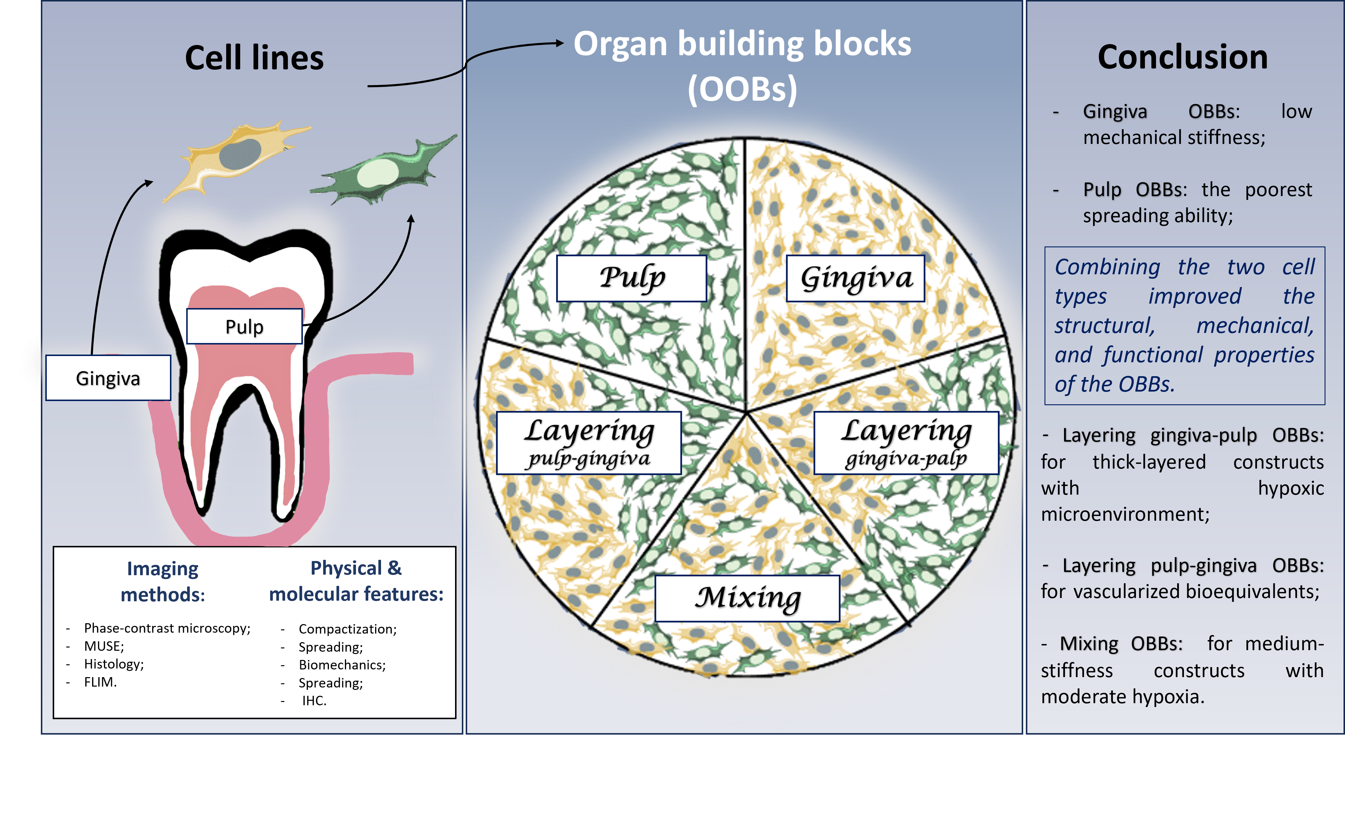
Today, organ building blocks (OBBs) serve as important tools for in vitro tissue modeling, personalized medicine, and regenerative approaches. Despite substantial advances in dental reconstruction methods, tissues of the oral cavity remain challenging to regenerate due to their complex structure and microenvironment. However, effective regeneration of the periodontal complex, e.g., in diseases such as periodontitis, persists, as current methods do not achieve complete tissue restoration. Cells from the gingiva and dental pulp are accessible sources of mesenchymal stem cells with high regenerative potential, making them promising materials for creating OBBs. These cells can serve as fundamental units for restoring the periodontal complex using techniques such as 3D bioprinting. This study aims to characterize and compare OBBs derived from gingival cells, pulp cells, and their combinations by assessing key parameters, including morphology, extracellular matrix composition, biomechanical properties, histology, and metabolic activity. Combining the two cell types improved the structural, mechanical, and functional properties of OBBs, making them more suitable for bioprinting than those derived from a single cell type. Moreover, all types of OBBs from the two cell cultures may be suitable as components of bioinks, depending on the specific purposes. The results provide insights into the potential use of these cell sources for tissue engineering and the development of personalized periodontal bio-constructs that may significantly improve treatment approaches for oral diseases.
- De Lauretis A, Ovrebo O, Romandini M, Lyngstadaas SP, Rossi F, Haugen HJ. From Basic Science to Clinical Practice: A Review of Current Periodontal/Mucogingival Regenerative Biomaterials. Adv Sci. 2024;11(17):2308848. doi: 10.1002/advs.202308848
- Wolf KJ, Weiss JD, Uzel SGM, Skylar-Scott MA, Lewis JA. Biomanufacturing human tissues via organ building blocks. Cell Stem Cell. 2022;29(5):667-677. doi: 10.1016/j.stem.2022.04.012
- Bhise NS, Manoharan V, Massa S, et al. A liver-on-a-chip platform with bioprinted hepatic spheroids. Biofabrication. 2016;8(1):014101. doi: 10.1088/1758-5090/8/1/014101
- Lee J, Van Der Valk WH, Serdy SA, et al. Generation and characterization of hair-bearing skin organoids from human pluripotent stem cells. Nat Protoc. 2022;17(5):1266-1305. doi: 10.1038/s41596-022-00681-y
- Birey F, Andersen J, Makinson CD, et al. Assembly of functionally integrated human forebrain spheroids. Nature. 2017;545(7652):54-59. doi: 10.1038/nature22330
- Hofer M, Lutolf MP. Engineering organoids. Nat Rev Mater. 2021;6(5):402-420. doi: 10.1038/s41578-021-00279-y
- Workman MJ, Mahe MM, Trisno S, et al. Engineered human pluripotent-stem-cell-derived intestinal tissues with a functional enteric nervous system. Nat Med. 2017;23(1):49- 59. doi: 10.1038/nm.4233
- Picado-Tejero D, Mendoza-Cerezo L, Rodríguez-Rego JM, Carrasco-Amador JP, Marcos-Romero AC. Recent Advances in 3D Bioprinting of Porous Scaffolds for Tissue Engineering: A Narrative and Critical Review. J Funct Biomater. 2025;16(9):328. doi: 10.3390/jfb16090328
- Michelutti L, Tel A, Robiony M, et al. The Properties and Applicability of Bioprinting in the Field of Maxillofacial Surgery. Bioengineering. 2025;12(3):251. doi: 10.3390/bioengineering12030251
- Xu H, Zhang Y, Zhang Y, et al. 3D bioprinting advanced biomaterials for craniofacial and dental tissue engineering – A review. Mater Des. 2024;241:112886. doi: 10.1016/j.matdes.2024.112886
- Smirani R, Médina C, Becker J, et al. In vivo vessel connection of pre-vascularised 3D-bioprinted gingival connective tissue substitutes. Biofabrication. 2025;17(2):025009. doi: 10.1088/1758-5090/adac90
- Almela T, Al-Sahaf S, Brook IM, et al. 3D printed tissue engineered model for bone invasion of oral cancer. Tissue Cell. 2018;52:71-77. doi: 10.1016/j.tice.2018.03.009
- Huang GTJ, Gronthos S, Shi S. Mesenchymal Stem Cells Derived from Dental Tissues vs. Those from Other Sources: Their Biology and Role in Regenerative Medicine. J Dent Res. 2009;88(9):792-806. doi: 10.1177/0022034509340867
- Stefańska K, Volponi AA, Kulus M, et al. Dental pulp stem cells – A basic research and future application in regenerative medicine. Biomed Pharmacother. 2024;178:116990. doi: 10.1016/j.biopha.2024.116990
- Miura M, Gronthos S, Zhao M, et al. SHED: Stem cells from human exfoliated deciduous teeth. Proc Natl Acad Sci USA. 2003;100(10):5807-5812. doi: 10.1073/pnas.0937635100
- Zhu W, Liang M. Periodontal Ligament Stem Cells: Current Status, Concerns, and Future Prospects. Stem Cells Int. 2015;2015:972313. doi: 10.1155/2015/972313
- Kang J, Fan W, Deng Q, He H, Huang F. Stem Cells from the Apical Papilla: A Promising Source for Stem Cell-Based Therapy. Biomed Res Int. 2019;2019:6104738. doi: 10.1155/2019/6104738
- Bi R, Lyu P, Song Y, et al. Function of Dental Follicle Progenitor/Stem Cells and Their Potential in Regenerative Medicine: From Mechanisms to Applications. Biomolecules. 2021;11(7):997. doi: 10.3390/biom11070997
- Cabaña-Muñoz ME, Pelaz Fernández MJ, Parmigiani- Cabaña JM, Parmigiani-Izquierdo JM, Merino JJ. Adult Mesenchymal Stem Cells from Oral Cavity and Surrounding Areas: Types and Biomedical Applications. Pharmaceutics. 2023;15(8):2109. doi: 10.3390/pharmaceutics15082109
- Li P, Ou Q, Shi S, Shao C. Immunomodulatory properties of mesenchymal stem cells/dental stem cells and their therapeutic applications. Cell Mol Immunol. 2023;20(6):558- 569. doi: 10.1038/s41423-023-00998-y
- Awais S, Balouch SS, Riaz N, Choudhery MS. Human Dental Pulp Stem Cells Exhibit Osteogenic Differentiation Potential. Open Life Sci. 2020;15(1):229-236. doi: 10.1515/biol-2020-0023
- Sabbagh J, Ghassibe-Sabbagh M, Fayyad-Kazan M, et al. Differences in osteogenic and odontogenic differentiation potential of DPSCs and SHED. J Dent. 2020;101:103413. doi: 10.1016/j.jdent.2020.103413
- Li Z, Zhao Z, Gu B, et al. Alteration of immunomodulatory properties of locally applied gingival-derived mesenchymal stem cells by the oral inflammatory environment via Caspase-3/8 in periodontitis. Int Immunopharmacol. 2025;161:114978. doi: 10.1016/j.intimp.2025.114978
- Shetty SS, Sowmya S, Pradeep A, Jayakumar R. Gingival Mesenchymal Stem Cells: A Periodontal Regenerative Substitute. Tissue Eng Regen Med. 2025;22(1):1-21. doi: 10.1007/s13770-024-00676-8
- Cacciamali A, Villa R, Dotti S. 3D Cell Cultures: Evolution of an Ancient Tool for New Applications. Front Physiol. 2022;13:836480. doi: 10.3389/fphys.2022.836480
- Ji Q, Tang R, Liu X, et al. From microtissues to organs: the future of reconstructive surgery with organ building block-based bioprinting. Biofabrication. 2025;17(3):032010. doi: 10.1088/1758-5090/aded37
- Bikmulina P, Kosheleva N, Efremov Y, et al. 3D or not 3D: a guide to assess cell viability in 3D cell systems. Soft Matter. 2022;18(11):2222-2233. doi: 10.1039/D2SM00018K
- Dominici M, Le Blanc K, Mueller I, et al. Minimal criteria for defining multipotent mesenchymal stromal cells. The International Society for Cellular Therapy position statement. Cytotherapy. 2006;8(4):315-317. doi: 10.1080/14653240600855905
- Fonseca LN, Bolívar-Moná S, Agudelo T, et al. Cell surface markers for mesenchymal stem cells related to the skeletal system: A scoping review. Heliyon. 2023;9(2):e13464. doi: 10.1016/j.heliyon.2023.e13464
- Lertkiatmongkol P, Liao D, Mei H, Hu Y, Newman PJ. Endothelial functions of platelet/endothelial cell adhesion molecule-1 (CD31). Curr Opin Hematol. 2016;23(3):253- 259. doi: 10.1097/MOH.0000000000000239
- Huang MS, Christakopoulos F, Roth JG, Heilshorn SC. Organoid bioprinting: from cells to functional tissues. Nat Rev Bioeng. 2025;3(2):126-142. doi: 10.1038/s44222-024-00268-0
- Kuntjoro M, Hendrijantini N, Agustono B, Rahmania PN, Al Bana FR, Gabriela N. Osteogenic differentiation enhancement of human dental pulp mesenchymal stem cells: A review. World J Adv Res Rev. 2023;19(2):146-151. doi: 10.30574/wjarr.2023.19.2.1525
- Nowwarote N, Petit S, Ferre FC, et al. Extracellular Matrix Derived From Dental Pulp Stem Cells Promotes Mineralization. Front Bioeng Biotechnol. 2022;9:740712. doi: 10.3389/fbioe.2021.740712
- Kornsuthisopon C, Nowwarote N, Chansaenroj A, et al. Human dental pulp stem cells derived extracellular matrix promotes mineralization via Hippo and Wnt pathways. Sci Rep. 2024;14(1):6777. doi: 10.1038/s41598-024-56845-1
- Martinez EF, Araújo VC. In vitro immunoexpression of extracellular matrix proteins in dental pulpal and gingival human fibroblasts. Int Endod J. 2004;37(11):749-755. doi: 10.1111/j.1365-2591.2004.00864.x
- Angelopoulos I, Brizuela C, Khoury M. Gingival Mesenchymal Stem Cells Outperform Haploidentical Dental Pulp-derived Mesenchymal Stem Cells in Proliferation Rate, Migration Ability, and Angiogenic Potential. Cell Transplant. 2018;27(6):967-978. doi: 10.1177/0963689718759649
- Roato I, Chinigò G, Genova T, Munaron L, Mussano F. Oral Cavity as a Source of Mesenchymal Stem Cells Useful for Regenerative Medicine in Dentistry. Biomedicines. 2021;9(9):1085. doi: 10.3390/biomedicines9091085
- Lee H, Hwa S, Cho S, et al. Impact of Polydeoxyribonucleotides on the Morphology, Viability, and Osteogenic Differentiation of Gingiva-Derived Stem Cell Spheroids. Medicina. 2024;60(10):1610. doi: 10.3390/medicina60101610
- Toyoda M, Fukuda T, Fujimoto R, et al. Scaffold-free bone-like 3D structure established through osteogenic differentiation from human gingiva-derived stem cells. Biochem Biophys Rep. 2024;38:101656. doi: 10.1016/j.bbrep.2024.101656
- Alfonso García SL, Mira Uribe LM, Castaño López S, Parada-Sanchez MT, Arboleda-Toro D. Ultrastructural Characterization of Human Gingival Fibroblasts in 3D Culture. Cells. 2022;11(22):3647. doi: 10.3390/cells11223647
- Guo L, Zou Z, Freytag M, et al. Human Dental Pulp Cells form Spheroids in the Presence of Serum When Seeded on a Low-Attachment Cultural Surface. Processes. 2022;10(5):1021. doi: 10.3390/pr10051021
- Zheng Y, Jiang L, Yan M, et al. Optimizing Conditions for Spheroid Formation of Dental Pulp Cells in Cell Culture. In Vivo. 2021;35(4):1965-1972. doi: 10.21873/invivo.12464
- Liu F, Wu Q, Liu Q, et al. Dental pulp stem cells-derived cannabidiol-treated organoid-like microspheroids show robust osteogenic potential via upregulation of WNT6. Commun Biol. 2024;7(1):972. doi: 10.1038/s42003-024-06655-y
- Van Der Geest AT, Jakobs CE, Ljubikj T, et al. Molecular pathology, developmental changes and synaptic dysfunction in (pre-) symptomatic human C9ORF72-ALS/FTD cerebral organoids. Acta Neuropathol Commun. 2024;12(1):152. doi: 10.1186/s40478-024-01857-1
- Yin J, VanDongen AM. Enhanced Neuronal Activity and Asynchronous Calcium Transients Revealed in a 3D Organoid Model of Alzheimer’s Disease. ACS Biomater Sci Eng. 2021;7(1):254-264. doi: 10.1021/acsbiomaterials.0c01583
- Kronemberger GS, Spagnuolo FD, Karam AS, Chattahy K, Storey KJ, Kelly DJ. Rapidly Degrading Hydrogels to Support Biofabrication and 3D Bioprinting Using Cartilage Microtissues. ACS Biomater Sci Eng. 2024;10(10):6441-6450. doi: 10.1021/acsbiomaterials.4c00819
- Tartagni O, Borók A, Mensà E, et al. Microstructured soft devices for the growth and analysis of populations of homogenous multicellular tumor spheroids. Cell Mol Life Sci. 2023;80(4):93. doi: 10.1007/s00018-023-04748-1
- Kosheleva NV, Efremov YM, Koteneva PI, et al. Building a tissue: Mesenchymal and epithelial cell spheroids mechanical properties at micro- and nanoscale. Acta Biomater. 2023;165:140-152. doi: 10.1016/j.actbio.2022.09.051
- Revokatova DP, Koteneva PI, Kosheleva NV, Shpichka AI, Timashev PS. Spheroids from Epithelial and Mesenchymal Cell Phenotypes as Building Blocks in Bioprinting (Review). Sovrem Tekhnol Med. 2025;17(1):121-132. doi: 10.17691/stm2025.17.1.11
- Rovere M, Reverberi D, Arnaldi P, Palamà MEF, Gentili C. Spheroid size influences cellular senescence and angiogenic potential of mesenchymal stromal cell-derived soluble factors and extracellular vesicles. Front Bioeng Biotechnol. 2023;11:1297644. doi: 10.3389/fbioe.2023.1297644
- Nilsson Hall G, Rutten I, Lammertyn J, et al. Cartilaginous spheroid-assembly design considerations for endochondral ossification: towards robotic-driven biomanufacturing. Biofabrication. 2021;13(4):045025. doi: 10.1088/1758-5090/ac2208
- Efremov YM, Zurina IM, Presniakova VS, et al. Mechanical properties of cell sheets and spheroids: the link between single cells and complex tissues. Biophys Rev. 2021;13(4):541- 561. doi: 10.1007/s12551-021-00821-w
- Banerjee D, Singh YP, Datta P, et al. Strategies for 3D bioprinting of spheroids: A comprehensive review. Biomaterials. 2022;291:121881. doi: 10.1016/j.biomaterials.2022.121881
- De Moor L, Fernandez S, Vercruysse C, et al. Hybrid Bioprinting of Chondrogenically Induced Human Mesenchymal Stem Cell Spheroids. Front Bioeng Biotechnol. 2020;8:484. doi: 10.3389/fbioe.2020.00484
- Bikmulina P, Kosheleva N, Efremov Y, et al. Building a tissue: gingiva- and adipose-derived mesenchymal cell spheroids’ survivability and functionality after 3D extrusion bioprinting. Bioprinting. 2023;32:e00279. doi: 10.1016/j.bprint.2023.e00279
- Skala MC, Riching KM, Bird DK, et al. In vivo multiphoton fluorescence lifetime imaging of protein-bound and free nicotinamide adenine dinucleotide in normal and precancerous epithelia. J Biomed Opt. 2007;12(2):024014. doi: 10.1117/1.2717503
- Stringari C, Edwards RA, Pate KT, Waterman ML, Donovan PJ, Gratton E. Metabolic trajectory of cellular differentiation in small intestine by Phasor Fluorescence Lifetime Microscopy of NADH. Sci Rep. 2012;2(1):568. doi: 10.1038/srep00568
58. Heikal AA. Intracellular Coenzymes as Natural Biomarkers for Metabolic Activities and Mitochondrial Anomalies. Biomark Med. 2010;4(2):241-263. doi: 10.2217/bmm.10.1