

The rise of callus organoids for skeletal repair: Embedding developmental biology principles in technology-based tissue engineering

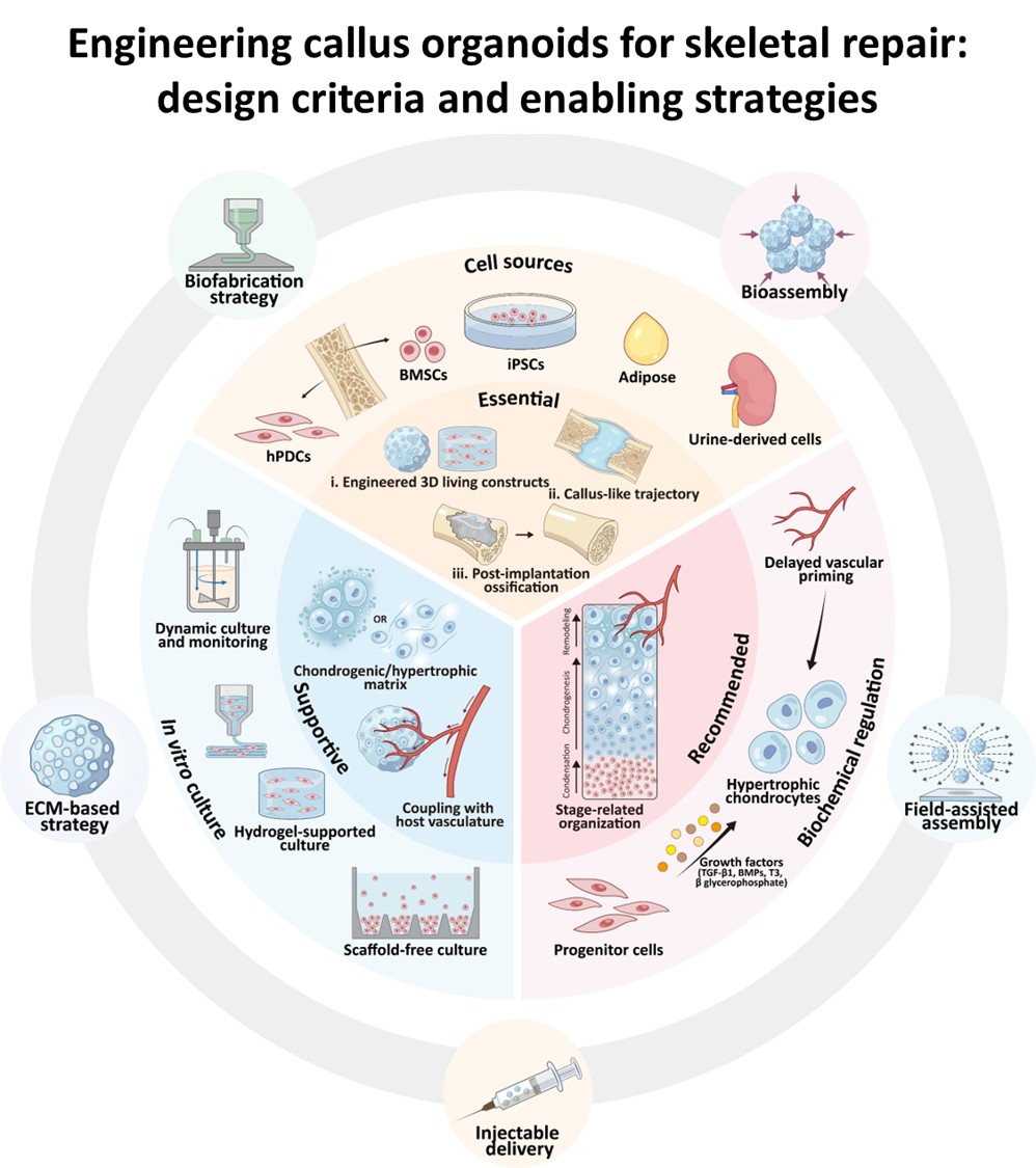
Treating critical-sized bone defects is challenging because successful repair relies on proper tissue transitions and timely vascular access, rather than on a single cell fate. Creating fully mature, defect-scale bone grafts in vitro remains constrained by scale, mass transport, and reproducibility, motivating strategies that deliver a programmed starting state and rely on in vivo progression. Organoid systems offer a useful paradigm in this context, as self-organizing microtissues can mimic developmental processes and produce consistent intermediate states. In this developmental engineering framework, callus organoids are cartilage-primed microtissues designed to follow an endochondral callus-to-bone path after implantation. This review synthesizes the mechanisms by which callus organoids are programmed to transition from chondrogenesis to hypertrophy, vascular invasion, ossification, and remodeling. It compares callus organoids to bone organoids and traditional scaffold-based bone tissue engineering, focusing on trajectory control, phase transitions, and timed integration with host transport and vascular systems. Key design variables include the endochondral potential of initial cells, the sequencing of biochemical and mechanical signals, and the timing of maturation and implantation to maintain vascular readiness. The review also discusses bioassembly and biofabrication in relation to diffusion limits and process-compatible potency assessment and quality attributes. Finally, donor variability, mass-transport limitations, incomplete multicellular complexity, and manufacturing standardization are key challenges driving priorities in perfusion and vascularization, architecture-informed fabrication, staged integration of vascular and immune components, and the development of extracellular matrix-based callus-mimetic templates. Overall, the emphasis shifts from building mature bone in vitro to manufacturing standardized callus-like building blocks whose potency is defined by their ability to execute orderly endochondral progression after implantation.
- Wu AM, Bisignano C, James SL, et al. Global, regional, and national burden of bone fractures in 204 countries and territories, 1990-2019: a systematic analysis from the Global Burden of Disease Study 2019. Lancet Healthy Longev. 2021;2(9):e580-e592. doi: 10.1016/S2666-7568(21)00172-0
- Mills LA, Aitken SA, Simpson A. The risk of non-union per fracture: current myths and revised figures from a population of over 4 million adults. Acta Orthop. 2017;88(4):434-439. doi: 10.1080/17453674.2017.1321351
- Bezstarosti H, Metsemakers WJ, van Lieshout EMM, et al. Management of critical-sized bone defects in the treatment of fracture-related infection: a systematic review and pooled analysis. Arch Orthop Trauma Surg. 2021;141(7):1215-1230. doi: 10.1007/s00402-020-03525-0
- Vanderkarr MF, Ruppenkamp JW, Vanderkarr M, Holy CE, Blauth M. Risk factors and healthcare costs associated with long bone fracture non-union: a retrospective US claims database analysis. J Orthop Surg Res. 2023;18(1):745. doi: 10.1186/s13018-023-04232-3
- Delloye C, Cornu O, Druez V, Barbier O. Bone allografts: What they can offer and what they cannot. J Bone Joint Surg Br. 2007;89(5):574-580. doi: 10.1302/0301-620X.89B5.19039
- Sen MK, Miclau T. Autologous iliac crest bone graft: should it still be the gold standard for treating nonunions? Injury. 2007;38 Suppl 1:S75-80. doi: 10.1016/j.injury.2007.02.012
- Koons GL, Diba M, Mikos AG. Materials design for bone-tissue engineering. Nat Rev Mater. 2020;5(8):584-603. doi: 10.1038/s41578-020-0204-2
- Maia FR, Bastos AR, Oliveira JM, Correlo VM, Reis RL. Recent approaches towards bone tissue engineering. Bone. 2022;154:116256. doi: 10.1016/j.bone.2021.116256
- Bai L, Zhou D, Li G, Liu J, Chen X, Su J. Engineering bone/ cartilage organoids: strategy, progress, and application. Bone Res. 2024;12(1):66. doi: 10.1038/s41413-024-00376-y
- Liu Y, Kuang B, Rothrauff BB, Tuan RS, Lin H. Robust bone regeneration through endochondral ossification of human mesenchymal stem cells within their own extracellular matrix. Biomaterials. 2019;218:119336. doi: 10.1016/j.biomaterials.2019.119336
- Sheehy EJ, Mesallati T, Kelly L, Vinardell T, Buckley CT, Kelly DJ. Tissue Engineering Whole Bones Through Endochondral Ossification: Regenerating the Distal Phalanx. Biores Open Access. 2015;4(1):229-241. doi: 10.1089/biores.2015.0014
- Thompson EM, Matsiko A, Kelly DJ, Gleeson JP, O’Brien FJ. An Endochondral Ossification-Based Approach to Bone Repair: Chondrogenically Primed Mesenchymal Stem Cell- Laden Scaffolds Support Greater Repair of Critical-Sized Cranial Defects Than Osteogenically Stimulated Constructs In Vivo. Tissue Eng Part A. 2016;22(5-6):556-567. doi: 10.1089/ten.TEA.2015.0457
- Lenas P, Moos M, Luyten FP. Developmental engineering: a new paradigm for the design and manufacturing of cell-based products. Part I: from three-dimensional cell growth to biomimetics of in vivo development. Tissue Eng Part B Rev. 2009;15(4):381-394. doi: 10.1089/ten.TEB.2008.0575
- Papantoniou I, Nilsson Hall G, Loverdou N, et al. Turning Nature’s own processes into design strategies for living bone implant biomanufacturing: a decade of Developmental Engineering. Adv Drug Deliv Rev. 2021;169:22-39. doi: 10.1016/j.addr.2020.11.012
- Bahney CS, Zondervan RL, Allison P, et al. Cellular biology of fracture healing. J Orthop Res. 2019;37(1):35-50. doi: 10.1002/jor.24170
- Lenas P, Moos M, Luyten FP. Developmental engineering: a new paradigm for the design and manufacturing of cell-based products. Part II: from genes to networks: tissue engineering from the viewpoint of systems biology and network science. Tissue Eng Part B Rev. 2009;15(4):395-422. doi: 10.1089/ten.TEB.2009.0461
- Nilsson Hall G, Mendes LF, Gklava C, Geris L, Luyten FP, Papantoniou I. Developmentally Engineered Callus Organoid Bioassemblies Exhibit Predictive In Vivo Long Bone Healing. Adv Sci. 2020;7(2):1902295. doi: 10.1002/advs.201902295
- Decoene I, Svitina H, Hamed MB, et al. Callus organoids reveal distinct cartilage to bone transition mechanisms across donors and a role for biological sex. Bone Res. 2025;13(1):41. doi: 10.1038/s41413-025-00418-z
- Decoene I, Nasello G, Madeiro de Costa RF, et al. Robotics- Driven Manufacturing of Cartilaginous Microtissues for Skeletal Tissue Engineering Applications. Stem Cells Transl Med. 2024;13(3):278-292. doi: 10.1093/stcltm/szad091
- Jukes JM, Both SK, Leusink A, Sterk LM, van Blitterswijk CA, de Boer J. Endochondral bone tissue engineering using embryonic stem cells. Proc Natl Acad Sci USA. 2008;105(19):6840-6845. doi: 10.1073/pnas.0711662105
- Scotti C, Tonnarelli B, Papadimitropoulos A, et al. Recapitulation of endochondral bone formation using human adult mesenchymal stem cells as a paradigm for developmental engineering. Proc Natl Acad Sci USA. 2010;107(16):7251-7256. doi: 10.1073/pnas.1000302107
- Berendsen AD, Olsen BR. Bone development. Bone. 2015;80:14-18. doi: 10.1016/j.bone.2015.04.035
- Kronenberg HM. Developmental regulation of the growth plate. Nature. 2003;423(6937):332-336. doi: 10.1038/nature01657
- Mackie EJ, Ahmed YA, Tatarczuch L, Chen KS, Mirams M. Endochondral ossification: how cartilage is converted into bone in the developing skeleton. Int J Biochem Cell Biol. 2008;40(1):46-62. doi: 10.1016/j.biocel.2007.06.009
- Herpelinck T, Ory L, Verbraeken T, et al. A single-cell atlas of the murine limb skeleton integrating the developmental and adult stages. Sci Rep. 2025;15(1):22514. doi: 10.1038/s41598-025-05277-6
- Zhou X, von der Mark K, Henry S, Norton W, Adams H, de Crombrugghe B. Chondrocytes transdifferentiate into osteoblasts in endochondral bone during development, postnatal growth and fracture healing in mice. PLoS Genet. 2014;10(12):e1004820. doi: 10.1371/journal.pgen.1004820
- Yang G, Zhu L, Hou N, et al. Osteogenic fate of hypertrophic chondrocytes. Cell Res. 2014;24(10):1266-1269. doi: 10.1038/cr.2014.111
- Aghajanian P, Mohan S. The art of building bone: emerging role of chondrocyte-to-osteoblast transdifferentiation in endochondral ossification. Bone Res. 2018;6(1):19. doi: 10.1038/s41413-018-0021-z
- Gerber HP, Vu TH, Ryan AM, Kowalski J, Werb Z, Ferrara N. VEGF couples hypertrophic cartilage remodeling, ossification and angiogenesis during endochondral bone formation. Nat Med. 1999;5(6):623-628. doi: 10.1038/9467
- Walma DAC, Yamada KM. The extracellular matrix in development. Development. 2020;147(10). doi: 10.1242/dev.175596
- Frantz C, Stewart KM, Weaver VM. The extracellular matrix at a glance. J Cell Sci. 2010;123(Pt 24):4195-4200. doi: 10.1242/jcs.023820
- Maes C, Kobayashi T, Selig MK, et al. Osteoblast precursors, but not mature osteoblasts, move into developing and fractured bones along with invading blood vessels. Dev Cell. 2010;19(2):329-344. doi: 10.1016/j.devcel.2010.07.010
- Gerstenfeld LC, Cullinane DM, Barnes GL, Graves DT, Einhorn TA. Fracture healing as a post-natal developmental process: molecular, spatial, and temporal aspects of its regulation. J Cell Biochem. 2003;88(5):873-884. doi: 10.1002/jcb.10435
- Marsell R, Einhorn TA. The biology of fracture healing. Injury. 2011;42(6):551-555. doi: 10.1016/j.injury.2011.03.031
- Schindeler A, McDonald MM, Bokko P, Little DG. Bone remodeling during fracture repair: The cellular picture. Semin Cell Dev Biol. 2008;19(5):459-466. doi: 10.1016/j.semcdb.2008.07.004
- Loi F, Cordova LA, Pajarinen J, Lin TH, Yao Z, Goodman SB. Inflammation, fracture and bone repair. Bone. 2016;86:119- 130. doi: 10.1016/j.bone.2016.02.020
- Stegen S, van Gastel N, Carmeliet G. Bringing new life to damaged bone: the importance of angiogenesis in bone repair and regeneration. Bone. 2015;70:19-27. doi: 10.1016/j.bone.2014.09.017
- Thompson EM, Matsiko A, Farrell E, Kelly DJ, O’Brien FJ. Recapitulating endochondral ossification: a promising route to in vivo bone regeneration. J Tissue Eng Regen Med. 2015;9(8):889-902. doi: 10.1002/term.1918
- Bahney CS, Hu DP, Miclau T, 3rd, Marcucio RS. The multifaceted role of the vasculature in endochondral fracture repair. Front Endocrinol. 2015;6:4. doi: 10.3389/fendo.2015.00004
- Wan C, Gilbert SR, Wang Y, et al. Activation of the hypoxia-inducible factor-1alpha pathway accelerates bone regeneration. Proc Natl Acad Sci USA. 2008;105(2):686-691. doi: 10.1073/pnas.0708474105
- Marcucio RS, Miclau T, 3rd, Bahney CS. A Shifting Paradigm: Transformation of Cartilage to Bone during Bone Repair. J Dent Res. 2023;102(1):13-20. doi: 10.1177/00220345221125401
- Kodama J, Wilkinson KJ, Iwamoto M, Otsuru S, Enomoto- Iwamoto M. The role of hypertrophic chondrocytes in regulation of the cartilage-to-bone transition in fracture healing. Bone Rep. 2022;17:101616. doi: 10.1016/j.bonr.2022.101616
- Hachemi Y, Perrin S, Ethel M, et al. Multimodal analyses of immune cells during bone repair identify macrophages as a therapeutic target in musculoskeletal trauma. Bone Res. 2024;12(1):56. doi: 10.1038/s41413-024-00347-3
- Bowers KM, Anderson DE. Delayed Union and Nonunion: Current Concepts, Prevention, and Correction: A Review. Bioengineering. 2024;11(6). doi: 10.3390/bioengineering11060525
- Maruyama M, Rhee C, Utsunomiya T, et al. Modulation of the Inflammatory Response and Bone Healing. Front Endocrinol. 2020;11:386. doi: 10.3389/fendo.2020.00386
- Sheehy EJ, Kelly DJ, O’Brien FJ. Biomaterial-based endochondral bone regeneration: a shift from traditional tissue engineering paradigms to developmentally inspired strategies. Mater Today Bio. 2019;3:100009. doi: 10.1016/j.mtbio.2019.100009
- Lenas P, Ikonomou L. Developmental engineering: design of clinically efficacious bioartificial tissues through developmental and systems biology. Sci China Life Sci. 2018;61(8):978-981. doi: 10.1007/s11427-017-9255-3
- Burgan J, Rahmati M, Lee M, Saiz AM. Innate immune response to bone fracture healing. Bone. 2025;190:117327. doi: 10.1016/j.bone.2024.117327
- Lancaster MA, Knoblich JA. Organogenesis in a dish: modeling development and disease using organoid technologies. Science. 2014;345(6194):1247125. doi: 10.1126/science.1247125
- Clevers H. Modeling Development and Disease with Organoids. Cell. 2016;165(7):1586-1597. doi: 10.1016/j.cell.2016.05.082
- Sato T, Vries RG, Snippert HJ, et al. Single Lgr5 stem cells build crypt-villus structures in vitro without a mesenchymal niche. Nature. 2009;459(7244):262-265. doi: 10.1038/nature07935
- Lancaster MA, Renner M, Martin CA, et al. Cerebral organoids model human brain development and microcephaly. Nature. 2013;501(7467):373-379. doi: 10.1038/nature12517
- Takebe T, Sekine K, Enomura M, et al. Vascularized and functional human liver from an iPSC-derived organ bud transplant. Nature. 2013;499(7459):481-484. doi: 10.1038/nature12271
- Drakhlis L, Biswanath S, Farr CM, et al. Human heart-forming organoids recapitulate early heart and foregut development. Nat Biotechnol. 2021;39(6):737-746. doi: 10.1038/s41587-021-00815-9
- Cheng D, Clark CT, Smith Q. Advances in engineered models of peri-gastrulation. iScience. 2025;28(6):112659. doi: 10.1016/j.isci.2025.112659
- Bondarenko V, Turco MY. Modeling the human maternal-fetal interface. Cell Stem Cell. 2025;32(9):1321-1345. doi: 10.1016/j.stem.2025.08.004
- Xie C, Liang R, Ye J, et al. High-efficient engineering of osteo-callus organoids for rapid bone regeneration within one month. Biomaterials. 2022;288:121741. doi: 10.1016/j.biomaterials.2022.121741
- Scotti C, Piccinini E, Takizawa H, et al. Engineering of a functional bone organ through endochondral ossification. Proc Natl Acad Sci USA. 2013;110(10):3997-4002. doi: 10.1073/pnas.1220108110
- Xiong J, Ma R, Xie K, et al. Recapitulation of endochondral ossification by hPSC-derived SOX9(+) sclerotomal progenitors. Nat Commun. 2025;16(1):2781. doi: 10.1038/s41467-025-58122-9
- Zhang X, Jiang W, Wu X, et al. Divide-and-conquer strategy with engineered ossification center organoids for rapid bone healing through developmental cell recruitment. Nat Commun. 2025;16(1):6200. doi: 10.1038/s41467-025-61619-y
- Ding H, Chen D, Tan X, et al. Enhanced Bone Repair using Callus Organoids Derived from Urine-Derived Stem Cells with Silk Fibroin. Adv Healthc Mater. 2025;14(23):e2501852. doi: 10.1002/adhm.202501852
- Pfister P, Lhospice E, Garcia-Garcia A, et al. Start, Stop, Rewind, Repeat-Cyclic Exposure of Adipose Stromal Cells-derived Cartilage Organoids to Chondrogenic and Proliferative Cues to Achieve Scaled-up and Customizable Bone Formation by Endochondral Ossification. Adv Healthc Mater. 2026:e04880. doi: 10.1002/adhm.202504880
- Dang PN, Herberg S, Varghai D, et al. Endochondral Ossification in Critical-Sized Bone Defects via Readily Implantable Scaffold-Free Stem Cell Constructs. Stem Cells Transl Med. 2017;6(7):1644-1659. doi: 10.1002/sctm.16-0222
- McDermott AM, Herberg S, Mason DE, et al. Recapitulating bone development through engineered mesenchymal condensations and mechanical cues for tissue regeneration. Sci Transl Med. 2019;11(495). doi: 10.1126/scitranslmed.aav7756
- Pitacco P, Sadowska JM, O’Brien FJ, Kelly DJ. 3D bioprinting of cartilaginous templates for large bone defect healing. Acta Biomater. 2023;156:61-74. doi: 10.1016/j.actbio.2022.07.037
- Peng L, Chandrakar A, Nilsson Hall G, et al. Structurally defined cartilaginous MEW-assembloids for critical-size long bone healing. Biomaterials. 2025;319:123202. doi: 10.1016/j.biomaterials.2025.123202
- Bancroft GN, Sikavitsas VI, van den Dolder J, et al. Fluid flow increases mineralized matrix deposition in 3D perfusion culture of marrow stromal osteoblasts in a dose-dependent manner. Proc Natl Acad Sci USA. 2002;99(20):12600-12605. doi: 10.1073/pnas.202296599
- Clark JR, Al Maruf DSA, Tomaskovic-Crook E, et al. Mechanobiologically-optimized non-resorbable artificial bone for patient-matched scaffold-guided bone regeneration. Nat Commun. 2025;16(1):9422. doi: 10.1038/s41467-025-64466-z
- Henkel J, Medeiros Savi F, Berner A, et al. Scaffold-guided bone regeneration in large volume tibial segmental defects. Bone. 2021;153:116163. doi: 10.1016/j.bone.2021.116163
- Yan C, Zhang P, Qin Q, et al. 3D-printed bone regeneration scaffolds modulate bone metabolic homeostasis through vascularization for osteoporotic bone defects. Biomaterials. 2024;311:122699. doi: 10.1016/j.biomaterials.2024.122699
- Bai JR, Zhang C, Li G, et al. Recapitulation of in vivo angiogenesis and osteogenesis within an ex vivo muscle pouch-based coral-derived macroporous construct organoid model. J Orthop Translat. 2025;52:478-491. doi: 10.1016/j.jot.2025.04.002
- Leukers B, Gulkan H, Irsen SH, et al. Hydroxyapatite scaffolds for bone tissue engineering made by 3D printing. J Mater Sci Mater Med. 2005;16(12):1121-1124. doi: 10.1007/s10856-005-4716-5
- Chen QZ, Thompson ID, Boccaccini AR. 45S5 Bioglass-derived glass-ceramic scaffolds for bone tissue engineering. Biomaterials. 2006;27(11):2414-2425. doi: 10.1016/j.biomaterials.2005.11.025
- Bonadio J, Smiley E, Patil P, Goldstein S. Localized, direct plasmid gene delivery in vivo: prolonged therapy results in reproducible tissue regeneration. Nat Med. 1999;5(7):753- 759. doi: 10.1038/10473
- Petite H, Viateau V, Bensaid W, et al. Tissue-engineered bone regeneration. Nat Biotechnol. 2000;18(9):959-963. doi: 10.1038/79449
- Kneser U, Polykandriotis E, Ohnolz J, et al. Engineering of vascularized transplantable bone tissues: induction of axial vascularization in an osteoconductive matrix using an arteriovenous loop. Tissue Eng. 2006;12(7):1721-1731. doi: 10.1089/ten.2006.12.1721
- Warnke PH, Springer IN, Wiltfang J, et al. Growth and transplantation of a custom vascularised bone graft in a man. Lancet. 2004;364(9436):766-770. doi: 10.1016/S0140-6736(04)16935-3
- Cao SS, Li SY, Geng YM, et al. Prefabricated 3D-Printed Tissue-Engineered Bone for Mandibular Reconstruction: A Preclinical Translational Study in Primate. ACS Biomater Sci Eng. 2021;7(12):5727-5738. doi: 10.1021/acsbiomaterials.1c00509
- Akiva A, Melke J, Ansari S, et al. An Organoid for Woven Bone. Adv Funct Mater. 2021;31(17):2010524. doi: 10.1002/adfm.202010524
- Kale S, Biermann S, Edwards C, Tarnowski C, Morris M, Long MW. Three-dimensional cellular development is essential for ex vivo formation of human bone. Nat Biotechnol. 2000;18(9):954-958. doi: 10.1038/79439
- Park Y, Cheong E, Kwak JG, Carpenter R, Shim JH, Lee J. Trabecular bone organoid model for studying the regulation of localized bone remodeling. Sci Adv. 2021;7(4). doi: 10.1126/sciadv.abd6495
- Gehlen J, Qiu W, Schadli GN, Muller R, Qin XH. Tomographic volumetric bioprinting of heterocellular bone-like tissues in seconds. Acta Biomater. 2023;156:49-60. doi: 10.1016/j.actbio.2022.06.020
- Wang J, Wu Y, Li G, et al. Engineering Large-Scale Self-Mineralizing Bone Organoids with Bone Matrix- Inspired Hydroxyapatite Hybrid Bioinks. Adv Mater. 2024;36(30):e2309875. doi: 10.1002/adma.202309875
- Iordachescu A, Amin HD, Rankin SM, et al. An In Vitro Model for the Development of Mature Bone Containing an Osteocyte Network. Adv Biosyst. 2018;2(2):1700156. doi: 10.1002/adbi.201700156
- Zhang J, Griesbach J, Ganeyev M, et al. Long-term mechanical loading is required for the formation of 3D bioprinted functional osteocyte bone organoids. Biofabrication. 2022;14(3). doi: 10.1088/1758-5090/ac73b9
- Chen S, Chen X, Geng Z, Su J. The horizon of bone organoid: A perspective on construction and application. Bioact Mater. 2022;18:15-25. doi: 10.1016/j.bioactmat.2022.01.048
- Zimmermann B, Wachtel HC, Somogyi H. Endochondral mineralization in cartilage organoid culture. Cell Differ Dev. 1990;31(1):11-22. doi: 10.1016/0922-3371(90)90086-c
- Johnstone B, Hering TM, Caplan AI, Goldberg VM, Yoo JU. In vitro chondrogenesis of bone marrow-derived mesenchymal progenitor cells. Exp Cell Res. 1998;238(1):265-272. doi: 10.1006/excr.1997.3858
- Farrell E, Both SK, Odorfer KI, et al. In-vivo generation of bone via endochondral ossification by in-vitro chondrogenic priming of adult human and rat mesenchymal stem cells. BMC Musculoskelet Disord. 2011;12(1):31. doi: 10.1186/1471-2474-12-31
- Knuth CA, Witte-Bouma J, Ridwan Y, Wolvius EB, Farrell E. Mesenchymal stem cell-mediated endochondral ossification utilising micropellets and brief chondrogenic priming. Eur Cell Mater. 2017;34:142-161. doi: 10.22203/eCM.v034a10
- Wiese DM, Wood CA, Braid LR. From Vial to Vein: Crucial Gaps in Mesenchymal Stromal Cell Clinical Trial Reporting. Front Cell Dev Biol. 2022;10:867426. doi: 10.3389/fcell.2022.867426
- Nadine S, Fernandes IJ, Correia CR, Mano JF. Close-to-native bone repair via tissue-engineered endochondral ossification approaches. iScience. 2022;25(11):105370. doi: 10.1016/j.isci.2022.105370
- Ji E, Leijsten L, Witte-Bouma J, et al. In Vitro Mineralisation of Tissue-Engineered Cartilage Reduces Endothelial Cell Migration, Proliferation and Tube Formation. Cells. 2023;12(8). doi: 10.3390/cells12081202
- Franco RAG, McKenna E, Shajib MS, et al. Microtissue Culture Provides Clarity on the Relative Chondrogenic and Hypertrophic Response of Bone-Marrow-Derived Stromal Cells to TGF-beta1, BMP-2, and GDF-5. Cells. 2023;13(1). doi: 10.3390/cells13010037
- Zhuang P, Chiang YH, Fernanda MS, He M. Using Spheroids as Building Blocks Towards 3D Bioprinting of Tumor Microenvironment. Int J Bioprint. 2021;7(4):444. doi: 10.18063/ijb.v7i4.444
- Licata JP, Schwab KH, Har-El YE, Gerstenhaber JA, Lelkes PI. Bioreactor Technologies for Enhanced Organoid Culture. Int J Mol Sci. 2023;24(14). doi: 10.3390/ijms241411427
- Nwokoye PN, Abilez OJ. Bioengineering methods for vascularizing organoids. Cell Rep Methods. 2024;4(6):100779. doi: 10.1016/j.crmeth.2024.100779
- Deckers T, Lambrechts T, Viazzi S, et al. High-throughput image-based monitoring of cell aggregation and microspheroid formation. PLoS ONE. 2018;13(6):e0199092. doi: 10.1371/journal.pone.0199092
- Ongenae S, Svitina H, Belpaire TER, et al. Active foam dynamics of tissue spheroid fusion. Nat Commun. 2025;16(1):10467. doi: 10.1038/s41467-025-65463-y
- Colnot C, Zhang X, Knothe Tate ML. Current insights on the regenerative potential of the periosteum: molecular, cellular, and endogenous engineering approaches. J Orthop Res. 2012;30(12):1869-1878. doi: 10.1002/jor.22181
- Jeffery EC, Mann TLA, Pool JA, Zhao Z, Morrison SJ. Bone marrow and periosteal skeletal stem/progenitor cells make distinct contributions to bone maintenance and repair. Cell Stem Cell. 2022;29(11):1547-1561 e6 doi: 10.1016/j.stem.2022.10.002
- Ortinau LC, Wang H, Lei K, et al. Identification of Functionally Distinct Mx1+alphaSMA+ Periosteal Skeletal Stem Cells. Cell Stem Cell. 2019;25(6):784-796.e5. doi: 10.1016/j.stem.2019.11.003
- Duchamp de Lageneste O, Julien A, Abou-Khalil R, et al. Periosteum contains skeletal stem cells with high bone regenerative potential controlled by Periostin. Nat Commun. 2018;9(1):773. doi: 10.1038/s41467-018-03124-z
- Perrin S, Ethel M, Bretegnier V, et al. Single-nucleus transcriptomics reveal the differentiation trajectories of periosteal skeletal/stem progenitor cells in bone regeneration. eLife. 2024;13. doi: 10.7554/eLife.92519
- Xing W, Feng H, Jiang B, et al. Itm2a expression marks periosteal skeletal stem cells that contribute to bone fracture healing. J Clin Invest. 2024;134(17). doi: 10.1172/JCI176528
- Liu YL, Tang XT, Shu HS, Zou W, Zhou BO. Fibrous periosteum repairs bone fracture and maintains the healed bone throughout mouse adulthood. Dev Cell. 2024;59(9):1192-1209.e6. doi: 10.1016/j.devcel.2024.03.019
- Roberts SJ, van Gastel N, Carmeliet G, Luyten FP. Uncovering the periosteum for skeletal regeneration: the stem cell that lies beneath. Bone. 2015;70:10-18. doi: 10.1016/j.bone.2014.08.007
- Bolander J, Ji W, Leijten J, et al. Healing of a Large Long-Bone Defect through Serum-Free In Vitro Priming of Human Periosteum-Derived Cells. Stem Cell Rep. 2017;8(3):758-772. doi: 10.1016/j.stemcr.2017.01.005
- Bolander J, Herpelinck T, Chaklader M, Gklava C, Geris L, Luyten FP. Single-cell characterization and metabolic profiling of in vitro cultured human skeletal progenitors with enhanced in vivo bone forming capacity. Stem Cells Transl Med. 2020;9(3):389-402. doi: 10.1002/sctm.19-0151
- Nilsson Hall G, Rutten I, Lammertyn J, et al. Cartilaginous spheroid-assembly design considerations for endochondral ossification: towards robotic-driven biomanufacturing. Biofabrication. 2021;13(4). doi: 10.1088/1758-5090/ac2208
- Herberg S, McDermott AM, Dang PN, et al. Combinatorial morphogenetic and mechanical cues to mimic bone development for defect repair. Sci Adv. 2019;5(8):eaax2476. doi: 10.1126/sciadv.aax2476
- Bourgine PE, Scotti C, Pigeot S, Tchang LA, Todorov A, Martin I. Osteoinductivity of engineered cartilaginous templates devitalized by inducible apoptosis. Proc Natl Acad Sci USA. 2014;111(49):17426-17431. doi: 10.1073/pnas.1411975111
- Mohamed-Ahmed S, Fristad I, Lie SA, et al. Adipose-derived and bone marrow mesenchymal stem cells: a donor-matched comparison. Stem Cell Res Ther. 2018;9(1):168. doi: 10.1186/s13287-018-0914-1
- Chang H, Knothe Tate ML. Concise review: the periosteum: tapping into a reservoir of clinically useful progenitor cells. Stem Cells Transl Med. 2012;1(6):480-491. doi: 10.5966/sctm.2011-0056
- van der Stok J, Koolen MK, Jahr H, et al. Chondrogenically differentiated mesenchymal stromal cell pellets stimulate endochondral bone regeneration in critical-sized bone defects. Eur Cell Mater. 2014;27:137-148; discussion 148. doi: 10.22203/ecm.v027a11
- Craft AM, Rockel JS, Nartiss Y, Kandel RA, Alman BA, Keller GM. Generation of articular chondrocytes from human pluripotent stem cells. Nat Biotechnol. 2015;33(6):638-645. doi: 10.1038/nbt.3210
- Tam WL, Freitas Mendes L, Chen X, et al. Human pluripotent stem cell-derived cartilaginous organoids promote scaffold-free healing of critical size long bone defects. Stem Cell Res Ther. 2021;12(1):513. doi: 10.1186/s13287-021-02580-7
- Adkar SS, Wu CL, Willard VP, et al. Step-Wise Chondrogenesis of Human Induced Pluripotent Stem Cells and Purification Via a Reporter Allele Generated by CRISPR-Cas9 Genome Editing. Stem Cells. 2019;37(1):65-76. doi: 10.1002/stem.2931
- Wei X, Qiu J, Lai R, et al. A human organoid drug screen identifies alpha2-adrenergic receptor signaling as a therapeutic target for cartilage regeneration. Cell Stem Cell. 2024;31(12):1813-1830.e8. doi: 10.1016/j.stem.2024.09.001
- Saxer F, Scherberich A, Todorov A, et al. Implantation of Stromal Vascular Fraction Progenitors at Bone Fracture Sites: From a Rat Model to a First-in-Man Study. Stem Cells. 2016;34(12):2956-2966. doi: 10.1002/stem.2478
- Di Maggio N, Martella E, Frismantiene A, et al. Extracellular matrix and alpha(5)beta(1) integrin signaling control the maintenance of bone formation capacity by human adipose-derived stromal cells. Sci Rep. 2017;7(1):44398. doi: 10.1038/srep44398
- Osinga R, Di Maggio N, Todorov A, et al. Generation of a Bone Organ by Human Adipose-Derived Stromal Cells Through Endochondral Ossification. Stem Cells Transl Med. 2016;5(8):1090-1097. doi: 10.5966/sctm.2015-0256
- Guerrero J, Pigeot S, Muller J, Schaefer DJ, Martin I, Scherberich A. Fractionated human adipose tissue as a native biomaterial for the generation of a bone organ by endochondral ossification. Acta Biomater. 2018;77:142-154. doi: 10.1016/j.actbio.2018.07.004
- Huang RL, Guerrero J, Senn AS, et al. Dispersion of ceramic granules within human fractionated adipose tissue to enhance endochondral bone formation. Acta Biomater. 2020;102:458-467. doi: 10.1016/j.actbio.2019.11.046
- Chaaban M, Moya A, Garcia-Garcia A, et al. Harnessing human adipose-derived stromal cell chondrogenesis in vitro for enhanced endochondral ossification. Biomaterials. 2023;303:122387. doi: 10.1016/j.biomaterials.2023.122387
- Brocher J, Janicki P, Voltz P, et al. Inferior ectopic bone formation of mesenchymal stromal cells from adipose tissue compared to bone marrow: rescue by chondrogenic pre-induction. Stem Cell Res. 2013;11(3):1393-406. doi: 10.1016/j.scr.2013.07.008
- Zhang Y, McNeill E, Tian H, et al. Urine derived cells are a potential source for urological tissue reconstruction. J Urol. 2008;180(5):2226-2233. doi: 10.1016/j.juro.2008.07.023
- Atia GA, Rashed F, Taher ES, et al. Challenges of therapeutic applications and regenerative capacities of urine based stem cells in oral, and maxillofacial reconstruction. Biomed Pharmacother. 2024;177:117005. doi: 10.1016/j.biopha.2024.117005
- Guan J, Zhang J, Li H, et al. Human Urine Derived Stem Cells in Combination with beta-TCP Can Be Applied for Bone Regeneration. PLoS ONE. 2015;10(5):e0125253. doi: 10.1371/journal.pone.0125253
- Usas A, Huard J. Muscle-derived stem cells for tissue engineering and regenerative therapy. Biomaterials. 2007;28(36):5401-5406. doi: 10.1016/j.biomaterials.2007.09.008
- Glass GE, Chan JK, Freidin A, Feldmann M, Horwood NJ, Nanchahal J. TNF-alpha promotes fracture repair by augmenting the recruitment and differentiation of muscle-derived stromal cells. Proc Natl Acad Sci USA. 2011;108(4):1585-1590. doi: 10.1073/pnas.1018501108
- Liu R, Birke O, Morse A, et al. Myogenic progenitors contribute to open but not closed fracture repair. BMC Musculoskelet Disord. 2011;12(1):288. doi: 10.1186/1471-2474-12-288
- Julien A, Kanagalingam A, Martinez-Sarra E, et al. Direct contribution of skeletal muscle mesenchymal progenitors to bone repair. Nat Commun. 2021;12(1):2860. doi: 10.1038/s41467-021-22842-5
- Ingber DE, Mow VC, Butler D, et al. Tissue engineering and developmental biology: going biomimetic. Tissue Eng. 2006;12(12):3265-3283. doi: 10.1089/ten.2006.12.3265
- Freeman FE, McNamara LM. Endochondral Priming: A Developmental Engineering Strategy for Bone Tissue Regeneration. Tissue Eng Part B Rev. 2017;23(2):128-141. doi: 10.1089/ten.TEB.2016.0197
- Fu R, Liu C, Yan Y, Li Q, Huang RL. Bone defect reconstruction via endochondral ossification: A developmental engineering strategy. J Tissue Eng. 2021;12:20417314211004211. doi: 10.1177/20417314211004211
- Bahney CS, Hu DP, Taylor AJ, et al. Stem cell-derived endochondral cartilage stimulates bone healing by tissue transformation. J Bone Miner Res. 2014;29(5):1269-1282. doi: 10.1002/jbmr.2148
- Einhorn TA, Gerstenfeld LC. Fracture healing: mechanisms and interventions. Nat Rev Rheumatol. 2015;11(1):45-54. doi: 10.1038/nrrheum.2014.164
- Pelttari K, Winter A, Steck E, et al. Premature induction of hypertrophy during in vitro chondrogenesis of human mesenchymal stem cells correlates with calcification and vascular invasion after ectopic transplantation in SCID mice. Arthritis Rheum. 2006;54(10):3254-3266. doi: 10.1002/art.22136
- Freeman FE, Allen AB, Stevens HY, Guldberg RE, McNamara LM. Effects of in vitro endochondral priming and pre-vascularisation of human MSC cellular aggregates in vivo. Stem Cell Res Ther. 2015;6(1):218. doi: 10.1186/s13287-015-0210-2
- Dang PN, Dwivedi N, Phillips LM, et al. Controlled Dual Growth Factor Delivery From Microparticles Incorporated Within Human Bone Marrow-Derived Mesenchymal Stem Cell Aggregates for Enhanced Bone Tissue Engineering via Endochondral Ossification. Stem Cells Transl Med. 2016;5(2):206-217. doi: 10.5966/sctm.2015-0115
- Herberg S, Varghai D, Cheng Y, et al. High-density human mesenchymal stem cell rings with spatiotemporally-controlled morphogen presentation as building blocks for engineering bone diaphyseal tissue. Nanotheranostics. 2018;2(2):128-143. doi: 10.7150/ntno.23354
- Nulty J, Burdis R, Kelly DJ. Biofabrication of Prevascularised Hypertrophic Cartilage Microtissues for Bone Tissue Engineering. Front Bioeng Biotechnol. 2021;9:661989. doi: 10.3389/fbioe.2021.661989
- Yamazaki S, Hirayama R, Ikeda Y, Iseki S, Yoda T, Ikeda MA. Hyaluronic acid hydrogels support to generate integrated bone formation through endochondral ossification in vivo using mesenchymal stem cells. PLoS ONE. 2023;18(2):e0281345. doi: 10.1371/journal.pone.0281345
- Schott NG, Kaur G, Coleman R, Stegemann JP. Modular, Vascularized Hypertrophic Cartilage Constructs for Bone Tissue Engineering Applications. Tissue Eng Part A. 2025;31(23-24):1297-1308. doi: 10.1089/ten.tea.2024.0367
- Herberg S, Varghai D, Alt DS, et al. Scaffold-free human mesenchymal stem cell construct geometry regulates long bone regeneration. Commun Biol. 2021;4(1):89. doi: 10.1038/s42003-020-01576-y
- Freeman FE, Brennan MA, Browe DC, et al. A Developmental Engineering-Based Approach to Bone Repair: Endochondral Priming Enhances Vascularization and New Bone Formation in a Critical Size Defect. Front Bioeng Biotechnol. 2020;8:230. doi: 10.3389/fbioe.2020.00230
- Knuth C, Kiernan C, Wolvius E, Narcisi R, Farrell E. Understanding tissue-engineered endochondral ossification; towards improved bone formation. Eur Cell Mater. 2019;37:277-291. doi: 10.22203/eCM.v037a17
- Yang W, Both SK, van Osch GJ, Wang Y, Jansen JA, Yang F. Effects of in vitro chondrogenic priming time of bone-marrow-derived mesenchymal stromal cells on in vivo endochondral bone formation. Acta Biomater. 2015;13:254- 265. doi: 10.1016/j.actbio.2014.11.029
- Freeman FE, Haugh MG, McNamara LM. Investigation of the optimal timing for chondrogenic priming of MSCs to enhance osteogenic differentiation in vitro as a bone tissue engineering strategy. J Tissue Eng Regen Med. 2016;10(4):E250-262. doi: 10.1002/term.1793
- Jeyakumar V, Amraish N, Niculescu-Morsza E, Bauer C, Pahr D, Nehrer S. Decellularized Cartilage Extracellular Matrix Incorporated Silk Fibroin Hybrid Scaffolds forEndochondral Ossification Mediated Bone Regeneration. Int J Mol Sci. 2021;22(8). doi: 10.3390/ijms22084055
- Bordbar S, Li Z, Lotfibakhshaiesh N, et al. Cartilage tissue engineering using decellularized biomatrix hydrogel containing TGF-beta-loaded alginate microspheres in mechanically loaded bioreactor. Sci Rep. 2024;14(1):11991. doi: 10.1038/s41598-024-62474-5
- Freeman FE, Haugh MG, McNamara LM. An in vitro bone tissue regeneration strategy combining chondrogenic and vascular priming enhances the mineralization potential of mesenchymal stem cells in vitro while also allowing for vessel formation. Tissue Eng Part A. 2015;21(7-8):1320-1332. doi: 10.1089/ten.TEA.2014.0249
- Lin Z, Zhang X, Fritch MR, et al. Engineering pre-vascularized bone-like tissue from human mesenchymal stem cells through simulating endochondral ossification. Biomaterials. 2022;283:121451. doi: 10.1016/j.biomaterials.2022.121451
- Decoene I, Herpelinck T, Geris L, Luyten FP, Papantoniou I. Engineering bone-forming callus organoid implants in a xenogeneic-free differentiation medium. Front Chem Eng. 2022;4:892190. doi: 10.3389/fceng.2022.892190
- Motoike S, Inada Y, Toguchida J, Kajiya M, Ikeya M. Jawbone-like organoids generated from human pluripotent stem cells. Nat Biomed Eng. 2025;9(11):1816-1834. doi: 10.1038/s41551-025-01419-3
- Kesharwani A, Tani S, Nishikawa M, et al. Modeling vascular dynamics at the initial stage of endochondral ossification on a microfluidic chip using a human embryonic-stem-cell-derived organoid. Regen Ther. 2025;28:90-100. doi: 10.1016/j.reth.2024.11.018
- Yamashita A, Morioka M, Yahara Y, et al. Generation of scaffoldless hyaline cartilaginous tissue from human iPSCs. Stem Cell Rep. 2015;4(3):404-418. doi: 10.1016/j.stemcr.2015.01.016
- Daly AC, Davidson MD, Burdick JA. 3D bioprinting of high cell-density heterogeneous tissue models through spheroid fusion within self-healing hydrogels. Nat Commun. 2021;12(1):753. doi: 10.1038/s41467-021-21029-2
- Albillos Sanchez A, Marks MP, Casademunt P, et al. Packed for Ossification: High-Density Bioprinting of hPDC Spheroids in HAMA for Endochondral Ossification. Adv Healthc Mater. 2026:e05855. doi: 10.1002/adhm.202505855
- Agten H, Van Hoven I, Van Hoorick J, Van Vlierberghe S, Luyten FP, Bloemen V. In vitro and in vivo evaluation of periosteum-derived cells and iPSC-derived chondrocytes encapsulated in GelMA for osteochondral tissue engineering. Front Bioeng Biotechnol. 2024;12:1386692. doi: 10.3389/fbioe.2024.1386692
- Huang D, Li Y, Ma Z, et al. Collagen hydrogel viscoelasticity regulates MSC chondrogenesis in a ROCK-dependent manner. Sci Adv. 2023;9(6):eade9497. doi: 10.1126/sciadv.ade9497
- Murphy KC, Fang SY, Leach JK. Human mesenchymal stem cell spheroids in fibrin hydrogels exhibit improved cell survival and potential for bone healing. Cell Tissue Res. 2014;357(1):91-99. doi: 10.1007/s00441-014-1830-z
- Decarli MC, Seijas-Gamardo A, Morgan FLC, et al. Bioprinting of Stem Cell Spheroids Followed by Post- Printing Chondrogenic Differentiation for Cartilage Tissue Engineering. Adv Healthc Mater. 2023;12(19):e2203021. doi: 10.1002/adhm.202203021
- Saravanou SF, Ioannidis K, Dimopoulos A, et al. Dually crosslinked injectable alginate-based graft copolymer thermoresponsive hydrogels as 3D printing bioinks for cell spheroid growth and release. Carbohydr Polym. 2023;312:120790. doi: 10.1016/j.carbpol.2023.120790
- Dong L, Chen W, Luo X, et al. Constructing bone organoids based on endochondral ossification model via endogenous enzyme-induced mineralization. Chem Eng J. 2024;502:157930. doi: 10.1016/j.cej.2024.157930
- Bolander J, Mota C, Ooi HW, et al. Bioinspired Development of an In Vitro Engineered Fracture Callus for the Treatment of Critical Long Bone Defects. Adv Funct Mater. 2021;31(46):2104159. doi: 10.1002/adfm.202104159
- Loverdou N, Cuvelier M, Nilsson Hall G, et al. Stirred culture of cartilaginous microtissues promotes chondrogenic hypertrophy through exposure to intermittent shear stress. Bioeng Transl Med. 2023;8(3):e10468. doi: 10.1002/btm2.10468
- Su Y, He Z, Li J, et al. Synergistic promotion of bone regeneration through co-culture of endothelial cells with mesenchymal stem cells in endochondral ossification organoids. Stem Cell Res Ther. 2025;16(1):647. doi: 10.1186/s13287-025-04733-4
- Ji E, Garmendia Urdalleta A, Witte-Bouma J, et al. Development of a Complex Human In Vitro Model of Endochondral Ossification. Tissue Eng Part C Methods. 2025. doi: 10.1177/19373341251378152
- Molitoris JM, Paliwal S, Sekar RB, et al. Precisely parameterized experimental and computational models of tissue organization. Integr Biol. 2016;8(2):230-242. doi: 10.1039/c5ib00270b
- Gao D, Li R, Pan J, et al. 3D bioprinting bone/cartilage organoids: construction, applications, and challenges. J Orthop Translat. 2025;55:75-93. doi: 10.1016/j.jot.2025.08.008
- Byun H, Lee S, Jang GN, Lee H, Park S, Shin H. Magnetism-controlled assembly of composite stem cell spheroids for the biofabrication of contraction-modulatory 3D tissue. Biofabrication. 2021;14(1). doi: 10.1088/1758-5090/ac318b
- Souza GR, Molina JR, Raphael RM, et al. Three-dimensional tissue culture based on magnetic cell levitation. Nat Nanotechnol. 2010;5(4):291-296. doi: 10.1038/nnano.2010.23
- Deng C, Li Z, Lu L, et al. Sophisticated Magneto-Mechanical Actuation Promotes In Situ Stem Cell Assembly and Chondrogenesis for Treating Osteoarthritis. ACS Nano. 2023;17(21):21690-21707. doi: 10.1021/acsnano.3c06909
- Ioannidis K, Dimopoulos A, Decoene I, et al. 4D Biofabrication of Magnetically Augmented Callus Assembloid Implants Enables Rapid Endochondral Ossification via Activation of Mechanosensitive Pathways. Adv Sci. 2025;12(15):e2413680. doi: 10.1002/advs.202413680
- Santos LF, Mendes MC, Pereira JA, et al. Remote-Controlled Magnetic Stimulation of Cell-Based Bioengineered Tissues for In Situ Bone Regeneration. Adv Mater. 2025;37(39):e2500657. doi: 10.1002/adma.202500657
- Sriphutkiat Y, Kasetsirikul S, Zhou Y. Formation of cell spheroids using Standing Surface Acoustic Wave (SSAW). Int J Bioprint. 2018;4(1):130. doi: 10.18063/IJB.v4i1.130
- Vuille-Dit-Bille E, Fonta CL, Heub S, Boder-Pasche S, Sakar MS, Weder G. An acoustic levitation platform for high-content histological analysis of 3D tissue culture. Lab Chip. 2025;25(11):2732-2743. doi: 10.1039/d5lc00153f
- Cooke ME, Rosenzweig DH. The rheology of direct and suspended extrusion bioprinting. APL Bioeng. 2021;5(1):011502. doi: 10.1063/5.0031475
- Burdis R, Kelly DJ. Biofabrication and bioprinting using cellular aggregates, microtissues and organoids for the engineering of musculoskeletal tissues. Acta Biomater. 2021;126:1-14. doi: 10.1016/j.actbio.2021.03.016
- Caprio ND, Burdick JA. Engineered biomaterials to guide spheroid formation, function, and fabrication into 3D tissue constructs. Acta Biomater. 2023;165:4-18. doi: 10.1016/j.actbio.2022.09.052
- Hinton TJ, Jallerat Q, Palchesko RN, et al. Three-dimensional printing of complex biological structures by freeform reversible embedding of suspended hydrogels. Sci Adv. 2015;1(9):e1500758. doi: 10.1126/sciadv.1500758
- Moroni L, Boland T, Burdick JA, et al. Biofabrication: A Guide to Technology and Terminology. Trends Biotechnol. 2018;36(4):384-402. doi: 10.1016/j.tibtech.2017.10.015
- Grigoryan B, Paulsen SJ, Corbett DC, et al. Multivascular networks and functional intravascular topologies within biocompatible hydrogels. Science. 2019;364(6439):458-464. doi: 10.1126/science.aav9750
- Bernal PN, Delrot P, Loterie D, et al. Volumetric Bioprinting of Complex Living-Tissue Constructs within Seconds. Adv Mater. 2019;31(42):e1904209. doi: 10.1002/adma.201904209
- Guillotin B, Souquet A, Catros S, et al. Laser assisted bioprinting of engineered tissue with high cell density and microscale organization. Biomaterials. 2010;31(28):7250- 7256. doi: 10.1016/j.biomaterials.2010.05.055
- Hall GN, Fan Y, Viellerobe B, et al. Laser-assisted bioprinting of targeted cartilaginous spheroids for high density bottom-up tissue engineering. Biofabrication. 2024;16(4). doi: 10.1088/1758-5090/ad6e1a
- Greiner A, Wendorff JH. Electrospinning: a fascinating method for the preparation of ultrathin fibers. Angew Chem Int Ed Engl. 2007;46(30):5670-5703. doi: 10.1002/anie.200604646
- Dai X, Nie W, Shen H, et al. Electrospinning based biomaterials for biomimetic fabrication, bioactive protein delivery and wound regenerative repair. Regen Biomater. 2025;12:rbae139. doi: 10.1093/rb/rbae139
- Mizraji G, Davidzohn A, Gursoy M, Gursoy U, Shapira L, Wilensky A. Membrane barriers for guided bone regeneration: An overview of available biomaterials. Periodontol 2000. 2023;93(1):56-76. doi: 10.1111/prd.12502
- King WE, 3rd, Bowlin GL. Near-Field Electrospinning and Melt Electrowriting of Biomedical Polymers-Progress and Limitations. Polymers. 2021;13(7). doi: 10.3390/polym13071097
- Kade JC, Dalton PD. Polymers for Melt Electrowriting. Adv Healthc Mater. 2021;10(1):e2001232. doi: 10.1002/adhm.202001232
- Dalton PD. Melt electrowriting with additive manufacturing principles. Curr Opin Biomed Eng. 2017;2:49-57. doi: 10.1016/j.cobme.2017.05.007
- Bertsch P, Diba M, Mooney DJ, Leeuwenburgh SCG. Self- Healing Injectable Hydrogels for Tissue Regeneration. Chem Rev. 2023;123(2):834-873. doi: 10.1021/acs.chemrev.2c00179
- Feng Q, Xu J, Zhang K, et al. Dynamic and Cell-Infiltratable Hydrogels as Injectable Carrier of Therapeutic Cells and Drugs for Treating Challenging Bone Defects. ACS Cent Sci. 2019;5(3):440-450. doi: 10.1021/acscentsci.8b00764
- Li D, Chen K, Tang H, et al. A Logic-Based Diagnostic and Therapeutic Hydrogel with Multistimuli Responsiveness to Orchestrate Diabetic Bone Regeneration. Adv Mater. 2022;34(11):e2108430. doi: 10.1002/adma.202108430
- Deng Y, Chen S, Tuerxun M, Xiong X, Tang J. Rapid neuralized and vascularized osteogenesis in infected bone defect using biomimetic biomineralized and antibacterial hydrogels. Front Bioeng Biotechnol. 2025;13:1611639. doi: 10.3389/fbioe.2025.1611639
- Pigeot S, Klein T, Gullotta F, et al. Manufacturing of Human Tissues as off-the-Shelf Grafts Programmed to Induce Regeneration. Adv Mater. 2021;33(43):e2103737. doi: 10.1002/adma.202103737
- Garcia-Garcia A, Prithiviraj S, Raina DB, et al. Engineered and decellularized human cartilage graft exhibits intrinsic immunosuppressive properties and full skeletal repair capacity. Appl Biol Sci. 2026;123(2):e2507185123. doi: 10.1073/pnas.2507185123
- Hussey GS, Dziki JL, Badylak SF. Extracellular matrix-based materials for regenerative medicine. Nat Rev Mater. 2018;3(7):159-173. doi: 10.1038/s41578-018-0023-x
- Crapo PM, Gilbert TW, Badylak SF. An overview of tissue and whole organ decellularization processes. Biomaterials. 2011;32(12):3233-3243. doi: 10.1016/j.biomaterials.2011.01.057
- Cunniffe GM, Vinardell T, Murphy JM, et al. Porous decellularized tissue engineered hypertrophic cartilage as a scaffold for large bone defect healing. Acta Biomater. 2015;23:82-90. doi: 10.1016/j.actbio.2015.05.031
- Pigeot S, Bourgine PE, Claude J, et al. Orthotopic Bone Formation by Streamlined Engineering and Devitalization of Human Hypertrophic Cartilage. Int J Mol Sci. 2020;21(19). doi: 10.3390/ijms21197233
- Longoni A, Utomo L, Robinson A, Levato R, Rosenberg A, Gawlitta D. Acceleration of Bone Regeneration Induced by a Soft-Callus Mimetic Material. Adv Sci. 2022;9(6):e2103284. doi: 10.1002/advs.202103284
- de Silva L, Longoni A, Staubli F, et al. Bone Regeneration in a Large Animal Model Featuring a Modular Off-the-Shelf Soft Callus Mimetic. Adv Healthc Mater. 2023;12(29):e2301717. doi: 10.1002/adhm.202301717
- Jones S, Tai M, Ayushman M, Peasah A, Johannsen J, Yang F. Donor Variability and 3D Culture Models Influence Human Mesenchymal Stem Cell Differentiation. Tissue Eng Part A. 2025. doi: 10.1089/ten.tea.2025.0028
- Younesi FS, Hinz B. The Myofibroblast Fate of Therapeutic Mesenchymal Stromal Cells: Regeneration, Repair, or Despair? Int J Mol Sci. 2024;25(16). doi: 10.3390/ijms25168712
- Payne KA, Didiano DM, Chu CR. Donor sex and age influence the chondrogenic potential of human femoral bone marrow stem cells. Osteoarthr Cartil. 2010;18(5):705-713. doi: 10.1016/j.joca.2010.01.011
- Breulmann FL, Berger SA, Della Bella E, Stoddart MJ. Donor-dependent regulation of type II and X collagen deposition by early modulation of miR-335-5p and miR- 1246 during chondrogenic commitment. Stem Cell Res Ther. 2025;16(1):473. doi: 10.1186/s13287-025-04589-8
- Zoneff E, Wang Y, Jackson C, et al. Controlled oxygen delivery to power tissue regeneration. Nat Commun. 2024;15(1):4361. doi: 10.1038/s41467-024-48719-x
- Malda J, Rouwkema J, Martens DE, et al. Oxygen gradients in tissue-engineered PEGT/PBT cartilaginous constructs: measurement and modeling. Biotechnol Bioeng. 2004;86(1):9- 18. doi: 10.1002/bit.20038
- Figueiredo L, Pace R, D’Arros C, et al. Assessing glucose and oxygen diffusion in hydrogels for the rational design of 3D stem cell scaffolds in regenerative medicine. J Tissue Eng Regen Med. 2018;12(5):1238-1246. doi: 10.1002/term.2656
- Li S, Oreffo RO, Sengers BG, Tare RS. The effect of oxygen tension on human articular chondrocyte matrix synthesis: integration of experimental and computational approaches. Biotechnol Bioeng. 2014;111(9):1876-1885. doi: 10.1002/bit.25241
- Lewis MC, Macarthur BD, Malda J, Pettet G, Please CP. Heterogeneous proliferation within engineered cartilaginous tissue: the role of oxygen tension. Biotechnol Bioeng. 2005;91(5):607-615. doi: 10.1002/bit.20508
- Gao B, Chen Z, Long Y, et al. Cartilage organoids: an emerging platform for novel osteoarthritis therapies. Front Cell Dev Biol. 2025;13:1668766. doi: 10.3389/fcell.2025.1668766
- Carroll SF, Buckley CT, Kelly DJ. Measuring and Modeling Oxygen Transport and Consumption in 3D Hydrogels Containing Chondrocytes and Stem Cells of Different Tissue Origins. Front Bioeng Biotechnol. 2021;9:591126. doi: 10.3389/fbioe.2021.591126
- Lou X, Zhou Q, Dong Z, Bai L, Su J, Yue H. Innovative strategies for bone organoid: Synergistic application and exploration of advanced technologies. J Orthop Translat. 2025;54:180-198. doi: 10.1016/j.jot.2025.07.010
- Chen Y, Zhang L, Li Z, et al. Urine-derived stem cells efficiently assemble into micro-bone organoids supported by decellularized bone matrix microparticles for rapidly repairing bone defects through direct filling and paracrine functions. Mater Today Bio. 2025;35:102533. doi: 10.1016/j.mtbio.2025.102533
- Buckley CT, Meyer EG, Kelly DJ. The influence of construct scale on the composition and functional properties of cartilaginous tissues engineered using bone marrow-derived mesenchymal stem cells. Tissue Eng Part A. 2012;18(3- 4):382-396. doi: 10.1089/ten.TEA.2011.0145
- Daly AC, Sathy BN, Kelly DJ. Engineering large cartilage tissues using dynamic bioreactor culture at defined oxygen conditions. J Tissue Eng. 2018;9:2041731417753718. doi: 10.1177/2041731417753718
- Chen Z, Bo Q, Wang C, Xu Y, Fei X, Chen R. Single BMSC-derived cartilage organoids for gradient heterogeneous osteochondral regeneration by leveraging native vascular microenvironment. J Nanobiotechnology. 2025;23(1):325. doi: 10.1186/s12951-025-03403-0
- Bergemann C, Elter P, Lange R, et al. Cellular Nutrition in Complex Three-Dimensional Scaffolds: A Comparison between Experiments and Computer Simulations. Int J Biomater. 2015;2015:584362. doi: 10.1155/2015/584362
- Wendt D, Stroebel S, Jakob M, John GT, Martin I. Uniform tissues engineered by seeding and culturing cells in 3D scaffolds under perfusion at defined oxygen tensions. Biorheology. 2006;43(3,4):481-488. doi: 10.1177/0006355X2006043003004030
- Peng Y, Zhuang Y, Liu Y, et al. Bioinspired gradient scaffolds for osteochondral tissue engineering. Exploration. 2023;3(4):20210043. doi: 10.1002/EXP.20210043
- Zhang Y, He SY, Wang P, et al. Impacts of permeability and effective diffusivity of porous scaffolds on bone ingrowth: In silico and in vivo analyses. Biomater Adv. 2024;161:213901. doi: 10.1016/j.bioadv.2024.213901
- Eghbali H, Nava MM, Leonardi G, et al. An experimental-numerical investigation on the effects of macroporous scaffold geometry on cell culture parameters. Int J Artif Organs. 2017;40(4):185-195. doi: 10.5301/ijao.5000554
- Sawyer SW, Shridhar SV, Zhang K, et al. Perfusion directed 3D mineral formation within cell-laden hydrogels. Biofabrication. 2018;10(3):035013. doi: 10.1088/1758-5090/aacb42
- Novak S, Tanigawa H, Singh V, et al. Endothelial to mesenchymal Notch signaling regulates skeletal repair. JCI Insight. 2024;9(12). doi: 10.1172/jci.insight.181073
- Bixel MG, Sivaraj KK, Timmen M, et al. Angiogenesis is uncoupled from osteogenesis during calvarial bone regeneration. Nat Commun. 2024;15(1):4575. doi: 10.1038/s41467-024-48579-5
- Rivera KO, Russo F, Boileau RM, et al. Local injections of beta- NGF accelerates endochondral fracture repair by promoting cartilage to bone conversion. Sci Rep. 2020;10(1):22241. doi: 10.1038/s41598-020-78983-y
- Xu M, Li Z, Thottappillil N, et al. Mapping somatosensory afferent circuitry to bone identifies neurotrophic signals required for fracture healing. Science. 2026;391(6781):eadr9608. doi: 10.1126/science.adr9608
- Biswas L, Chen J, De Angelis J, et al. Lymphatic vessels in bone support regeneration after injury. Cell. 2023;186(2):382-397. e24. doi: 10.1016/j.cell.2022.12.031
- Zheng Y, Cong L, Zhao L, et al. Lymphatic platelet thrombosis limits bone repair by precluding lymphatic transporting DAMPs. Nat Commun. 2025;16(1):829. doi: 10.1038/s41467-025-56147-8
- Zhang T, Huang L, Zhang X, Guo J, Shen Y, Luo Y. 3D printed bioceramic scaffolds with fully interconnected channel networks for enhanced vascularized bone regeneration. Biomater Sci. 2025;13(17):4830-4845. doi: 10.1039/d5bm00824g
- Duan J, Fang Y, Tian Y, Wang Z, Yang B, Xiong Z. 3D Bioprinting of Prevascularized Bone Organoids for Rapid In Situ Cranial Bone Reconstruction. Adv Healthc Mater. 2025;14(16):e2501376. doi: 10.1002/adhm.202501376
- Wu M, Chen G, Li YP. TGF-beta and BMP signaling in osteoblast, skeletal development, and bone formation, homeostasis and disease. Bone Res. 2016;4(1):16009. doi: 10.1038/boneres.2016.9
- Wrublewsky S, Schultz J, Ammo T, et al. Biofabrication of prevascularized spheroids for bone tissue engineering by fusion of microvascular fragments with osteoblasts. Front Bioeng Biotechnol. 2024;12:1436519. doi: 10.3389/fbioe.2024.1436519
- Kim W, Jang CH, Kim G. Bone tissue engineering supported by bioprinted cell constructs with endothelial cell spheroids. Theranostics. 2022;12(12):5404-5417. doi: 10.7150/thno.74852
- Zhang J, Suttapreyasri S, Leethanakul C, Samruajbenjakun B. Fabrication of vascularized tissue-engineered bone models using triaxial bioprinting. J Biomed Mater Res A. 2024;112(7):1093-1106. doi: 10.1002/jbm.a.37694
- Liu H, Chen H, Han Q, et al. Recent advancement in vascularized tissue-engineered bone based on materials design and modification. Mater Today Bio. 2023;23:100858. doi: 10.1016/j.mtbio.2023.100858
- Musthafa H-SN, Walker J, Domagala M. Computational Modelling and Simulation of Scaffolds for Bone Tissue Engineering. Computation. 2024;12(4):74. doi: 10.3390/computation12040074
- Heylman CM, Santoso S, Krebs MD, Saidel GM, Alsberg E, Muschler GF. Modeling and experimental methods to predict oxygen distribution in bone defects following cell transplantation. Med Biol Eng Comput. 2014;52(4):321-330. doi: 10.1007/s11517-013-1133-7
- Florczak S, Grossbacher G, Ribezzi D, et al. Adaptive and context-aware volumetric printing. Nature. 2025;645(8079):108-114. doi: 10.1038/s41586-025-09436-7
- Wang M, Li W, Hao J, et al. Biomaterial-minimalistic photoactivated bioprinting of cell-dense tissues. Cell. 2026;189(1):106-122 e26. doi: 10.1016/j.cell.2025.11.012
- Lawlor KT, Vanslambrouck JM, Higgins JW, et al. Cellular extrusion bioprinting improves kidney organoid reproducibility and conformation. Nat Mater. 2021;20(2):260-271. doi: 10.1038/s41563-020-00853-9
- Cooke ME, Caprio ND, Killgore J, Burdick JA. Tailoring agarose fluid gels for use in suspension bath bioprinting and culture of spheroid-based bioinks. Biofabrication. 2025;17(4). doi: 10.1088/1758-5090/ae0aff
- Kim MH, Singh YP, Celik N, et al. High-throughput bioprinting of spheroids for scalable tissue fabrication. Nat Commun. 2024;15(1):10083. doi: 10.1038/s41467-024-54504-7
- Kim MH, Ozbolat IT. Aspiration-assisted bioprinting of spheroids. Nat Protoc. 2025. doi: 10.1038/s41596-025-01240-x
- Paggi CA, Teixeira LM, Le Gac S, Karperien M. Joint-on-chip platforms: entering a new era of in vitro models for arthritis. Nat Rev Rheumatol. 2022;18(4):217-231. doi: 10.1038/s41584-021-00736-6
- Smith KWY, Fung SL, Wu HF, et al. Developing an in vitro osteochondral micro-physiological system for modeling cartilage-bone crosstalk in arthritis. Front Immunol. 2025;16:1495613. doi: 10.3389/fimmu.2025.1495613
- Palma C, Salehi S, Polidoro MA, et al. A Compartmentalized Joint-on-chip (JoC) Model to Unravel the Contribution of Cartilage and Synovium to Osteoarthritis Pathogenesis. Adv Sci. 2025;12(42):e00374. doi: 10.1002/advs.202500374
- Xu G, Zhou J, Liu K, et al. Macrophage-augmented intestinal organoids model virus-host interactions in enteric viral diseases and facilitate therapeutic development. Nat Commun. 2025;16(1):4475. doi: 10.1038/s41467-025-59639-9
- Mirazi H, Wood ST. Microfluidic chip-based co-culture system for modeling human joint inflammation in osteoarthritis research. Front Pharmacol. 2025;16:1579228. doi: 10.3389/fphar.2025.1579228
- Xue li ZZ, Tian Tian, Chen Chen,et al. High-throughput microfluidic generation of self-assembled, uniform 3D vascularized and mineralized bone organoids without matrix biomaterials. Talanta Open. 2025;12:100531. doi: 10.1016/j.talo.2025.100531
- Petta D, D’Arrigo D, Salehi S, et al. A personalized osteoarthritic joint-on-a-chip as a screening platform for biological treatments. Mater Today Bio. 2024;26:101072. doi: 10.1016/j.mtbio.2024.101072
- Bahmaee H, Owen R, Boyle L, et al. Design and Evaluation of an Osteogenesis-on-a-Chip Microfluidic Device Incorporating 3D Cell Culture. Front Bioeng Biotechnol. 2020;8:557111. doi: 10.3389/fbioe.2020.557111