
Lactylation-orchestrated Tprolif dynamics in skin cutaneous melanoma: Prognostic significance and therapeutic implications

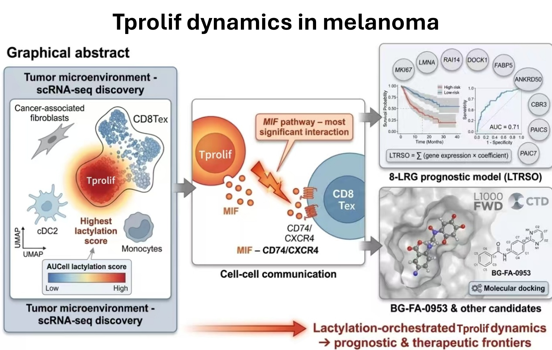
Skin cutaneous melanoma is a highly aggressive malignancy with poor prognosis, particularly in advanced stages. Lactylation, a post-translational modification, plays a crucial role in shaping the tumor microenvironment and contributing to immune suppression, making it a potential therapeutic target. In this study, single-cell RNA sequencing data from the Tumor Immune Single-cell Hub database and bulk RNA sequencing data from The Cancer Genome Atlas and Gene Expression Omnibus were analyzed. Lactylation-related genes were obtained from the Molecular Signatures Database, and lactylation scores for each cell type were computed using the AUCell R package. Cell–cell communication networks were constructed using CellChat, and a prognostic model was developed based on machine learning—including least absolute shrinkage and selection operator, random forest, and XGBoost. Drug candidates were identified via the L1000 Fireworks Display database and the Comparative Toxicogenomics Database, followed by molecular docking for drug–target interaction assessment. Results revealed that Tprolif cells exhibited the highest lactylation levels, indicating their immunosuppressive role in the tumor microenvironment, with the macrophage migration inhibitory factor receptor–ligand pathway identified as a key interaction hub. The eight lactylation-related genes–Tprolif prognostic model demonstrated moderate predictive ability for patient survival, and BG-FA-0953 emerged as a potential therapeutic candidate that may target lactylation-related pathways, warranting further investigation. This study provides novel insights into lactylation-driven immune modulation in skin cutaneous melanoma, with potential implications for risk stratification and therapeutic intervention in advanced-stage melanoma.
- Long GV, Swetter SM, Menzies AM, et al. Cutaneous melanoma. Lancet. 2023;402(10400):485-502. doi: 10.1016/S0140-6736(23)00821-8
- Leonardi G, Falzone L, Salemi R, et al. Cutaneous melanoma: From pathogenesis to therapy (Review). Int J Oncol. 2018;52(4):1071-1080. doi: 10.3892/ijo.2018.4287
- Gordon D, Hansson J, Eloranta S, et al. Primary tumor sites in relation to ultraviolet radiation exposure and skin visibility correlate with survival in cutaneous melanoma. Int J Cancer. 2017;141(7):1345-1354. doi: 10.1002/ijc.30843
- Davis LE, Shalin SC, Tackett AJ. Current state of melanoma diagnosis and treatment. Cancer Biol Ther. 2019;20(11):1366- 1379. doi: 10.1080/15384047.2019.1640032
- Eriksson H, Utjés D, Olofsson Bagge R, et al. The Proportion Cured of Patients with Resected Stage II-III Cutaneous Melanoma in Sweden. Cancers. 2021;13(10):2456. doi: 10.3390/cancers13102456
- Țăpoi DA, Derewicz D, Gheorghișan-Gălățeanu AA, et al. The Impact of Clinical and Histopathological Factors on Disease Progression and Survival in Thick Cutaneous Melanomas. Biomedicines. 2023;11(10):2616. doi: 10.3390/biomedicines11102616
- Larkin J, Chiarion-Sileni V, Gonzalez R, et al. Five-Year Survival with Combined Nivolumab and Ipilimumab in Advanced Melanoma. N Engl J Med. 2019;381(16):1535-1546. doi: 10.1056/NEJMoa1910836
- Klobuch S, Seijkens TTP, Schumacher TN, et al. Tumour-infiltrating lymphocyte therapy for patients with advanced-stage melanoma. Nat Rev Clin Oncol. 2024;21(3):173-184. doi: 10.1038/s41571-023-00848-w
- Gide TN, Wilmott JS, Scolyer RA, et al. Primary and Acquired Resistance to Immune Checkpoint Inhibitors in Metastatic Melanoma. Clin Cancer Res. 2018;24(6):1260-1270. doi: 10.1158/1078-0432.CCR-17-2267
- Jenkins RW, Barbie DA, Flaherty KT. Mechanisms of resistance to immune checkpoint inhibitors. Br J Cancer. 2018;118(1):9-16. doi: 10.1038/bjc.2017.434
- Pitt JM, Vétizou M, Daillère R, et al. Resistance Mechanisms to Immune-Checkpoint Blockade in Cancer: Tumor- Intrinsic and -Extrinsic Factors. Immunity. 2016;44(6):1255-1269. doi: 10.1016/j.immuni.2016.06.001
- Durante MA, Rodriguez DA, Kurtenbach S, et al. Single-cell analysis reveals new evolutionary complexity in uveal melanoma. Nat Commun. 2020;11(1). doi: 10.1038/s41467-019-14256-1
- Quek C, Pratapa A, Bai X, et al. Single-cell spatial multiomics reveals tumor microenvironment vulnerabilities in cancer resistance to immunotherapy. Cell Rep. 2024;43(7):114392. doi: 10.1016/j.celrep.2024.114392
- Marzagalli M, Ebelt ND, Manuel ER. Unraveling the crosstalk between melanoma and immune cells in the tumor microenvironment. Semin Cancer Biol. 2019;59:236-250. doi: 10.1016/j.semcancer.2019.08.002
- Yang Z, Zheng Y, Gao Q. Lysine lactylation in the regulation of tumor biology. Trends Endocrinol Metab. 2024;35(8):720- 731. doi: 10.1016/j.tem.2024.01.011
- Li H, Sun L, Gao P, et al. Lactylation in cancer: Current understanding and challenges. Cancer Cell. 2024;42(11):1803-1807. doi: 10.1016/j.ccell.2024.09.006
- Zhang D, Tang Z, Huang H, et al. Metabolic regulation of gene expression by histone lactylation. Nature. 2019;574(7779):575-580. doi: 10.1038/s41586-019-1678-1
- Gu J, Xu X, Li X, et al. Tumor-resident microbiota contributes to colorectal cancer liver metastasis by lactylation and immune modulation. Oncogene. 2024;43(31):2389-2404. doi: 10.1038/s41388-024-03080-7
- Xiong J, He J, Zhu J, et al. Lactylation-driven METTL3- mediated RNA m(6)A modification promotes immunosuppression of tumor-infiltrating myeloid cells. Mol Cell. 2022;82(9):1660-1677.e10. doi: 10.1016/j.molcel.2022.02.033
- Li G, Wang D, Zhai Y, et al. Glycometabolic reprogramming-induced XRCC1 lactylation confers therapeutic resistance in ALDH1A3-overexpressing glioblastoma. Cell Metab. 2024;36(8):1696-1710.e10. doi: 10.1016/j.cmet.2024.07.011
- Han Y, Wang Y, Dong X, et al. TISCH2: expanded datasets and new tools for single-cell transcriptome analyses of the tumor microenvironment. Nucleic Acids Res. 2023;51(D1):D1425-D1431. doi: 10.1093/nar/gkac959
- Subramanian A, Tamayo P, Mootha VK, et al. Gene set enrichment analysis: a knowledge-based approach for interpreting genome-wide expression profiles. Proc Natl Acad Sci U S A. 2005;102(43):15545-15550. doi: 10.1073/pnas.0506580102
- Hao Y, Hao S, Andersen-Nissen E, et al. Integrated analysis of multimodal single-cell data. Cell. 2021;184(13):3573- 3587.e29. doi: 10.1016/j.cell.2021.04.048
- Wickham H. Navarro D, Pedersen TL, ggplot2: Elegant Graphics for Data Analysis. 2016. https://ggplot2-book.org/
- Jin S, Guerrero-Juarez CF, Zhang L, et al. Inference and analysis of cell-cell communication using CellChat. Nat Commun. 2021;12(1). doi: 10.1038/s41467-021-21246-9
- Colaprico A, Silva TC, Olsen C, et al. TCGAbiolinks: an R/Bioconductor package for integrative analysis of TCGA data. Nucleic Acids Res. 2016;44(8):e71. doi: 10.1093/nar/gkv1507
- Ritchie ME, Phipson B, Wu D, et al. limma powers differential expression analyses for RNA-sequencing and microarray studies. Nucleic Acids Res. 2015;43(7):e47. doi: 10.1093/nar/gkv007
- Yu G, Wang LG, Han Y, et al. clusterProfiler: an R package for comparing biological themes among gene clusters. OMICS: A Journal of Integrative Biology. 2012;16(5):284-287. doi: 10.1089/omi.2011.0118
- Xie Y, Zhang J, Li M, et al. Identification of Lactate- Related Gene Signature for Prediction of Progression and Immunotherapeutic Response in Skin Cutaneous Melanoma. Front. Oncol. 2022;12. doi: 10.3389/fonc.2022.818868
- Friedman J, Hastie T, Tibshirani R. Regularization Paths for Generalized Linear Models via Coordinate Descent. J Stat Soft. 2010;33(1). doi: 10.18637/jss.v033.i01
- Strobl C, Boulesteix AL, Zeileis A, et al. Bias in random forest variable importance measures: illustrations, sources and a solution. BMC Bioinformatics. 2007;8(1). doi: 10.1186/1471-2105-8-25
- Chen T, Guestrin C. XGBoost. In: Proceedings of the 22nd ACM SIGKDD International Conference on Knowledge Discovery and Data Mining. ACM; 2016:785-794. doi: 10.1145/2939672.2939785
- Morris GM, Huey R, Lindstrom W, et al. AutoDock4 and AutoDockTools4: Automated docking with selective receptor flexibility. J Comput Chem. 2009;30(16):2785-2791. doi: 10.1002/jcc.21256
- Rauf MA, Zubair S, Azhar A. Ligand docking and binding site analysis with pymol and autodock/vina. IJBAS. 2015;4(2):168-177. doi: 10.14419/ijbas.v4i2.4123
- Sumaiya K, Langford D, Natarajaseenivasan K, Shanmughapriya S. Macrophage migration inhibitory factor (MIF): A multifaceted cytokine regulated by genetic and physiological strategies. Pharmacol Ther. 2022;233:108024. doi: 10.1016/j.pharmthera.2021.108024
- Gaillard T. Evaluation of AutoDock and AutoDock Vina on the CASF-2013 Benchmark. J Chem Inf Model. 2018;58(8):1697-1706. doi: 10.1021/acs.jcim.8b00312
- Schwartz RH. T cell anergy. Annu Rev Immunol. 2003;21(1):305-334. doi: 10.1146/annurev.immunol.21.120601.141110
- Pardoll DM. The blockade of immune checkpoints in cancer immunotherapy. Nat Rev Cancer. 2012;12(4):252-264. doi: 10.1038/nrc3239
- Wherry EJ, Kurachi M. Molecular and cellular insights into T cell exhaustion. Nat Rev Immunol. 2015;15(8):486-499. doi: 10.1038/nri3862
- Brizel DM, Schroeder T, Scher RL, et al. Elevated tumor lactate concentrations predict for an increased risk of metastases in head-and-neck cancer. Int J Radiat Oncol Biol Phys. 2001;51(2):349-353. doi: 10.1016/s0360-3016(01)01630-3
- Pucino V, Cucchi D, Mauro C. Lactate transporters as therapeutic targets in cancer and inflammatory diseases. Expert Opinion on Therapeutic Targets. 2018;22(9):735-743. doi: 10.1080/14728222.2018.1511706
- Pérez-Tomás R, Pérez-Guillén I. Lactate in the Tumor Microenvironment: An Essential Molecule in Cancer Progression and Treatment. Cancers. 2020;12(11):3244. doi: 10.3390/cancers12113244
- Fischer K, Hoffmann P, Voelkl S, et al. Inhibitory effect of tumor cell-derived lactic acid on human T cells. Blood. 2007;109(9):3812-3819. doi: 10.1182/blood-2006-07-035972
- Raychaudhuri D, Singh P, Chakraborty B, et al. Histone lactylation drives CD8(+) T cell metabolism and function. Nat Immunol. 2024;25(11):2140-2151. doi: 10.1038/s41590-024-01985-9
- Kaanane H, Senhaji N, Berradi H, et al. The influence of Interleukin-6, Interleukin-8, Interleukin-10, Interleukin-17, TNF-A, MIF, STAT3 on lung cancer risk in Moroccan population. Cytokine. 2022;151:155806. doi: 10.1016/j.cyto.2022.155806
- de la Cruz-López KG, Castro-Muñoz LJ, Reyes- Hernández DO, et al. Lactate in the Regulation of Tumor Microenvironment and Therapeutic Approaches. Front Oncol. 2019;9. doi: 10.3389/fonc.2019.01143
- Lin S, Wang M, Liu X, et al. Association of genetic polymorphisms in MIF with breast cancer risk in Chinese women. Clin Exp Med. 2017;17(3):395-401. doi: 10.1007/s10238-016-0439-9
- Zhu GQ, Tang Z, Huang R, et al. CD36(+) cancer-associated fibroblasts provide immunosuppressive microenvironment for hepatocellular carcinoma via secretion of macrophage migration inhibitory factor. Cell Discov. 2023;9(1). doi: 10.1038/s41421-023-00529-z
- Jia X, Xi J, Tian B, et al. The Tautomerase Activity of Tumor Exosomal MIF Promotes Pancreatic Cancer Progression by Modulating MDSC Differentiation. Cancer Immunol Res. 2023;12(1):72-90. doi: 10.1158/2326-6066.CIR-23-0205
- Cohen Shvefel S, Pai JA, Cao Y, et al. Temporal genomic analysis of homogeneous tumor models reveals key regulators of immune evasion in melanoma. Cancer Discov. 2024;15(3):553-577. doi: 10.1158/2159-8290.CD-23-1422
- Lin T, Ren Q, Zuo W, et al. Valproic acid exhibits anti-tumor activity selectively against EGFR/ErbB2/ErbB3- coexpressing pancreatic cancer via induction of ErbB family members-targeting microRNAs. J Exp Clin Cancer Res. 2019;38(1). doi: 10.1186/s13046-019-1160-9
- Cai Z, Lim D, Liu G, et al. Valproic Acid-Like Compounds Enhance and Prolong the Radiotherapy Effect on Breast Cancer by Activating and Maintaining Anti-Tumor Immune Function. Front Immunol. 2021;12. doi: 10.3389/fimmu.2021.646384
- Chavez A, Quesenberry CP, Jr., Darbinian J, et al. Association of Valproic Acid Use, a Potent Histone Deacetylase Inhibitor, and Melanoma Risk. J Invest Dermatol. 2020;140(12):2353-2358. doi: 10.1016/j.jid.2020.04.007
- Han W, Shi L, Ren L, et al. A nanomedicine approach enables co-delivery of cyclosporin A and gefitinib to potentiate the therapeutic efficacy in drug-resistant lung cancer. Signal Transduct Target Ther. 2018;3(1). doi: 10.1038/s41392-018-0019-4
- Liu H, Chai Z, Gao Y, et al. Ivermectin inhibits the growth of ESCC by activating the ATF4-mediated endoplasmic reticulum stress-autophagy pathway. Acta Biochim Biophys Sin. 2024;57(6):995-1005. doi: 10.3724/abbs.2024210
- Que ZJ, Yao JL, Zhou ZY, et al. Jinfukang inhibits lung cancer metastasis by upregulating CX3CL1 to recruit NK cells to kill CTCs. J Ethnopharmacol. 2021;275:114175. doi: 10.1016/j.jep.2021.114175