

A quantitative calibration strategy for reproducible extrusion-based bioprinting using gelatin methacryloyl hydrogels

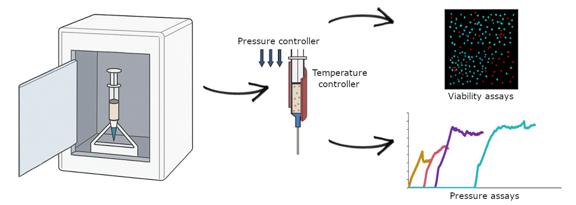
Extrusion-based three-dimensional bioprinting remains limited by the absence of standardized methods to define the pressure conditions required for stable hydrogel flow. As a result, most workflows still rely on empirical tuning, which compromises reproducibility, structural fidelity, and interlaboratory comparability. Addressing this gap requires quantitative tools capable of identifying the minimum pressure for extrusion and the pressure range in which continuous, defect-free flow is maintained. This work presents a modular characterization platform integrating real-time pressure sensing, together with nozzle temperature regulation and environmental monitoring (temperature, humidity, and carbon dioxide) to ensure controlled and reproducible extrusion conditions. Using 10% gelatin methacryloyl, force–displacement curves revealed a stabilization pressure of 165 kPa, associated with continuous extrusion and minimal geometric deviation, and bounded by experimentally validated under-extrusion (155 kPa) and over-extrusion (185 kPa) conditions. Bioprinting tests confirmed that operating within this stabilization zone improves filament uniformity, print fidelity, and dimensional accuracy compared with under- and over-extrusion regimes. Quantitative metrics, including deflection analysis, printability parameter, void–area similarity, and structural similarity index, demonstrated superior reproducibility at the stabilization pressure. Biological validation using MCF-7 cells showed that 165 kPa preserved high viability (97.9%), whereas extrusion at 185 kPa reduced survival to 88.6%, confirming the sensitivity of cell integrity to excess shear stress. Together, these findings establish a sensor-driven calibration strategy that replaces trial-and-error parameter selection with quantitative and reproducible pre-print optimization. The device is compatible with commercial bioprinters and provides a practical framework for improving process standardization, structural fidelity, and biological safety in extrusion-based bioprinting. The platform is conceived as an independent, modular device for experimental calibration of extrusion parameters prior to bioprinting.
- Jiang T, Munguia-Lopez JG, Flores-Torres S, Kort- Mascort J, Kinsella JM. Extrusion bioprinting of soft materials: an emerging technique for biological model fabrication. Appl Phys Rev. 2019;6(1):011310. doi: 10.1063/1.5059393
- Askari M, Afzali Naniz M, Kouhi M, Saberi A, Zolfagharian A, Bodaghi M. Recent progress in extrusion 3D bioprinting of hydrogel biomaterials for tissue regeneration: a comprehensive review with focus on advanced fabrication techniques. Biomater Sci. 2021;9(3): 535-573. doi: 10.1039/D0BM00973C
- He C, He J, Wu C, et al. 3D printing for tissue/organ regeneration in China. Bio Des Manuf. 2025;8:169-244. doi: 10.1631/BDM.2400309
- Murphy SV, Atala A. 3D bioprinting of tissues and organs. Nat Biotechnol. 2014;32(8):773-785. doi: 10.1038/nbt.2958
- Ozbolat IT, Hospodiuk M. Current advances and future perspectives in extrusion-based bioprinting. Biomaterials. 2016;76:321-343. doi: 10.1016/J.BIOMATERIALS.2015.10.076
- Geckil H, Xu F, Zhang X, Moon S, Demirci U. Engineering hydrogels as extracellular matrix mimics. Nanomedicine (Lond). 2010;5(3):469-484. doi: 10.2217/NNM.10.12
- Gopinathan J, Noh I. Recent trends in bioinks for 3D printing. Biomater Res. 2018;22(11):1-15. doi: 10.1186/S40824-018-0122-1
- Wang H, Bi S, Shi B, et al. Recent advances in engineering bioinks for 3D bioprinting. Adv Eng Mater. 2023;25(19):2300648. doi: 10.1002/ADEM.202300648
- Gasperini L, Mano JF, Reis RL. Natural polymers for the microencapsulation of cells. J R Soc Interface. 2014;11(100):20140817. doi: 10.1098/RSIF.2014.0817
- Olabisi RM. Cell microencapsulation with synthetic polymers. J Biomed Mater Res A. 2015;103(2):846-859. doi: 10.1002/JBM.A.35205
- Esfahani RR, Jun H, Rahmani S, Miller A, Lahann J. Microencapsulation of live cells in synthetic polymer capsules. ACS Omega. 2017;2(6):2839-2847. doi: 10.1021/acsomega.7b00570
- Cardoso LM da F, Alves LA, Barreto T, Gama JFG. Natural biopolymers as additional tools for cell microencapsulation applied to cellular therapy. Polymers. 2022;14(13):2641. doi: 10.3390/POLYM14132641
- Leberfinger AN, Ravnic DJ, Dhawan A, Ozbolat IT. Concise review: bioprinting of stem cells for transplantable tissue fabrication. Stem Cells Transl Med. 2017;6(10):1940. doi: 10.1002/SCTM.17-0148
- Gu Q, Tomaskovic-Crook E, Lozano R, et al. Functional 3D neural mini-tissues from printed gel-based bioink and human neural stem cells. Adv Healthc Mater. 2016;5(12):1429-1438. doi: 10.1002/ADHM.201600095
- Jeon O, Lee Y Bin, Jeong H, Lee SJ, Wells D, Alsberg E. Individual cell-only bioink and photocurable supporting medium for 3D printing and generation of engineered tissues with complex geometries. Mater Horiz. 2019;6(8):1625. doi: 10.1039/C9MH00375D
- Boularaoui S, Al Hussein G, Khan KA, Christoforou N, Stefanini C. An overview of extrusion-based bioprinting with a focus on induced shear stress and its effect on cell viability. Bioprinting. 2020;20:e00093. doi: 10.1016/J.BPRINT.2020.E00093
- Rossi A, Pescara T, Gambelli AM, et al. Biomaterials for extrusion-based bioprinting and biomedical applications. Front Bioeng Biotechnol. 2024;12:1393641. doi: 10.3389/fbioe.2024.1393641
- Mirshafiei M, Rashedi H, Yazdian F, Rahdar A, Baino F. Advancements in tissue and organ 3D bioprinting: current techniques, applications, and future perspectives. Mater Des. 2024;240:112853. doi: 10.1016/J.MATDES.2024.112853
- Jang J, Park JY, Gao G, Cho DW. Biomaterials-based 3D cell printing for next-generation therapeutics and diagnostics. Biomaterials. 2018;156:88-106. doi: 10.1016/J.BIOMATERIALS.2017.11.030
- Malda J, Visser J, Melchels FP, et al. 25th anniversary article: engineering hydrogels for biofabrication. Adv Mater. 2013;25(36):5011-5028. doi: 10.1002/ADMA.201302042
- Schwab A, Levato R, D’Este M, Piluso S, Eglin D, Malda J. Printability and shape fidelity of bioinks in 3D bioprinting. Chem Rev. 2020;120(19):11028-11055. doi: 10.1021/ACS.CHEMREV.0C00084
- Mendoza Cerezo L, Romero ACM, García AM, Amador JPC, Rego JMR. Recent advances in 3D bioprinting. Compendium of 3D Bioprinting Technology. Boca Raton: CRC Press; 2025:69-85. doi: 10.1201/9781003505198-4
- Rego JMR, Cendal AIR, Prado SMD, García AM, Romero ACM, Cerezo LM. 3D bioprinting of cartilage. Compendium of 3D Bioprinting Technology. Boca Raton: CRC Press; 2025:469-481. doi: 10.1201/9781003505198-25
- Datta P, Ayan B, Ozbolat IT. Bioprinting for vascular and vascularized tissue biofabrication. Acta Biomater. 2017;51:1-20. doi: 10.1016/J.ACTBIO.2017.01.035
- Strauß S, Schroth B, Hubbuch J. Evaluation of the reproducibility and robustness of extrusion-based bioprinting processes applying a flow sensor. Front Bioeng Biotechnol. 2022;10:831350. doi: 10.3389/FBIOE.2022.831350/BIBTEX
- Strauß S, Garces DG, Hubbuch J. Analytics in extrusion-based bioprinting: standardized methods improving quantification and comparability of the performance of bioinks. Polymers. 2023;15(8):1829. doi: 10.3390/POLYM15081829
- Grijalva Garces D, Strauß S, Gretzinger S, et al. On the reproducibility of extrusion-based bioprinting: round robin study on standardization in the field. Biofabrication. 2023;16(1):015002. doi: 10.1088/1758-5090/ACFE3B
- Armstrong AA, Pfeil A, Alleyne AG, Wagoner Johnson AJ. Process monitoring and control strategies in extrusion-based bioprinting to fabricate spatially graded structures. Bioprinting. 2021;21:e00126. doi: 10.1016/J.BPRINT.2020.E00126
- Mũnoz Z, Shih H, Lin CC. Gelatin hydrogels formed by orthogonal thiol–norbornene photochemistry for cell encapsulation. Biomater Sci. 2014;2(8):1063-1072. doi: 10.1039/C4BM00070F
- Eslahi N, Abdorahim M, Simchi A. Smart polymeric hydrogels for cartilage tissue engineering: a review on the chemistry and biological functions. Biomacromolecules. 2016;17(11):3441-3463. doi: 10.1021/ACS.BIOMAC.6B01235
- Xiao S, Zhao T, Wang J, et al. Gelatin methacrylate (GelMA)-based hydrogels for cell transplantation: an effective strategy for tissue engineering. Stem Cell Rev Rep. 2019;15(5):664-679. doi: 10.1007/S12015-019-09893-4
- Yue K, Trujillo-de Santiago G, Alvarez MM, Tamayol A, Annabi N, Khademhosseini A. Synthesis, properties, and biomedical applications of gelatin methacryloyl (GelMA) hydrogels. Biomaterials. 2015;73:254-271. doi: 10.1016/J.BIOMATERIALS.2015.08.045
- Zhao X, Lang Q, Yildirimer L, et al. Photocrosslinkable gelatin hydrogel for epidermal tissue engineering. Adv Healthc Mater. 2016;5(1):108-118. doi: 10.1002/ADHM.201500005
- Lee BH, Shirahama H, Cho NJ, Tan LP. Efficient and controllable synthesis of highly substituted gelatin methacrylamide for mechanically stiff hydrogels. RSC Adv. 2015;5(128):106094-106097. doi: 10.1039/C5RA22028A
- Sun M, Sun X, Wang Z, Guo S, Yu G, Yang H. Synthesis and properties of gelatin methacryloyl (GelMA) hydrogels and their recent applications in load-bearing tissue. Polymers (Basel). 2018;10(11):1290. doi: 10.3390/POLYM10111290
- Arguchinskaya NV, Isaeva EV, Kisel AA, et al. Properties and printability of the synthesized hydrogel based on GelMA. Int J Mol Sci. 2023;24(3):2121. doi: 10.3390/IJMS24032121
- Han S, Kim CM, Jin S, Kim TY. Study of the process-induced cell damage in forced extrusion bioprinting. Biofabrication. 2021;13(3). doi: 10.1088/1758-5090/AC0415
- Paxton N, Smolan W, Böck T, Melchels F, Groll J, Jungst T. Proposal to assess printability of bioinks for extrusion-based bioprinting and evaluation of rheological properties governing bioprintability. Biofabrication. 2017;9(4):044107. doi: 10.1088/1758-5090/AA8DD8
- Ribeiro A, Blokzijl MM, Levato R, et al. Assessing bioink shape fidelity to aid material development in 3D bioprinting. Biofabrication. 2017;10(1):014102. doi: 10.1088/1758-5090/AA90E2
- Rodríguez-Rego JM, Mendoza-Cerezo L, Macías-García A, Carrasco-Amador JP, Marcos-Romero AC. Methodology for characterizing the printability of hydrogels. Int J Bioprint. 2023;9(2):280-291. doi: 10.18063/IJB.V9I2.667
- Ouyang L, Yao R, Zhao Y, Sun W. Effect of bioink properties on printability and cell viability for 3D bioplotting of embryonic stem cells. Biofabrication. 2016;8(3):035020. doi: 10.1088/1758-5090/8/3/035020
- Ángeles Pérez M, Compaired PM, García-Gareta E. An experimental workflow for bioprinting optimization: application to a custom-made biomaterial ink. Int J Bioprint. 2025;11(3):397-415. doi: 10.36922/IJB025120094
- Naghieh S, Chen X. Printability–a key issue in extrusion-based bioprinting. J Pharm Anal. 2021;11(5):564-579. doi: 10.1016/J.JPHA.2021.02.001
- Malekpour A, Chen X. Printability and cell viability in extrusion-based bioprinting from experimental, computational, and machine learning views. J Funct Biomater. 2022;13(2):40. doi: 10.3390/JFB13020040
- Ning L, Betancourt N, Schreyer DJ, Chen X. Characterization of cell damage and proliferative ability during and after bioprinting. ACS Biomater Sci Eng. 2018;4(11): 3906-3918. doi: 10.1021/acsbiomaterials.8b00714