

Three-dimensional-printed bionic dual crosslinked drug-loaded hydrogel composite scaffolds for large bone defect repair

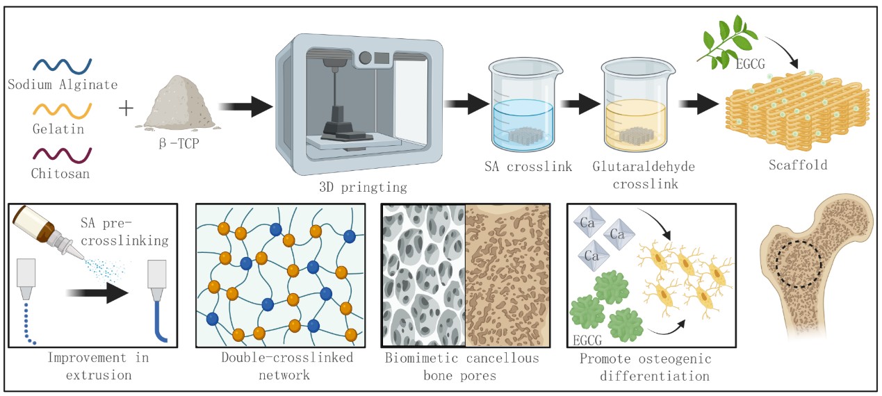
The structure, composition, and function of natural bone have long been the focus of bone tissue engineering. However, existing organic–inorganic three-dimensional (3D) printing systems are limited by the stability of hydrogels and the content of inorganic salts, hindering the fabrication of robust 3D scaffolds. In this study, we developed a hydrogel–inorganic particle bioink and implemented a multi-step crosslinking strategy. The organic phase, composed of sodium alginate, gelatin, and chitosan, was combined with β-tricalcium phosphate and crosslinked via ionic pre-crosslinking followed by a Schiff base reaction to form a dual-crosslinked network. The resulting scaffolds exhibited excellent mechanical properties and biomimetic microarchitecture while maintaining shape stability under physiological conditions. Furthermore, the tunable swelling behavior of the hydrogel enabled efficient loading and controlled release of the small molecule epigallocatechin gallate. This composite scaffold demonstrated adjustable swelling, controllable degradation, and significantly enhanced cellular compatibility, providing a novel, efficient, and scalable strategy for repairing complex bone defects and offering new insights for the design and application of 3D-printed bone scaffolds.
- Wang Y, Zhang H, Hu Y, Jing Y, Geng Z, Su J. Bone repair biomaterials: a perspective from immunomodulation. Adv Funct Mater. 2022;32(51):2208639. doi: 10.1002/adfm.202208639
- Batra P, Das S, Jain S. Correlation of radiovisuographic analysis of interdental and interradicular bone loss in furcation involvement of mandibular first molars: a retrospective study. Indian J Dent Res. 2018;29(3):329-332. doi: 10.4103/ijdr.IJDR_52_16. PMID: 29900917
- Chang HY, Park SY, Kim JA, Kim YK, Lee HJ. Early radiographic diagnosis of peri-implantitis enhances the outcome of peri-implantitis treatment: a 5-year retrospective study after non-surgical treatment. J Periodontal Implant Sci. 2015;45(3):82-93. doi: 10.5051/jpis.2015.45.3.82
- Kironde E, Sekimpi P, Kajja I, Mubiri P. Prevalence and patterns of traumatic bone loss following open long bone fractures at Mulago Hospital. OTA Int. 2019.2(1): e015-e015. doi: 10.1097/OI9.0000000000000015
- Puisys A, Auzbikaviciute V, Minkauskaite A, et al. Early crestal bone loss: is it really loss? Clin Case Rep. 2019;7(10):1913-1915. doi: 10.1002/ccr3.2376
- Zhou H, Liang B, Jiang H, Deng Z, Yu K. Magnesium-based biomaterials as emerging agents for bone repair and regeneration: from mechanism to application. J Magnes Alloy. 2021;9(3):779-804. doi: 10.1016/j.jma.2021.03.004
- Du M, Chen J, Liu K, Xing H, Song C. Recent advances in biomedical engineering of nano-hydroxyapatite including dentistry, cancer treatment and bone repair. Compos Part B Eng. 2021;215:108790. doi: 10.1016/j.compositesb.2021.108790
- Feng P, Zhao R Y, Tang W M, et al. Structural and functional adaptive artificial bone: materials, fabrications, and properties. Adv Funct Mater. 2023;33(23):2214726. doi: 10.1002/adfm.202214726
- Nissen FI, Andreasen C, Borgen TT, Bjørnerem Å, Hansen AK. Cortical bone structure of the proximal femur and incident fractures. Bone. 2022;155:116284. doi: 10.1016/j.bone.2021.116284
- Rodriguez-Palomo A, Ostergaard M, Birkedal H. Bone hierarchical structure: heterogeneity and uniformity. Adv Funct Mater. 2023;34(35):2307026. doi: 10.1002/adfm.202307026
- Zhou C, Zhang XL, Ai J, et al. Chiral hierarchical structure of bone minerals. Nano Res. 2022;15(2):1295-1302. doi: 10.1007/s12274-021-3653-z
- Yusof F, Sha’ban M, Azhim A. Decellularization strategies to engineer fibrocartilage bioscaffolds: a review. J Biomater Tissue Eng. 2021;11(8):1435-1451. doi: 10.1166/jbt.2021.2740
- He J, Fang J, Wei P, et al. Cancellous bone-like porous Fe@Zn scaffolds with core-shell-structured skeletons for biodegradable bone implants. Acta Biomater. 2021;121:665-681. doi: 10.1016/j.actbio.2020.11.032
- Yoon SJ, Kim SH, Choi JW, Chun HJ, Yang DH. Guided cortical and cancellous bone formation using a minimally invasive technique of BMSC- and BMP-2-laden visible light-cured carboxymethyl chitosan hydrogels. Int J Biol Macromol. 2023;227:641-653. doi: 10.1016/j.ijbiomac.2022.12.13
- Zhao ZY, Li G, Ruan HT, et al. Capturing magnesium ions via microfluidic hydrogel microspheres for promoting cancellous bone regeneration. ACS Nano. 2021;15(8):13041-130540. doi: 10.1021/acsnano.1c02147
- Hu X, Lin Z, He J, et al. Recent progress in 3D printing degradable polylactic acid‐based bone repair scaffold for the application of cancellous bone defect. MedComm Biomater Appl. 2022;1(1):e14. doi: 10.1002/mba2.14
- Riggs C, Goodship A. Bone structure and function. Fractures in the Horse. 2022:11-27. doi: 10.1002/9781119431749.ch2
- Li C, Du YW, Zhang TT, et al. “Genetic scissors” CRISPR/ Cas9 genome editing cutting-edge biocarrier technology for bone and cartilage repair. Bioact Mater. 2023;22:254-273. doi: 10.1016/j.bioactmat.2022.09.026
- Salhotra A, Shah HN, Levi B, Longaker MT. Mechanisms of bone development and repair. Nat Rev Mol Cell Biol. 2020;21(11):696-711. doi: 10.1038/s41580-020-00279-w
- Seidi A, Ramalingam M. Engineering of gradient biomaterials as biomimetic systems for tissue engineering. J Biomater Tissue Eng. 2011;1(2):139-148. doi: 10.1166/jbt.2011.1020
- Li J, Chen Q, Zhang Q, et al. Improving mechanical properties and biocompatibilities by highly oriented long chain branching poly(lactic acid) with bionic surface structures. ACS Appl Mater Interfaces. 2020;12(12): 14365-14375. doi: 10.1021/acsami.9b20264
- Qi T, Zhang X, Gu X, Cui S. Experimental study on repairing peripheral nerve defects with novel bionic tissue engineering. Adv Healthc Mater. 2023;12(17):e2203199. doi: 10.1002/adhm.202203199
- Wang Y, Jiang X, Li X, et al. Bionic ordered structured hydrogels: structure types, design strategies, optimization mechanism of mechanical properties and applications. Mater Horiz. 2023;10(10):4033-4058. doi: 10.1039/d3mh00326d
- Wang L, Li AF, Zhang D, et al. Injectable double-network hydrogel for corneal repair. Chem Eng J. 2023;455:140698. doi: 10.1016/j.cej.2022.140698.
- Zhu WX, Zhou Z, Huang YT, et al. A versatile 3D-printable hydrogel for antichondrosarcoma, antibacterial, and tissue repair. J Mater Sci Technol. 2023;136:200-211. doi: 10.1016/j.jmst.2022.07.010
- Ahmed EM. Hydrogel: preparation, characterization, and applications: a review. J Adv Res. 2015;6(2):105-121. doi: 10.1016/j.jare.2013.07.006
- Lee KY, Mooney DJ. Alginate: properties and biomedical applications. Prog Polym Sci. 2012;37(1):106-126. doi: 10.1016/j.progpolymsci.2011.06.003
- Liang YP, He JH, Guo BL. Functional hydrogels as wound dressing to enhance wound healing. ACS Nano. 2021;15(8):12687-12722. doi: 10.1021/acsnano.1c04206
- Correa CS, Grosskopf AK, Hernandez HL, Chan D, Appel EA. Translational applications of hydrogels. Chem Rev. 2021;121(18):11385-11457. doi: 10.1021/acs.chemrev.0c01177
- Nie R, Sun Y, Lv H, et al. 3D printing of MXene composite hydrogel scaffolds for photothermal antibacterial activity and bone regeneration in infected bone defect models. Nanoscale. 2022;14(22):8112-8129. doi: 10.1039/D2NR02176E
- Gong JP, Katsuyama Y, Kurokawa T, Osada Y. Double‐network hydrogels with extremely high mechanical strength. Adv Mater. 2003;15(14):1155-1158. doi: 10.1002/adma.200304907
- Aldana AA, Houben S, Moroni L, Baker MB, Pitet LM. Trends in double networks as bioprintable and injectable hydrogel scaffolds for tissue regeneration. ACS Biomater Sci Eng. 2021;7(9):4077-4101. doi: 10.1021/acsbiomaterials.0c01749
- Xu X, Jerca VV, Hoogenboom R. Bioinspired double network hydrogels: from covalent double network hydrogels via hybrid double network hydrogels to physical double network hydrogels. Mater Horiz. 2021;8(4):1173-1188. doi: 10.1039/d0mh01514h
- Yang J, Li K, Tang C, et al. Recent progress in double network elastomers: one plus one is greater than two. Adv Funct Mater. 2022;32(19):2110244. doi: 10.1002/adfm.202110244
- Gao N, Zhang Y, Yang Z, et al. Ba2+/Ca2+ co-crosslinked alginate hydrogel filtration membrane with high strength, high flux and stability for dye/salt separation. Chin Chem Lett. 2023;35(5):108820. doi: 10.1016/j.cclet.2023.108820
- Zhang A, Wang F, Chen L, et al. 3D printing hydrogels for actuators: a review. Chin Chem Lett. 2021;32(10):2923-2932. doi: 10.1016/j.cclet.2021.03.073
- Wang Y, Jiao Y, Zeng Z, Chang J, Yang C, Dong Z. Three‐dimensional printed Zn2SiO4/sodium alginate composite scaffold with multiple biological functions for tendon‐to‐bone repair. MedComm Biomater Appl. 2023;2(4):e61. doi: 10.1002/mba2.61
- Kankariya Y, Chatterjee B. Biomedical application of chitosan and chitosan derivatives: a comprehensive review. Curr Pharm Des. 2023;29(17):1311-1325. doi: 10.2174/1381612829666230524153002
- Manna S, Seth A, Gupta P, et al. Chitosan derivatives as carriers for drug delivery and biomedical applications. ACS Biomater Sci Eng. 2023;9(5):2181-2202. doi: 10.1007/978-981-15-0263-7
- Sacco P, Pedroso-Santana S, Kumar Y, Joly N, Martin P, Bocchetta P. Ionotropic gelation of chitosan flat structures and potential applications. Molecules. 2021;26(3):660. doi: 10.3390/molecules26030660
- Yan D, Li Y, Liu Y, Li N, Zhang X, Yan C. Antimicrobial properties of chitosan and chitosan derivatives in the treatment of enteric infections. Molecules. 2021;26(23):7136. doi: 10.3390/molecules26237136
- Xulin H, Hu L, Liang Q, et al. 369Fabrication of 3D gel-printed β-tricalcium phosphate/titanium dioxide porous scaffolds for cancellous bone tissue engineering. Int J Bioprint. 2023;9(2):673. Published 2023 Jan 19. doi: 10.18063/ijb.v9i2.673
- Peres I, Rocha S, do Carmo Pereira M, Coelho M, Rangel M, Ivanova G. NMR structural analysis of epigallocatechin gallate loaded polysaccharide nanoparticles. Carbohydr Polym. 2010;82(3):861-866. doi: 10.1016/j.carbpol.2010.06.007
- Zhang S, Mao B, Cui S, et al. Absorption, metabolism, bioactivity, and biotransformation of epigallocatechin gallate. Crit Rev Food Sci Nutr. 2024;64(19):6546-6566. doi: 10.1080/10408398.2023.2170972
- Guo Z, Liu W, Liu T, et al. Engineered exosome hybrid copper nanoscale antibiotics facilitate simultaneous self-assembly imaging and elimination of intracellular multidrug-resistant superbugs. Chin Chem Lett. 2023;35(7):109060. doi: 10.1016/j.cclet.2023.109060
- Baranwal A, Aggarwal P, Rai A, Kumar N. Pharmacological actions and underlying mechanisms of catechin: a review. Mini Rev Med Chem. 2022;22(5):821-833. doi: 10.2174/1389557521666210902162120
- Cabezas Perez RJ, Ávila Rodríguez MF, Rosero Salazar DH. Exogenous antioxidants in remyelination and skeletal muscle recovery. Biomedicines. 2022;10(10):2557. doi: 10.3390/biomedicines10102557
- Spicer PP, Kretlow JD, Young S, Jansen JA, Kasper FK, Mikos AG. Evaluation of bone regeneration using the rat critical size calvarial defect. Nat Protoc. 2012;7(10):1918-1929. doi: 10.1038/nprot.2012.113