

Synergistic bone regeneration by surface-modified 3D-printed PCL/β-TCP scaffolds in various animal defect models

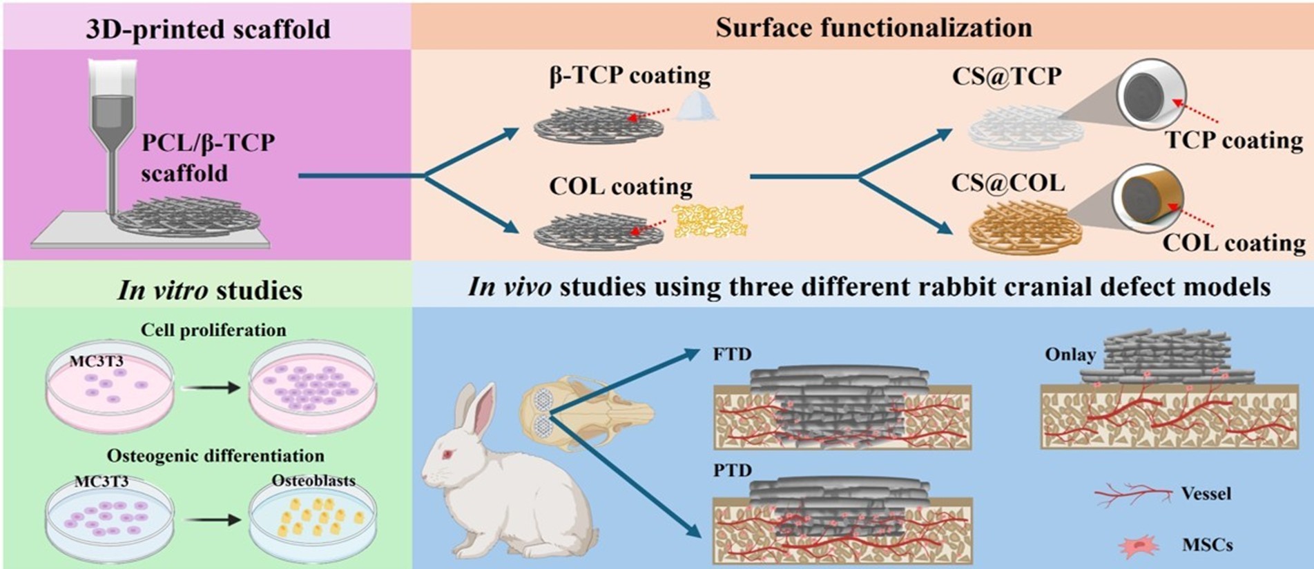
The regeneration of large-segmental bone defects remains a significant clinical challenge due to their complex microenvironments. Three-dimensional (3D)-printed polycaprolactone (PCL) scaffolds offer a potential solution but exhibit limited osteoinductive capacity. In this study, 3D-printed PCL/β-tricalcium phosphate (TCP) composite scaffolds were pretreated with NaOH, followed by functionalization with bioactive collagen and β‑TCP. These modifications markedly improved the scaffolds’ hydrophilicity without compromising mechanical integrity. In vitro studies with MC3T3-E1 cells demonstrated that the CS@TCP scaffolds significantly enhanced early osteogenic differentiation compared to C, CS, and CS@COL scaffolds, as indicated by the alkaline phosphatase activity assay. In vivo evaluation using three different rabbit cranial defect models revealed superior new bone formation in the partial-thickness cranial defect (PTD) groups compared to the full-thickness cranial defect (FTD) and intact cranial bone onlay (Onlay) groups, potentially due to the increased vascularization and abundant endogenous stem cells in the PTD groups. Despite reduced new bone formation in the Onlay group, its bone integration advantages may be advantageous for cosmetic surgery applications. This study investigated how β‑TCP surface modification interacts with clinical application-specific microenvironments to maximize the regenerative potential of 3D-printed scaffolds, providing crucial guidance for scaffold design in effective bone defect repair across various clinical scenarios.
- Yelin E, Weinstein S, King T. The burden of musculoskeletal diseases in the United States. Semin Arthritis Rheum. 2016;46(3):259-260. doi:10.1016/j.semarthrit.2016.07.013
- Ong KL, Mowat FS, Chan N, et al. Economic burden of revision hip and knee arthroplasty in Medicare enrollees. Clin Orthop Relat Res. 2006;446:22-28. doi:10.1097/01.blo.0000214439.95268.59
- Huang D, Li Z, Li G, et al. Biomimetic structural design in 3D-printed scaffolds for bone tissue engineering. Mater Today Bio. 2025;32:101664. doi:10.1016/j.mtbio.2025.101664
- Tsiklin IL, Shabunin AV, Kolsanov AV, Volova LT. In vivo bone tissue engineering strategies: advances and prospects. Polymers (Basel). 2022;14(15):3222. doi:10.3390/polym14153222
- Wu Y, Ji Y, Lyu Z. 3D printing technology and its combination with nanotechnology in bone tissue engineering. Biomed Eng Lett. 2024;14(3):451-464. doi:10.1007/s13534-024-00350-x
- Wen Y, Xun S, Haoye M, et al. 3D printed porous ceramic scaffolds for bone tissue engineering: a review. Biomater Sci. 2017;5(9):1690-1698. doi:10.1039/c7bm00315c
- Meng F, Yin Z, Ren X, Geng Z, Su J. Construction of local drug delivery system on titanium-based implants to improve osseointegration. Pharmaceutics. 2022;14(5):1069. doi:10.3390/pharmaceutics14051069
- Sun S, Liu H, Hu Y, et al. Selection and identification of a novel ssDNA aptamer targeting human skeletal muscle. Bioact Mater. 2023;20:166-178. doi:10.1016/j.bioactmat.2022.05.016
- Wang Q, Ye W, Ma Z, et al. 3D printed PCL/β-TCP cross-scale scaffold with high-precision fiber for providing cell growth and forming bones in the pores. Mater Sci Eng C Mater Biol Appl. 2021;127:112197. doi:10.1016/j.msec.2021.112197
- Weingärtner L, Latorre SH, Velten D, et al. The effect of collagen-I coatings of 3D printed PCL scaffolds for bone replacement on three different cell types. Appl Sci. 2021;11(22):11063. doi:10.3390/app112211063
- Jiang Y, Zhou C, Yang X, Ke D. 3D printed bioactive coated scaffolds boost osteogenesis and angiogenesis via the regulation of scaffold microstructure. Biofabrication. 2025;17(3):1-18. doi:10.1088/1758-5090/addc9c
- Furtado ASA, Cunha MHS, Sousa LMR, et al. 3D-printed PCL-based scaffolds with high nanosized synthetic smectic clay content: fabrication, mechanical properties, and biological evaluation for bone tissue engineering. Int J Nanomedicine. 2025;20:53-69. doi:10.2147/IJN.S497539
- Hao S, Wang M, Yin Z, et al. Microenvironment-targeted strategy steers advanced bone regeneration. Materials Today Bio. 2023;22:100741. doi:10.1016/j.mtbio.2023.100741
- Zhu G, Zhang T, Chen M, et al. Bone physiological microenvironment and healing mechanism: basis for future bone-tissue engineering scaffolds. Bioact Mater. 2021;6(11):4110-4140. doi:10.1016/j.bioactmat.2021.03.043
- Koushik TM, Miller CM, Antunes E. Bone tissue engineering scaffolds: function of multi-material hierarchically structured scaffolds. Adv Healthc Mater. 2023;12(9):2202766. doi:10.1002/adhm.202202766
- Riau AK, Venkatraman SS, Mehta JS. Biomimetic vs. direct approach to deposit hydroxyapatite on the surface of low melting point polymers for tissue engineering. Nanomaterials. 2020;10(11):2162. doi:10.3390/nano10112162
- Shen HY, Xing F, Shang SY, et al. Biomimetic mineralized 3D-printed polycaprolactone scaffold induced by self-adaptive nanotopology to accelerate bone regeneration. ACS Appl Mater Interfaces. 2024;16(15):18658-18670. doi:10.1021/acsami.4c02636
- Heo SY, Ko SC, Oh GW, et al. Fabrication and characterization of the 3D-printed polycaprolactone/fish bone extract scaffolds for bone tissue regeneration. J Biomed Mater Res B Appl Biomater. 2019;107(6):1937-1944. doi:10.1002/jbm.b.34286
- Tawfick S, De Volder M, Copic D, et al. Engineering of micro- and nanostructured surfaces with anisotropic geometries and properties. Adv Mater. 2012;24(13): 1628-1674. doi:10.1002/adma.201103796
- Kocagöz M, Tıhmınlıoğlu F, Aldemir Dikici B. Surface modification via alkali treatment and its effect on the physicochemical and biological properties of emulsion templated scaffolds. Polymer. 2025;330:128475. doi:10.1016/j.polymer.2025.128475
- Sherman VR, Yang W, Meyers MA. The materials science of collagen. J Mech Behav Biomed Mater. 2015;52:22-50. doi:10.1016/j.jmbbm.2015.05.023
- Gresita A, Raja I, Petcu E, Hadjiargyrou M. Collagen-coated hyperelastic bone promotes osteoblast adhesion and proliferation. Materials (Basel). 2023;16(21):6996. doi:10.3390/ma16216996
- Vandrovcová M, Douglas T, Hauk D, et al. Influence of collagen and chondroitin sulfate (CS) coatings on poly- (lactide-co-glycolide) (PLGA) on MG 63 osteoblast-like cells. Physiol Res. 2011;60(5):797-813. doi:10.33549/physiolres.931994
- Valdoz JC, Johnson BC, Jacobs DJ, et al. The ECM: to scaffold, or not to scaffold, that is the question. Int J Mol Sci. 2021;22(23):12690. doi:10.3390/ijms222312690
- Jeong J, Kim JH, Shim JH, Hwang NS, Heo CY. Bioactive calcium phosphate materials and applications in bone regeneration. Biomater Res. 2019;23:4. doi:10.1186/s40824-018-0149-3
- Boda R, Lázár I, Keczánné-Üveges A, et al. β-Tricalcium phosphate-modified aerogel containing PVA/Chitosan hybrid nanospun scaffolds for bone regeneration. Int J Mol Sci. 2023;24(8):7562. doi:10.3390/ijms24087562
- Silva-Barroso AS, Cabral CSD, Ferreira P, Moreira AF, Correia IJ. Lignin-enriched tricalcium phosphate/sodium alginate 3D scaffolds for application in bone tissue regeneration. Int J Biol Macromol. 2023;239:124258. doi:10.1016/j.ijbiomac.2023.124258
- Lu Q, Diao J, Wang Y, et al. 3D printed pore morphology mediates bone marrow stem cell behaviors via RhoA/ROCK2 signaling pathway for accelerating bone regeneration. Bioact Mater. 2023;26:413-424. doi:10.1016/j.bioactmat.2023.02.025
- Zheng J, Nozaki K, Hashimoto K, Yamashita K, Wakabayashi N. Exploring the biological impact of β-TCP surface polarization on osteoblast and osteoclast activity. Int J Mol Sci. 2024;26(1):141. doi:10.3390/ijms26010141
- Lu J, Yu H, Chen C. Biological properties of calcium phosphate biomaterials for bone repair: a review. RSC Adv. 2018;8(4):2015-2033. doi:10.1039/c7ra11278e
- Deng W, Yang X, Yu J, Omari-Siaw E, Xu X. Recent advances of physiochemical cues on surfaces for directing cell fates. Colloids Surf B Biointerfaces. 2025;250:114550. doi:10.1016/j.colsurfb.2025.114550
- Gupta D, Singh AK, Kar N, Dravid A, Bellare J. Modelling and optimization of NaOH-etched 3-D printed PCL for enhanced cellular attachment and growth with minimal loss of mechanical strength. Mater Sci Eng C. 2019;98:602-611. doi:10.1016/j.msec.2018.12.084
- Jiang L, Jin S, Geng S, et al. Maintenance and restoration effect of the surface hydrophilicity of pure titanium by sodium hydroxide treatment and its effect on the bioactivity of osteoblasts. Coatings. 2019;9(4):222. doi:10.3390/coatings9040222
- Witek L, Shi Y, Smay J. Controlling calcium and phosphate ion release of 3D printed bioactive ceramic scaffolds: an in vitro study. J Adv Ceram. 2017;6(2):157-164. doi:10.1007/s40145-017-0228-2
- Luo F, Yang Y, Li D, et al. Low-temperature plasma effect-induced enhancement of osteogenic activity in calcium phosphate ceramics. Acta Biomater. 2025;200:667-685. doi:10.1016/j.actbio.2025.04.048
- González Díaz EC, Shih YRV, Nakasaki M, Liu M, Varghese S. Mineralized biomaterials mediated repair of bone defects through endogenous cells. Tissue Eng Part A. 2018;24(13–14):1148-1156. doi:10.1089/ten.tea.2017.0297
- Weng Y, Jian Y, Huang W, et al. Alkaline earth metals for osteogenic scaffolds: from mechanisms to applications. J Biomed Mater Res B Appl Biomater. 2023;111(7):1447-1474. doi:10.1002/jbm.b.35246
- Wang K, Jiang K, Luo C, et al. An osteoimmunomodulatory Ca2+/Zn2+-doped scaffold promotes M2 macrophage polarization via the src-mediated chemoking signaling pathway to enhance osteoinduction. Compos Part B: Eng. 2024;284:111653. doi:10.1016/j.compositesb.2024.111653
- Ghiacci G, Graiani G, Ravanetti F, et al. “Over-inlay” block graft and differential morphometry: a novel block graft model to study bone regeneration and host-to-graft interfaces in rats. J Periodontal Implant Sci. 2016;46(4):220-233. doi:10.5051/jpis.2016.46.4.220
- Selvaraj V, Sekaran S, Dhanasekaran A, Warrier S. Type 1 collagen: synthesis, structure and key functions in bone mineralization. Differentiation. 2024;136:100757. doi:10.1016/j.diff.2024.100757
- Pilliar RM, Lee JM, Maniatopoulos C. Observations on the effect of movement on bone ingrowth into porous-surfaced implants. Clin Orthop Relat Res. 1986;7(208):108-113.
- Sosakul T, Tuchpramuk P, Suvannapruk W, et al. Evaluation of tissue ingrowth and reaction of a porous polyethylene block as an onlay bone graft in rabbit posterior mandible. J Periodontal Implant Sci. 2020;50(2):106-120. doi:10.5051/jpis.2020.50.2.106
- Canto FRT, Garcia SB, Issa JPM, et al. Influence of decortication of the recipient graft bed on graft integration and tissue neoformation in the graft-recipient bed interface. Eur Spine J. 2008;17(5):706-714. doi:10.1007/s00586-008-0642-9
- Wu S, Luo S, Cen Z, et al. All-in-one porous membrane enables full protection in guided bone regeneration. Nat Commun. 2024;15(1):119. doi:10.1038/s41467-023-43476-9
- Ku JK, Lee KG, Ghim MS, et al. Onlay-graft of 3D printed Kagome-structure PCL scaffold incorporated with rhBMP-2 based on hyaluronic acid hydrogel. Biomed Mater. 2021;16(5):055004. doi:10.1088/1748-605X/ac0f47