

3D-printed perfusable tumor model for evaluating perfusion and chemotherapeutic response in ovarian cancer cells

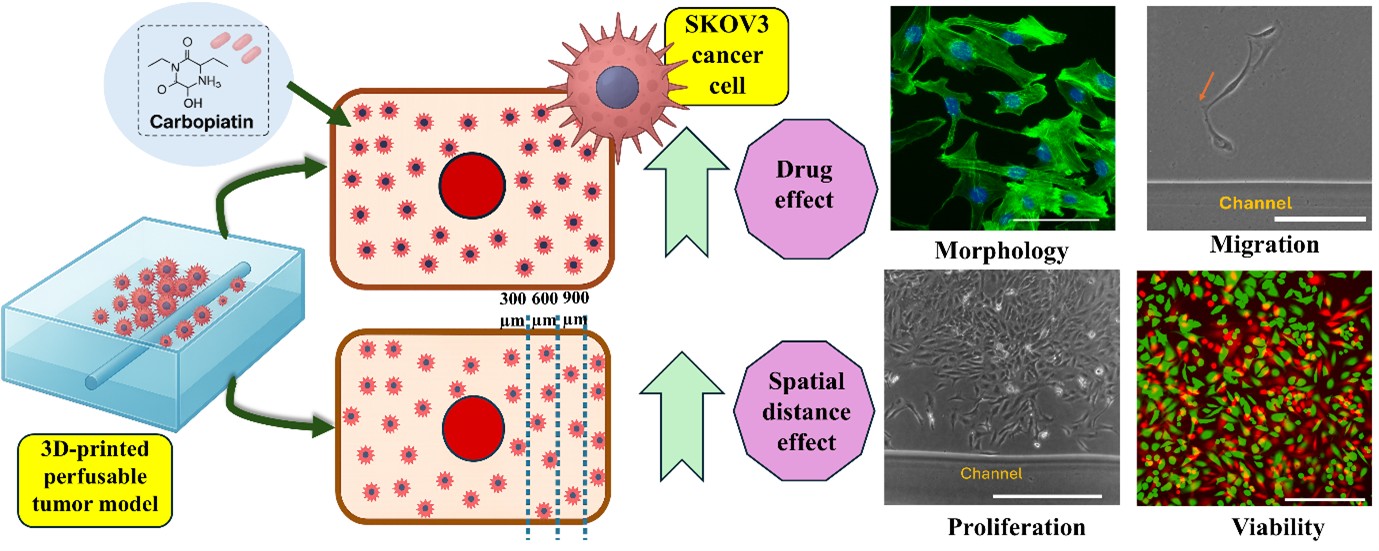
Understanding the role of perfusion and chemotherapeutic response in solid tumors requires advanced in vitro models that closely recapitulate the tumor microenvironment. Addressing this need, we developed a perfusable three-dimensional (3D) gelatin methacrylate (GelMA)-based tumor model embedded with a hollow microchannel to investigate spatial variations in SKOV3 ovarian cancer cell behavior and their response to carboplatin. This study aims to overcome the limitations of conventional two-dimensional and non-perfused 3D cultures by introducing controlled perfusion and directional drug delivery, thereby providing a more physiologically relevant platform for cancer research and drug testing. Using extrusion- and inkjet-based bioprinting, SKOV3 cells were cultured within the GelMA matrix and exposed to continuous medium flow. We observed that cell behavior varied significantly with distance from the perfusion channel. Cells closer to the channel (0–300 μm) showed increased elongation (aspect ratio: 3.5), faster migration (28.98 μm/day), higher viability (96%), and elevated proliferation (index: 3.8), which progressively declined with increasing distance. Upon carboplatin exposure (0–50 μM), SKOV3 cells exhibited dose-dependent reductions in viability, proliferation, migration, and elongation, with the aspect ratio dropping to 1.17 and the viability to 5% at 50 μM. Matrix degradation analysis revealed increased pore enlargement under perfusion (87–190 μm), suggesting higher matrix metalloproteinase activity. This perfused 3D model enables precise evaluation of chemotherapeutic efficacy and tumor cell heterogeneity, offering a powerful tool for preclinical drug screening, tumor biology research, and future integration of vascular and immune components.
- Shi X, Wang X, Yao W, et al. Mechanism insights and therapeutic intervention of tumor metastasis: latest developments and perspectives. Signal Transduct Target Ther. 2024;9(1):192. doi: 10.1038/s41392-024-01885-2
- Molinares M, Wolpert N, Gollahon L, Xu C. Effect of micropillar density on morphology and migration of low and high metastatic potential breast cancer cells. Colloids Surf B Biointerfaces. 2025;245:114214. doi: 10.1016/j.colsurfb.2024.114214
- Ge R, Wang Z, Cheng L. Tumor microenvironment heterogeneity an important mediator of prostate cancer progression and therapeutic resistance. NPJ Precis Oncol. 2022;6(1):31. doi: 10.1038/s41698-022-00272-w
- Herrmann D, Conway JR, Vennin C, et al. Three-dimensional cancer models mimic cell–matrix interactions in the tumour microenvironment. Carcinogenesis. 2014;35(8):1671-1679. doi: 10.1093/carcin/bgu108
- Cordeiro S, Oliveira BB, Valente R, et al. Breaking the mold: 3D cell cultures reshaping the future of cancer research. Front Cell Dev Biol. 2024;12:1507388. doi: 10.3389/fcell.2024.1507388
- Shukla P, Yeleswarapu S, Heinrich MA, Prakash J, Pati F. Mimicking tumor microenvironment by 3D bioprinting: 3D cancer modeling. Biofabrication. 2022;14(3):032002. doi: 10.1088/1758-5090/ac6d11
- Datta P, Dey M, Ataie Z, Unutmaz D, Ozbolat IT. 3D bioprinting for reconstituting the cancer microenvironment. NPJ Precis Oncol. 2020;4(1):18. doi: 10.1038/s41698-020-0121-2
- Gupta D, Derman ID, Xu C, Huang Y, Ozbolat IT. Droplet-based bioprinting. Nat Rev Methods Primers. 2025;5(1):25. doi: 10.1038/s43586-025-00394-y
- Sharifi S, Sharifi H, Akbari A, Chodosh J. Systematic optimization of visible light-induced crosslinking conditions of gelatin methacryloyl (GelMA). Sci Rep. 2021; 11(1):23276. doi: 10.1038/s41598-021-02830-x
- Zhou M, Lee BH, Tan LP. A dual crosslinking strategy to tailor rheological properties of gelatin methacryloyl. Int J Bioprint. 2017;3(2):003. doi: 10.18063/IJB.2017.02.003
- Wang C, Li J, Sinha S, Peterson A, Grant GA, Yang F. Mimicking brain tumor-vasculature microanatomical architecture via co-culture of brain tumor and endothelial cells in 3D hydrogels. Biomaterials. 2019;202:35-44. doi: 10.1016/j.biomaterials.2019.02.024
- Ertekin Ö, Monavari M, Krueger R, et al. 3D hydrogel-based microcapsules as an in vitro model to study tumorigenicity, cell migration and drug resistance. Acta Biomater. 2022;142:208-220. doi: 10.1016/j.actbio.2022.02.010
- Maggiotto F, Bova L, Micheli S, et al. 3D bioprinting for the production of a perfusable vascularized model of a cancer niche. Front Bioeng Biotechnol. 2025;13:1484738. doi: 10.3389/fbioe.2025.1484738
- Hong M, Hong S, Song JM. 3D bioprinted multidrug resistance (MDR)-dependent tumor spheroids. ACS Appl Mater Interfaces. 2025;17(5):7377-7394. doi: 10.1021/acsami.4c19291
- Fang T, Xie X, Lu W, et al. Patient-derived organoids on a microarray for drug resistance study in breast cancer. Anal Chem. 2024;96(46):18384-18391. doi: 10.1021/acs.analchem.4c02691
- Krishnamoorthy S, Wadnap S, Noorani B, Xu H, Xu C. Investigation of gelatin methacrylate working curves in dynamic optical projection stereolithography of vascular-like constructs. Eur Polym J. 2020;124:109487. doi: 10.1016/j.eurpolymj.2020.109487
- Ren B, Song K, Sanikommu AR, et al. Study of sacrificial ink-assisted embedded printing for 3D perfusable channel creation for biomedical applications. Appl Phys Rev. 2022;9(1):011408. doi: 10.1063/5.0068329
- Schmolka IR. Artificial skin I. Preparation and properties of pluronic F-127 gels for treatment of burns. J Biomed Mater Res. 1972;6(6):571-582. doi: 10.1002/jbm.820060609
- Liu J, Xu C. Improving uniformity of cell distribution in post-inkjet-based bioprinting. J Manuf Sci Eng. 2024;146(1):014501. doi: 10.1115/1.4063134
- Liu J, Xu H, Shahriar M, Xu C. Modeling of cell distribution dynamics in cell-laden bioink with active circulation. Addit Manuf. 2023;73:103669. doi: 10.1016/j.addma.2023.103669
- Xu H, Liu J, Shahriar M, and Xu C. Investigation of cell aggregation on the printing performance in inkjet-based bioprinting of cell-laden bioink. Langmuir. 2022;39(1):545-555. doi: 10.1021/acs.langmuir.2c02817
- Hyler AR, Baudoin NC, Brown MS, et al. Fluid shear stress impacts ovarian cancer cell viability, subcellular organization, and promotes genomic instability. PLoS One. 2018;13(3):e0194170. doi: 10.1371/journal.pone.0194170.
- Galpayage Dona KNU, Hale JF, Salako T, et al. The use of tissue engineering to fabricate perfusable 3D brain microvessels in vitro. Front Physiol. 2021;12:715431. doi: 10.3389/fphys.2021.715431.
- Nichol JW, Koshy ST, Bae H, Hwang CM, Yamanlar S, Khademhosseini A. Cell-laden microengineered gelatin methacrylate hydrogels. Biomaterials. 2010;31(21):5536-5544. doi: 10.1016/j.biomaterials.2010.03.064.
- Zhao X, Lang Q, Yildirimer L, et al. Photocrosslinkable gelatin hydrogel for epidermal tissue engineering. Adv Healthc Mater. 2016;5(1):108-118. doi: 10.1002/adhm.201500005.
- Krishnamoorthy S, Noorani B, Xu C. Effects of encapsulated cells on the physical–mechanical properties and microstructure of gelatin methacrylate hydrogels. Int J Mol Sci. 2019;20(20):5061. doi: 10.3390/ijms20205061.
- Friedl P, Wolf K. Tumour-cell invasion and migration: diversity and escape mechanisms. Nat Rev Cancer. 2003;3(5):362-374. doi: 10.1038/nrc1075.
- Helmy IM, AbdelAzim AM. Efficacy of ImageJ in the assessment of apoptosis. Diagn Pathol. 2012;7(1):15. doi: 10.1186/1746-1596-7-15.
- Liu J, Xu C. Spatial distribution of encapsulated cells dramatically alters biomechanical properties and microstructure of 3D printed cellular structures. J Manuf Processes. 2024;120:1203-1212. doi: 10.1016/j.jmapro.2024.05.029.
- Figueiredo L, Leisage C, Weiss P, Yang J. Quantifying oxygen levels in 3D bioprinted cell-laden thick constructs with perfusable microchannel networks. Polymers. 2020;12(6):1260. doi: 10.3390/polym12061260.
- Radisic M, Malda J, Epping E, Geng W, Langer R, Vunjak‐ Novakovic G. Oxygen gradients correlate with cell density and cell viability in engineered cardiac tissue. Biotechnol Bioeng. 2006;93(2):332-343. doi: 10.1002/bit.20722.
- Novak CM, Horst EN, Taylor CC, Liu CZ, Mehta G. Fluid shear stress stimulates breast cancer cells to display invasive and chemoresistant phenotypes while upregulating PLAU in a 3D bioreactor. Biotechnol Bioeng. 2019; 116(11):3084-3097. doi: 10.1002/bit.27119.
- Helmlinger G, Yuan F, Dellian M, Jain RK. Interstitial pH and pO2 gradients in solid tumors in vivo: high-resolution measurements reveal a lack of correlation. Nat Med. 1997;3(2):177-182. doi: 10.1038/nm0297-177.
- Liverani C, De Vita A, Minardi S, et al. A biomimetic 3D model of hypoxia-driven cancer progression. Sci Rep. 2019;9(1):12263. doi: 10.1038/s41598-019-48701-4.
- Lewis DM, Park KM, Tang V, et al. Intratumoral oxygen gradients mediate sarcoma cell invasion. Proc Natl Acad Sci USA. 2016;113(33):9292-9297. doi: 10.1073/pnas.1605317113.
- Polacheck WJ, German AE, Mammoto A, Ingber DE, Kamm RD. Mechanotransduction of fluid stresses governs 3D cell migration. Proc Natl Acad Sci USA. 2014;111(7):2447-2452. doi: 10.1073/pnas.1316848111.
- Avraham-Chakim L, Elad D, Zaretsky U, Kloog Y, Jaffa A, Grisaru D. Fluid-flow induced wall shear stress and epithelial ovarian cancer peritoneal spreading. PLoS One. 2013;8(4):e60965. doi: 10.1371/journal.pone.0060965.
- Gilmore AC, Flaherty SJ, Somasundaram V, et al. An in vitro tumorigenesis model based on live-cell-generated oxygen and nutrient gradients. Commun Biol. 2021;4(1):477. doi: 10.1038/s42003-021-01954-0.
- Chen Z, Han F, Du Y, Shi H, Zhou W. Hypoxic microenvironment in cancer: molecular mechanisms and therapeutic interventions. Signal Trans Targeted Ther. 2023;8(1):70. doi: 10.1038/s41392-023-01332-8.
- Pasini A, Lovecchio J, Cortesi M, et al. Perfusion flow enhances viability and migratory phenotype in 3D-cultured breast cancer cells. Ann Biomed Eng. 2021;49(9):2103-2113. doi: 10.1007/s10439-021-02727-w.
- Worsley CM, Veale RB, Mayne ES. The acidic tumour microenvironment: manipulating the immune response to elicit escape. Hum Immunol. 2022;83(5):399-408. doi: 10.1016/j.humimm.2022.01.014.
- Tannock IF. Oxygen diffusion and the distribution of cellular radiosensitivity in tumours. Br J Radiol. 1972;45(535):515-524. doi: 10.1259/0007-1285-45-535-515.
- Ayuso JM, Virumbrales-Munoz M, McMinn PH, et al. Tumor-on-a-chip: a microfluidic model to study cell response to environmental gradients. Lab Chip. 2019;19(20):3461-3471. doi: 10.1039/c9lc00270g.
- Lee D, Jeong HS, Hwang SY, Lee YG, Kang YJ. ABCB1 confers resistance to carboplatin by accumulating stem-like cells in the G2/M phase of the cell cycle in p53null ovarian cancer. Cell Death Discov. 2025;11(1):132. doi: 10.1038/s41420-025-02435-7.
- Alhalhooly L, Mamnoon B, Kim J, Mallik S, Choi Y. Dynamic cellular biomechanics in responses to chemotherapeutic drug in hypoxia probed by atomic force spectroscopy. Oncotarget. 2021;12(12):1165. doi: 10.18632/oncotarget.27974.
- Povea-Cabello S, Oropesa-Ávila M, De la Cruz-Ojeda P, et al. Dynamic reorganization of the cytoskeleton during apoptosis: the two coffins hypothesis. Int J Mol Sci. 2017;18(11):2393. doi: 10.3390/ijms18112393.
- He PJ, Ge RF, Mao WJ, et al. Oxidative stress induced by carboplatin promotes apoptosis and inhibits migration of HN-3 cells. Oncol Lett. 2018;16(6):7131-7138. doi: 10.3892/ol.2018.9563.
- Alamdari SG, Mohammadzadeh R, Amini M, et al. Improvement of carboplatin chemosensitivity in lung cancer cells by siRNA-mediated downregulation of DLGAP1-AS2 expression. Sci Rep. 2025; 15(1):7971. doi: 10.1038/s41598-025-87649-6.
- Vaidžiulytė K, Macé AS, Battistella A, Beng W, Schauer K, Coppey M. Persistent cell migration emerges from a coupling between protrusion dynamics and polarized trafficking. Elife. 2022;11:e69229. doi: 10.7554/eLife.69229.
- Shields JD, Fleury ME, Yong C, et al. Autologous chemotaxis as a mechanism of tumor cell homing to lymphatics via interstitial flow and autocrine CCR7 signaling. Cancer Cell. 2007;11(6):526-538. doi: 10.1016/j.ccr.2007.04.020.
- Waldeland JO, Evje S. Competing tumor cell migration mechanisms caused by interstitial fluid flow. J Biomech. 2018;81:22-35. doi: 10.1016/j.jbiomech.2018.09.011.
- Nath S, Pigula M, Khan AP, et al. Flow-induced shear stress confers resistance to carboplatin in an adherent three-dimensional model for ovarian cancer: a role for EGFR-targeted photoimmunotherapy informed by physical stress. J Clin Med. 2020;9(4):924. doi: 10.3390/jcm9040924.
- Banerji U, Sain N, Sharp SY, et al. An in vitro and in vivo study of the combination of the heat shock protein inhibitor 17-allylamino-17-demethoxygeldanamycin and carboplatin in human ovarian cancer models. Cancer Chemother Pharmacol. 2008;62(5):769-778. doi: 10.1007/s00280-007-0662-x.
- Sousa GFD, Wlodarczyk SR, Monteiro G. Carboplatin: molecular mechanisms of action associated with chemoresistance. Braz J Pharm Sci. 2014;50(4):693-701. doi: 10.1590/S1984-82502014000400004.
- Patra B, Lateef MA, Brodeur MN, et al. Carboplatin sensitivity in epithelial ovarian cancer cell lines: the impact of model systems. PLoS One. 2020;15(12):e0244549. doi: 10.1371/journal.pone.0244549.
- Lieberthal TJ, Sahakyants T, Szabo-Wexler NR, et al. Implantable 3D printed hydrogels with intrinsic channels for liver tissue engineering. Proc Natl Acad Sci U S A. 2024;121(47):e2403322121. doi: 10.1073/pnas.2403322121.
- Zhang N, Qavi I, Halder S, Tan G. Biomimetic hydrogel scaffolds embedded with porous microtubes as perfusion channels. Manuf Lett. 2023;35:184-193. doi: 10.1016/j.mfglet.2023.08.009.
- Grebenyuk S, Abdel Fattah AR, Kumar M, et al. Large-scale perfused tissues via synthetic 3D soft microfluidics. Nat Commun. 2023;14(1):193. doi: 10.1038/s41467-022-35619-1.
- Blanco-Fernandez B, Gaspar VM, Engel E, Mano JF. Proteinaceous hydrogels for bioengineering advanced 3D tumor models. Adv Sci (Weinh). 2021;8(4):2003129. doi: 10.1002/advs.202003129
- Zhu M, Wang Y, Ferracci G, Zheng J, Cho NJ, Lee BH. Gelatin methacryloyl and its hydrogels with an exceptional degree of controllability and batch-to-batch consistency. Sci Rep. 2019;9(1):6863. doi: 10.1038/s41598-019-42186-x
- Didwischus N, Guduru A, Badylak SF, Modo M. In vitro dose-dependent effects of matrix metalloproteinases on ECM hydrogel biodegradation. Acta Biomater. 2024;174:104-115. doi: 10.1016/j.actbio.2023₹.12.003
- Saggioro M, D’Agostino S, Veltri G, et al. A perfusion-based three-dimensional cell culture system to model alveolar rhabdomyosarcoma pathological features. Sci Rep. 2023;13(1):9444. doi: 10.1038/s41598-023-36210-4
- Huang Q, Hu X, He W, et al. Fluid shear stress and tumor metastasis. Am J Cancer Res. 2018;8(5):763-775.
- Marrella A, Varani G, Aiello M, et al. 3D fluid-dynamic ovarian cancer model resembling systemic drug administration for efficacy assay. Altex. 2021;38(1):82-94. doi: 10.14573/altex.2003131