

Multiscale vascularized tumor-on-a-chip via bioprinting for drug research

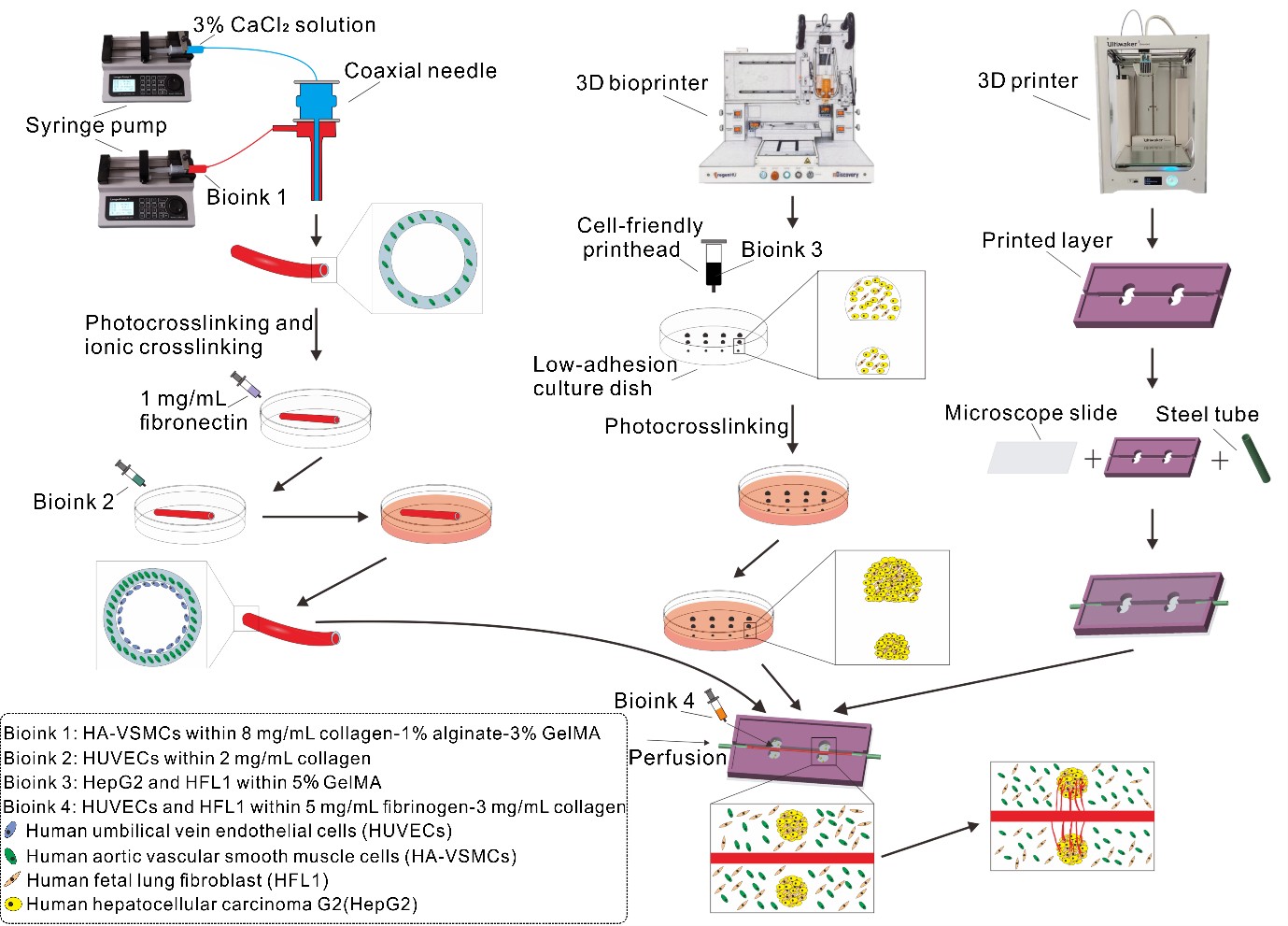
Current in vitro tumor models often fail to recapitulate the hierarchical vascular architecture and dynamic interactions of the tumor microenvironment (TME), limiting their utility in cancer research. In this study, we present a multiscale vascularized tumor model integrating coaxial bioprinting, inkjet printing, and fused deposition modeling (FDM) to address this challenge. Firstly, coaxial bioprinting enabled the fabrication of dual-layered vasculature with an endothelium layer and a smooth muscle layer. Secondly, tumor spheroids with precise size control (±10 μm) were generated via inkjet printing by modulating Methacrylate Gelatin (GelMA) concentration and valve actuation time. An FDM-printed chip was designed to co-culture these components under perfusion, facilitating the self-organization of a microvascular network around tumor spheroids. After 11 days of dynamic culture, the model demonstrated tumor-driven angiogenic sprouting and early metastatic behavior, validated by the upregulation of metastasis-related genes (CD44, MMP2, N-cadherin) in vascularized cohorts. Drug testing with paclitaxel revealed dose-dependent suppression of tumor proliferation and invasion. This platform not only mimics the structural and functional complexity of the TME but also provides a scalable, physiologically relevant tool for investigating tumor–vascular crosstalk and evaluating anti-cancer therapeutics.
- Lunt N. The global challenge of cancer governance. World Med Health Pol. 2023;15(4):672-681. doi: 10.1002/wmh3.577
- Sung H, Ferlay J, Siegel RL, et al. Global cancer statistics 2020: GLOBOCAN estimates of incidence and mortality worldwide for 36 cancers in 185 countries. CA Cancer J Clin. 2021;71(3):209-249. doi: 10.3322/caac.21660
- Sontheimer-Phelps A, Hassell BA, Ingber DE. Modelling cancer in microfluidic human organs-on-chips. Nat Rev Cancer. 2019;19(2):65-81. doi: 10.1038/s41568-018-0104-6
- Li CP, Li SB, Du K, Li P, Qiu BS, Ding WP. On-chip replication of extremely early-stage tumor behavior. Acs Appl Mater Inter. 2021;13(17):19768-19777. doi: 10.1021/acsami.1c03740
- Stock K, Estrada MF, Vidic S, et al. Capturing tumor complexity: comparative analysis of 2D and 3D tumor models for drug discovery. Sci Rep-Uk. 2016;6:28951. doi: 10.1038/srep28951
- Nie J, Gao Q, Fu JZ, He Y. Grafting of 3D bioprinting to in vitro drug screening: a review. Adv Healthc Mater. 2020;9(7):e190177310. doi: 10.1002/adhm.201901773
- Li JZ, Zhou YJ, Chen WL, et al. A novel 3D in vitro tumor model based on silk fibroin/chitosan scaffolds to mimic the tumor microenvironment. Acs Appl Mater Inter. 2018;10(43):36641-36651. doi: 10.1021/acsami.8b10679
- Lee G, Kim SJ, Park JK. Fabrication of a self-assembled and vascularized tumor array bioprinting on a microfluidic chip. Lab Chip. 2023;23(18):4079-4091. doi: 10.1039/d3lc00275f
- Nashimoto Y, Okada R, Hanada S, et al. Vascularized cancer on a chip: the effect of perfusion on growth and drug delivery of tumor spheroid. Biomaterials. 2020;229:119547. doi: 10.1016/j.biomaterials.2019.119547
- Lee VK, Lanzi AM, Ngo H, Yoo SS, Vincent PA, Dai GH. Generation of multi-scale vascular network system within 3D hydrogel using 3D bio-printing technology. Cell Mol Bioeng. 2014;7(3):460-472. doi: 10.1007/s12195-014-0340-0
- Eckermann CW, Lehle K, Schmid SA, Wheatley DN, Kunz-Schughart LA. Characterization and modulation of fibroblast/endothelial cell co-cultures for the preformation of three-dimensional tubular networks. Cell Biol Int. 2011;35(11):1097-1110. doi: 10.1042/Cbi20100718
- Enzerink A, Rantanen V, Vaheri A. Fibroblast nemosis induces angiogenic responses of endothelial cells. Exp Cell Res. 2010;316(5):826-835. doi: 10.1016/j.yexcr.2009.11.012
- Friedrich J, Ebner R, Kunz-Schughart LA. Experimental anti-tumor therapy in 3-D: spheroids - old hat or new challenge? Int J Radiat Biol. 2007;83(11–12):849-871. doi: 10.1080/09553000701727531
- Amann A, Zwierzina M, Gamerith G, et al. Development of an Innovative 3D cell culture system to study tumour - stroma interactions in non-small cell lung cancer cells. Plos One. 2014;9(3):e92511. doi: 10.1371/journal.pone.0092511
- Jing JF, Xiao H, Lin XW, et al. Cutting and Bonding Parafilm® to fast prototyping flexible hanging drop chips for 3d spheroid cultures. Cell Mol Bioeng. 2021;14(2):187-199. doi: 10.1007/s12195-020-00660-x
- Hurrell T, Ellero AA, Masso ZF, Cromarty AD. Characterization and reproducibility of HepG2 hanging drop spheroids toxicology. Toxicol in Vitro. 2018;50:86-94. doi: 10.1016/j.tiv.2018.02.013
- Meenach SA, Tsoras AN, McGarry RC, Mansour HM, Hilt JZ, Anderson KW. Development of three-dimensional lung multicellular spheroids in air- and liquid-interface culture for the evaluation of anticancer therapeutics. Int J Oncol. 2016;48(4):1701-1709. doi: 10.3892/ijo.2016.3376
- Costa EC, Gaspar VM, Coutinho P, Correia IJ. Optimization of liquid overlay technique to formulate heterogenic 3D co-cultures models. Biotechnol Bioeng. 2014;111(8):1672-1685. doi: 10.1002/bit.25210
- Fennema E, Rivron N, Rouwkema J, van Blitterswijk C, de Boer J. Spheroid culture as a tool for creating 3D complex tissues. Trends Biotechnol. 2013;31(2):108-115. doi: 10.1016/j.tibtech.2012.12.003
- Habanjar O, Diab-Assaf M, Caldefie-Chezet F, Delort L. 3D cell culture systems: tumor application, advantages, and disadvantages. Int J Mol Sci. 2021;22(22):12200. doi: 10.3390/ijms222212200
- Gao W, Wu D, Wang Y, et al. Development of a novel and economical agar-based non-adherent three-dimensional culture method for enrichment of cancer stem-like cells. Stem Cell Res Ther. 2018;9(1):243. doi: 10.1186/s13287-018-0987-x
- Jeong Y, Tin A, Irudayaraj J. Flipped well-plate hanging-drop technique for growing three-dimensional tumors. Front Bioeng Biotechnol. 2022;10:898699. doi: 10.3389/fbioe.2022.898699
- de Barros NR, Gomez A, Ermis M, et al. Gelatin methacryloyl and Laponite bioink for 3D bioprinted organotypic tumor modeling. Biofabrication. 2023;15(4):045005. doi: 10.1088/1758-5090/ace0db
- Wu L, Li H, Liu Y, et al. Research progress of 3D-bioprinted functional pancreas and in vitro tumor models. Int J Bioprint. 2024;10(1):1256. doi: 10.36922/ijb.1256
- Singh S, Ray LA, Shahi Thakuri P, et al. Organotypic breast tumor model elucidates dynamic remodeling of tumor microenvironment. Biomaterials. 2020;238:119853. doi: 10.1016/j.biomaterials.2020.119853
- Zhou X, Nowicki M, Sun H, et al. 3D bioprinting-tunable small-diameter blood vessels with biomimetic biphasic cell layers. ACS Appl Mater Inter. 2020;12(41):45904-45915. doi: 10.1021/acsami.0c14871
- Meng FB, Meyer CM, Joung D, Vallera DA, McAlpine MC, Panoskaltsis-Mortari A. 3D bioprinted in vitro metastatic models via reconstruction of tumor microenvironments. Adv Mater. 2019;31(10):1806899. doi: 10.1002/adma.201806899
- Nie J, Gao Q, Xie CQ, et al. Construction of multi-scale vascular chips and modelling of the interaction between tumours and blood vessels. Mater Horizons. 2020;7(1):82-92. doi: 10.1039/c9mh01283d
- Gao G, Kim H, Kim BS, et al. Tissue-engineering of vascular grafts containing endothelium and smooth-muscle using triple-coaxial cell printing. Appl Phys Rev. 2019;6(4):041402. doi: 10.1063/1.5099306
- Duong V, Dang TT, Hwang CH, Back SH, Koo KI. Coaxial printing of double-layered and free-standing blood vessel analogues without ultraviolet illumination for high-volume vascularised tissue. Biofabrication. 2020;12(4): 045033. doi: 10.1088/1758-5090/abafc6
- Kwak TJ, Lee E. In vitro modeling of solid tumor interactions with perfused blood vessels. Sci Rep-Uk. 2020;10(1): 20142. doi: 10.1038/s41598-020-77180-1
- Ahn J, Kim D, Koo DJ, et al. 3D microengineered vascularized tumor spheroids for drug delivery and efficacy testing. Acta Biomater. 2023;165:153-167. doi: 10.1016/j.actbio.2022.10.009
- Dey M, Kim MH, Dogan M, et al. Chemotherapeutics and CAR-T cell-based immunotherapeutics screening on a 3D bioprinted vascularized breast tumor model. Adv Funct Mater. 2022;32(52):2203966. doi: 10.1002/adfm.202203966
- Ozturk MS, Lee VK, Zou HY, Friedel RH, Intes X, Dai GH. High-resolution tomographic analysis of in vitro 3D glioblastoma tumor model under long-term drug treatment. Sci Adv. 2020;6(10):eaay7513. doi: 10.1126/sciadv.aay7513
- Hwang DG, Choi YM, Jang J. 3D bioprinting-based vascularized tissue models mimicking tissue-specific architecture and pathophysiology for studies. Front Bioeng Biotech. 2021;9:685507. doi: 10.3389/fbioe.2021.685507
- Nashimoto Y, Hayashi T, Kunita I, et al. Integrating perfusable vascular networks with a three-dimensional tissue in a microfluidic device. Integr Biol-Uk. 2017;9(6):506-518. doi: 10.1039/c7ib00024c
- Velez C, Cheng K, Crosby C. Synthesis and Characterization of Gelatin Methacryloyl: Introducing Chemistry Students to the Applications of Hydrogels in Medicine. J Chem Educ. 2024;101(3):1171-1179. doi: 10.26434/chemrxiv-2023-qbrh6
- Millik SC, Dostie AM, Karis DG, et al. 3D printed coaxial nozzles for the extrusion of hydrogel tubes toward modeling vascular endothelium. Biofabrication. 2019;11(4):045009. doi: 10.1088/1758-5090/ab2b4d
- Siminska-Stanny J, Nicolas L, Chafai A, et al. Advanced PEG-tyramine biomaterial ink for precision engineering of perfusable and flexible small-diameter vascular constructs via coaxial printing. Bioact Mater. 2024;36:168-184. doi: 10.1016/j.bioactmat.2024.02.019
- Elliott MB, Gerecht S. Three-dimensional culture of small-diameter vascular grafts. J Mater Chem B. 2016;4(20):3443-3453. doi: 10.1039/c6tb00024j
- Xia Y, Zhou HY, Ou JS, Liu YQ. The potential of a new natural vessel source: decellularized intercostal arteries as sufficiently long small-diameter vascular grafts. Bioengineering (Basel). 2024;11(7): 700. doi: 10.3390/bioengineering11070700
- Carrabba M, Fagnano M, Ghorbel MT, et al. Development of a novel hierarchically biofabricated blood vessel mimic decorated with three vascular cell populations for the reconstruction of small-diameter arteries. Adv Funct Mater. 2024;34(7):adfm.202300621. doi: 10.1002/adfm.202300621
- Haase K, Offeddu GS, Gillrie MR, Kamm RD. Endothelial regulation of drug transport in a 3D vascularized tumor model. Adv Funct Mater. 2020;30(48):2002444. doi: 10.1002/adfm.202002444
- Marei I, Abu Samaan T, Al-Quradaghi MA, et al. 3D tissue-engineered vascular drug screening platforms: promise and considerations. Front Cardiovasc Med. 2022;9:847554. doi: 10.3389/fcvm.2022.847554
- Ma DF, Liang D, Zhu CY, et al. The breakup dynamics and mechanism of viscous droplets in Y-shaped microchannels. Chem Eng Sci. 2021;231:116300. doi: 10.1016/j.ces.2020.116300
- Sur S, Rothstein J. Drop breakup dynamics of dilute polymer solutions: effect of molecular weight, concentration, and viscosity. J Rheol. 2018;62(5):1245-1259. doi: 10.1122/1.5038000
- Cho WJ, Elbasiony E, Singh A, Mittal SK, Chauhan SK. IL-36 gamma augments ocular angiogenesis by promoting the vascular endothelial growth factor-vascular endothelial growth factor receptor axis. Am J Pathol. 2023;193(11):1740-1749. doi: 10.1016/j.ajpath.2023.01.003
- Sitohy B, Chang S, Sciuto TE, et al. Early actions of anti-vascular endothelial growth factor/vascular endothelial growth factor receptor drugs on angiogenic blood vessels. Am J Pathol. 2017;187(10):2337-2347. doi: 10.1016/j.ajpath.2017.06.010
- Truelsen SLB, Mousavi N, Wei HC, et al. The cancer angiogenesis co-culture assay: quantification of the angiogenic potential of tumoroids. Plos One. 2021;16(7):e0253258. doi: 10.1371/journal.pone.0253258
- Rodoplu D, Matahum JS, Hsu CH. A microfluidic hanging drop-based spheroid co-culture platform for probing tumor angiogenesis. Lab Chip. 2022;22(7):1275-1285. doi: 10.1039/d1lc01177d
- Chang CW, Seibel AJ, Avendano A, Cortes-Medina MG, Song JW. Distinguishing specific CXCL12 isoforms on their angiogenesis and vascular permeability promoting properties. Adv Healthc Mater. 2020;9(4):e1901399. doi: 10.1002/adhm.201901399
- Zhou L, Wang L, Song X, Zhang X, Xiao Y. A tetrazine-based ratiometric sensor quantifying ph gradient in tumorspheres through bio-orthogonal labeling. Anal Chem. 2025;97(22):11486-11495. doi: 10.1021/acs.analchem.5c00291
- Lee S, Park J, Cho S, et al. Hyaluronan network remodeling by ZEB1 and ITIH2 enhances the motility and invasiveness of cancer cells. J Clin Invest. 2025;135(11):e180570. doi: 10.1172/JCI18057