

3D-bioprinted asymmetric bilayer scaffolds with anti-infection and pro-regeneration characteristics for chronic diabetic wound healing

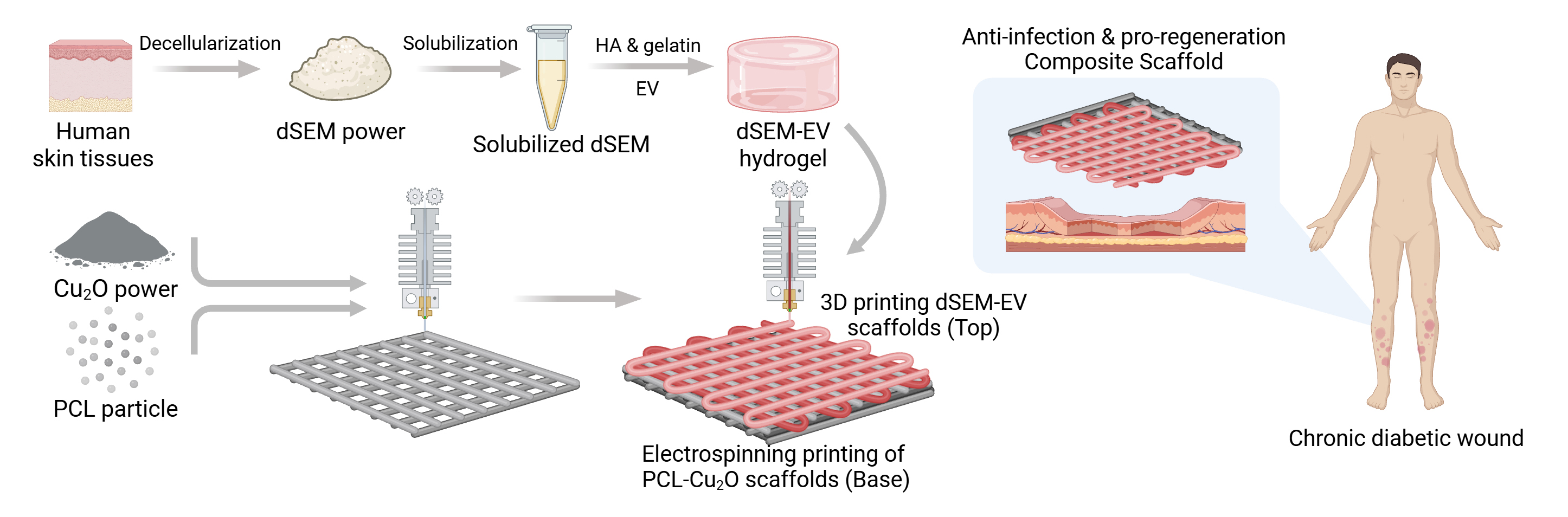
Chronic diabetic wounds, characterized by persistent infection and impaired tissue regeneration, remain a formidable clinical challenge. In the present study, a 3D-bioprinted asymmetric bilayer scaffold was developed by integrating electrospinning and 3D bioprinting technologies to achieve anti-infective and pro-regenerative functions. The scaffold features a distinctive asymmetric architecture comprising a superficial layer (Layer S) and a basal layer (Layer B). Layer B, consisting of electrospun copper(I) oxide (Cu2O)–poly-ε-caprolactone nanofibers, serves as an effective antibacterial barrier specifically targeting methicillin-resistant Staphylococcus aureus (MRSA), while Layer S employs a 3D-bioprinted decellularized extracellular matrix hydrogel loaded with human bone marrow mesenchymal stem cell-derived extracellular vesicles (hBMSC-EVs) to facilitate tissue repair. Experimental results demonstrated that hBMSC-EVs significantly augmented fibroblast proliferation and migration, and the Cu2O-doped layer exhibited potent bactericidal activity against MRSA. In db/db diabetic mice, this asymmetric composite scaffold significantly accelerated wound closure compared to standalone treatments. Histological analysis further confirmed enhanced neovascularization and accelerated extracellular matrix reconstruction. Overall, this synergistic 3D-bioprinted bilayer system provides a high-performance strategy for the targeted management of MRSA-infected chronic diabetic wounds.
- Slominski A, Wortsman J. Neuroendocrinology of the skin. Endocr Rev. 2000;21(5):457-487. doi: 10.1210/edrv.21.5.0410
- Proksch E, Brandner JM, Jensen JM. The skin: an indispensable barrier. Exp Dermatol. 2008;17(12):1063- 1072. doi: 10.1111/j.1600-0625.2008.00786.x
- Sen CK. Human Wound and Its Burden: Updated 2020 Compendium of Estimates. Adv Wound Care (New Rochelle). 2021;10(5):281-292. doi: 10.1089/wound.2021.0026
- Armstrong DG, Boulton AJM, Bus SA. Diabetic Foot Ulcers and Their Recurrence. N Engl J Med. 2017;376(24):2367- 2375. doi: 10.1056/NEJMra1615439
- Jones RE, Foster DS, Longaker MT. Management of Chronic Wounds-2018. JAMA. 2018;320(14):1481-1482. doi: 10.1001/jama.2018.12426
- Morton LM, Phillips TJ. Wound healing and treating wounds: Differential diagnosis and evaluation of chronic wounds. J Am Acad Dermatol. 2016;74(4):589-605. doi: 10.1016/j.jaad.2015.08.068
- Fu X. Wound healing center establishment and new technology application in improving the wound healing quality in China. Burns Trauma. 2020;8:tkaa038. doi: 10.1093/burnst/tkaa038
- Jiang Y, Huang S, Fu X, et al. Epidemiology of chronic cutaneous wounds in China. Wound Repair Regen. 2011;19(2):181-188. doi: 10.1111/j.1524-475X.2010.00666.x
- Wang Q, Luo Z, Li Z, et al. In-situ oxygen-supplying ROS nanopurifier for enhanced healing of MRSA-infected diabetic wounds via microenvironment modulation. Acta Biomater. 2025;193:334-347. doi: 10.1016/j.actbio.2024.12.044
- Lu Y, Xu J, Su Y, et al. A biocompatible double-crosslinked gelatin/sodium alginate/dopamine/quaternized chitosan hydrogel for wound dressings based on 3D bioprinting technology. Int J Bioprint. 2023;9(2):689. doi: 10.18063/ijb.689
- Fang H, Xu J, Ma H, et al. Functional materials of 3D bioprinting for wound dressings and skin tissue engineering applications: A review. Int J Bioprint. 2023;9(5):757. doi: 10.18063/ijb.757
- Mao S, Man J, Wang J, et al. Research progress and challenges of bioprinting in wound dressing and healing: Bibliometrics-based analysis and perspectives. Int J Bioprint. 2023;9(2):653. doi: 10.18063/ijb.v9i2.653
- Keirouz A, Chung M, Kwon J, Fortunato G, Radacsi N. 2D and 3D electrospinning technologies for the fabrication of nanofibrous scaffolds for skin tissue engineering: A review. Wiley Interdiscip Rev Nanomed Nanobiotechnol. 2020;12(4):e1626. doi: 10.1002/wnan.1626
- Wang X, Wang Y, Teng Y, et al. 3D bioprinting: opportunities for wound dressing development. Biomed Mater. 2023;18(5):052001. doi: 10.1088/1748-605X/ace228
- D A G, Adhikari J, Debnath P, Ghosh S, et al. 3D printing of bacterial cellulose for potential wound healing applications: Current trends and prospects. Int J Biol Macromol. 2024;279:135213. doi: 10.1016/j.ijbiomac.2024.135213
- Han Y, Lian M, Zhang C, et al. Study on bioactive PEGDA/ ECM hybrid bi-layered hydrogel scaffolds fabricated by electro-writing for cartilage regeneration. Appl Mater Today. 2022;28:101547. doi: 10.1016/j.apmt.2022.101547
- Xu J, Yang S, Su Y, et al. A 3D bioprinted tumor model fabricated with gelatin/sodium alginate/decellularized extracellular matrix bioink. Int J Bioprint. 2023;9(1):630. doi: 10.18063/ijb.v9i1.630
- Malta MD, Cerqueira MT, Marques AP. Extracellular matrix in skin diseases: The road to new therapies. J Adv Res. 2023;51:149-160. doi: 10.1016/j.jare.2022.11.008
- Durr HA, Abri S, Salinas SD, et al. Extracellular matrix repair and organization of chronic infected diabetic wounds treated with methacrylated chitosan-based hydrogels. Acta Biomater. 2025;199:166-177. doi: 10.1016/j.actbio.2025.04.062
- Song YT, Liu PC, Zhou XL, et al. Extracellular matrix-based biomaterials in burn wound repair: A promising therapeutic strategy. Int J Biol Macromol. 2024;283:137633. doi: 10.1016/j.ijbiomac.2024.137633
- Rueda-Gensini L, Serna JA, Cifuentes J, Cruz JC, Munoz- Camargo C. Graphene Oxide-Embedded Extracellular Matrix-Derived Hydrogel as a Multiresponsive Platform for 3D Bioprinting Applications. Int J Bioprint. 2021;7(3):353. doi: 10.18063/ijb.v7i3.353
- Lu Z, Miao X, Zhang C, et al. An osteosarcoma-on-a-chip model for studying osteosarcoma matrix-cell interactions and drug responses. Bioact Mater. 2024;34:1-16. doi: 10.1016/j.bioactmat.2023.12.005
- Lu Z, Jiang W, Zhao W, Zhao J, Dai K. Fabrication of 3D matrix microenvironment by two-photon lithography for mechanobiology study. Mechanobiol Med. 2023;1(1):100010. doi: 10.1016/j.mbm.2023.100010
- Carranza T, Uranga J, Irastorza A, et al. Combination of 3D printing and electrospinning to develop chitin/gelatin/PVA scaffolds. Int J Bioprint. 2023;9(3):701. doi: 10.18063/ijb.701
- Luo Y, Li D, Lin C, et al. 3D bioprinting of mechanically graded GelMA hydrogels with tri-layered vascularized architecture for full-thickness skin regeneration. Int J Bioprint. 2025;11(4):328-349. doi: 10.36922/ijb025090069
- Zhang C, Ma P, Qin A, et al. Current Immunotherapy Strategies for Rheumatoid Arthritis: The Immunoengineering and Delivery Systems. Research (Wash D C). 2023;6:0220. doi: 10.34133/research.0220
- Wang Y, Kong B, Chen X, et al. BMSC exosome-enriched acellular fish scale scaffolds promote bone regeneration. J Nanobiotechnology. 2022;20(1):444. doi: 10.1186/s12951-022-01646-9
- Su X, Yang J, Xu Z, et al. Fibrous scaffolds loaded with BMSC-derived apoptotic vesicles promote wound healing by inducing macrophage polarization. Genes Dis. 2025;12(2):101388. doi: 10.1016/j.gendis.2024.101388
- Wu J, Li S, Wang H, et al. High-yield BMSC-derived exosomes by the 3D culture system to enhance the skin wound repair. Regen Biomater. 2025;12:rbaf022. doi: 10.1093/rb/rbaf022
- Selvam S, Midhun BT, Bhowmick T, Chandru A. Bioprinting of exosomes: Prospects and challenges for clinical applications. Int J Bioprint. 2023;9(2):690. doi: 10.18063/ijb.690
- Han L, Liu Z, Li M, et al. 3D bioprinting of a dermal scaffold for full-thickness skin tissue regeneration. Bio-Des Manuf. 2025;8(1):68-84. doi: 10.1631/bdm.2400058
- Abaci A, Guvendiren M. Designing Decellularized Extracellular Matrix-Based Bioinks for 3D Bioprinting. Adv Healthc Mater. 2020;9(24):e2000734. doi: 10.1002/adhm.202000734
- Zhao H, Yang Y, An J, et al. Cu2O Nanocubes Embedded in Polycaprolactone Nanofibers for Photo–chemotherapeutic Wound Disinfection and Regeneration. ACS Appl Nano Mater. 2024;7(15):17707-17718. doi: 10.1021/acsanm.4c02928
- Falanga V. Wound healing and its impairment in the diabetic foot. Lancet. 2005;366(9498):1736-1743. doi: 10.1016/S0140-6736(05)67700-8
- Brem H, Tomic-Canic M. Cellular and molecular basis of wound healing in diabetes. J Clin Invest. 2007;117(5):1219- 1222. doi: 10.1172/JCI32169
- Liang Y, He J, Guo B. Functional Hydrogels as Wound Dressing to Enhance Wound Healing. ACS Nano. 2021;15(8):12687-12722. doi: 10.1021/acsnano.1c04206
- Zeng Q, Qi X, Shi G, Zhang M, Haick H. Wound Dressing: From Nanomaterials to Diagnostic Dressings and Healing Evaluations. ACS Nano. 2022;16(2):1708-1733. doi: 10.1021/acsnano.1c08411
- Liu C, Cheng C, Cheng K, et al. Precision exosome engineering for enhanced wound healing and scar revision. J Transl Med. 2025;23(1):578. doi: 10.1186/s12967-025-06578-0
- Rani S, Ryan AE, Griffin MD, Ritter T. Mesenchymal Stem Cell-derived Extracellular Vesicles: Toward Cell-free Therapeutic Applications. Mol Ther. 2015;23(5):812-823. doi: 10.1038/mt.2015.44
- Shabbir A, Cox A, Rodriguez-Menocal L, Salgado M, Van Badiavas E. Mesenchymal Stem Cell Exosomes Induce Proliferation and Migration of Normal and Chronic Wound Fibroblasts, and Enhance Angiogenesis In Vitro. Stem Cells Dev. 2015;24(14):1635-1647. doi: 10.1089/scd.2014.0316
- Zhang J, Guan J, Niu X, et al. Exosomes released from human induced pluripotent stem cells-derived MSCs facilitate cutaneous wound healing by promoting collagen synthesis and angiogenesis. J Transl Med. 2015;13:49. doi: 10.1186/s12967-015-0417-0
- Zhang S, Lu C, Zheng S, Hong G. Hydrogel loaded with bone marrow stromal cell-derived exosomes promotes bone regeneration by inhibiting inflammatory responses and angiogenesis. World J Stem Cells. 2024;16(5):499-511. doi: 10.4252/wjsc.v16.i5.499
- Bae M, Kim JJ, Kim J, Cho DW. Decellularized extracellular matrix for three-dimensional bioprinted in vitro disease modeling. Int J Bioprint. 2024;10(2):1970. doi: 10.36922/ijb.1970
- Liu W, Gao R, Yang C, et al. ECM-mimetic immunomodulatory hydrogel for methicillin-resistant Staphylococcus aureus-infected chronic skin wound healing. Sci Adv. 2022;8(27):eabn7006. doi: 10.1126/sciadv.abn7006
- Fu H, Zhang D, Zeng J, et al. Application of 3D-printed tissue-engineered skin substitute using innovative biomaterial loaded with human adipose-derived stem cells in wound healing. Int J Bioprint. 2023;9(2):674. doi: 10.18063/ijb.v9i2.674
- Dong R, Li Y, Chen M, et al. In Situ Electrospinning of Aggregation-Induced Emission Nanofibrous Dressing for Wound Healing. Small Methods. 2022;6(5):e2101247. doi: 10.1002/smtd.202101247
- Lipsky BA, Hoey C. Topical antimicrobial therapy for treating chronic wounds. Clin Infect Dis. 2009;49(10):1541- 1549. doi: 10.1086/644732
- Pino PAO, Bosco FAO, Mollea C, Onida BAO. Antimicrobial Nano-Zinc Oxide Biocomposites for Wound Healing Applications: A Review. Pharmaceutics. 2023;15(3):970. doi: 10.3390/pharmaceutics15030970
- Chatterjee AK, Chakraborty R, Basu T. Mechanism of antibacterial activity of copper nanoparticles. Nanotechnology. 2014;25(13):135101. doi: 10.1088/0957-4484/25/13/135101
- Zeng X, Gan J, Huang D, Zhao Y, Sun L. Recombinant human collagen hydrogels with different stem cell-derived exosomes encapsulation for wound treatment. J Nanobiotechnology. 2025;23(1):241. doi: 10.1186/s12951-025-03319-9
- Sheng Z, Fu X, Cai S, et al. Regeneration of functional sweat gland-like structures by transplanted differentiated bone marrow mesenchymal stem cells. Wound Repair Regen. 2009;17(3):427-435. doi: 10.1111/j.1524-475X.2009.00474.x
- Deng C, Dong K, Liu Y, et al. Hypoxic mesenchymal stem cell-derived exosomes promote the survival of skin flaps after ischaemia-reperfusion injury via mTOR/ULK1/ FUNDC1 pathways. J Nanobiotechnology. 2023;21(1):340. doi: 10.1186/s12951-023-02098-5
- Zhou S, Zhang X, Ni W, et al. An Immune-Regulating Polysaccharide Hybrid Hydrogel with Mild Photothermal Effect and Anti-Inflammatory for Accelerating Infected Wound Healing. Adv Healthc Mater. 2024;13(20):e2400003. doi: 10.1002/adhm.202400003
- Krzyszczyk P, Schloss R, Palmer A, Berthiaume F. The Role of Macrophages in Acute and Chronic Wound Healing and Interventions to Promote Pro-wound Healing Phenotypes. Front Physiol. 2018;9:419. doi: 10.3389/fphys.2018.00419
- Mirza RE, Fang MM, Ennis WJ, Koh TJ. Blocking interleukin-1beta induces a healing-associated wound macrophage phenotype and improves healing in type 2 diabetes. Diabetes. 2013;62(7):2579-2587. doi: 10.2337/db12-1450
- Chen T, Zhang X, Zhou S, et al. A versatile and double cross-linked hydrogel with potent antibacterial and immunomodulatory Zn@Met nanocomplexes for enhanced diabetic-infected wound healing. Chem Eng J. 2025;516:163942. doi: 10.1016/j.cej.2025.163942
- Xu N, Gao Y, Li Z, et al. Immunoregulatory hydrogel decorated with Tannic acid/Ferric ion accelerates diabetic wound healing via regulating Macrophage polarization. Chem Eng J. 2023;466:143173. doi: 10.1016/j.cej.2023.143173
- Zhao G, Usui ML, Lippman SI, et al. Biofilms and Inflammation in Chronic Wounds. Adv Wound Care (New Rochelle). 2013;2(7):389-399. doi: 10.1089/wound.2012.0381
- Sullivan TP, Eaglstein WH, Davis SC, Mertz P. The pig as a model for human wound healing. Wound Repair Regen. 2001;9(2):66-76. doi: 10.1046/j.1524-475x.2001.00066.x