

3D printing technology: Driving pioneering innovations in anti-cancer drug delivery systems

With the rising global incidence of cancer and the limitations of traditional treatment methods, the integration of three-dimensional (3D) printing technology with drug delivery systems offers a promising solution for precision medicine. 3D printing, with its high flexibility and precise production control, allows for the accurate modulation of drug delivery systems, particularly in terms of targeted delivery and controlled drug release rates, thus significantly enhancing therapeutic efficacy and reducing side effects. This review focuses on the applications of various drug delivery forms, such as microneedle patches, implants, and tablets, in the treatment of cancers including breast cancer, melanoma, osteosarcoma, cervical cancer, colorectal cancer, and prostate cancer. Furthermore, the review explores the synergistic effects of combination therapies, such as photothermal therapy, chemotherapy, and immunotherapy, within 3D-printed drug delivery systems, and assesses their potential in addressing tumor recurrence, drug resistance, and treatment-related side effects. Despite the substantial promise of 3D printing technology in cancer treatment, challenges remain in material selection, process optimization, and production standardization. As technology continues to evolve and multidisciplinary collaborations deepen, 3D printing is expected to play an increasingly significant role in the future of precision medicine and personalized cancer therapy.
1. Introduction
According to statistics from the International Agency for Research on Cancer (IARC) 2022 statistics, the global number of new cancer cases is close to 20 million, with approximately 9.7 million deaths.1 Currently, the main treatment options for cancer include surgery, radiotherapy, chemotherapy, immunotherapy, and hormone therapy. Despite the clinical use of various chemotherapy drugs with different mechanisms of action, the cure rate relying solely on chemotherapy remains at only 5%. Moreover, conventional chemotherapy drugs lack sufficient targeting ability and may accumulate in non-cancerous organs, causing severe toxicity to healthy tissues, such as hematopoietic cells in bone marrow and epithelial cells in the gastric mucosa.2 Traditional delivery methods (such as oral formulations or intravenous injection) often suffer from poor drug solubility, leading to ineffective outcomes and significant side effects. Additionally, tumor heterogeneity further exacerbates the side effects and recurrence risks associated with treatment.3 This heterogeneity exists not only between different cancer types but also among individuals with the same cancer type, resulting in limited treatment efficacy, increased patient suffering, and restricted therapeutic outcomes.4 To address this issue, the design and optimization of drug delivery systems (DDS) are of paramount importance. An ideal DDS should ensure precise drug delivery to the tumor site, improving therapeutic outcomes while significantly reducing both local and systemic side effects, thereby enhancing the quality of life for cancer patients through intelligent and personalized treatments.5 This goal can potentially be achieved through the integration of three-dimensional (3D) printing technology and nanotechnology.6
Since the US Food and Drug Administration (FDA) approved the first 3D-printed tablet, Spritam® (levetiracetam), in 2015, 3D printing technology has gained significant attention in the medical field.7 With continuous advancements in 3D printing, researchers have explored new possibilities in medical applications such as drug delivery systems, tissue engineering, prosthetics manufacturing, and organ modeling, propelling it to become one of the fastest-growing technologies in the biomedical and pharmaceutical markets.8 The introduction of 3D printing technology has revolutionized traditional drug manufacturing and delivery methods. With its high design flexibility and on-demand production capabilities,
3D printing significantly reduces research and development costs and time, offering customized doses and personalized solutions for drug manufacturing.9,10 This technology enables precise customization of drug doses, shapes, sizes, and release characteristics according to patient needs, particularly for drugs with narrow therapeutic windows that require drug monitoring, thus providing personalized treatment plans.11,12 This revolutionary production method is transforming treatment from the traditional “one-size-fits-all” model to a more precise and individualized approach. Through innovative strategies, it fosters the development and improvement of novel drug delivery systems, presenting unprecedented opportunities and challenges for the evolution of drug delivery systems.13,14
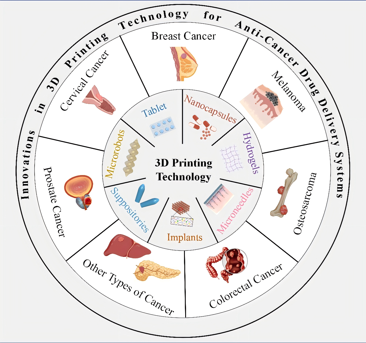
In this review, we begin by exploring various 3D printing technologies and their diverse applications in the development of drug delivery systems, particularly for anti-cancer treatment. We then discuss the innovations in the design and formulation of these delivery systems, focusing on their ability to target specific tumor sites, controlling drug release rates, and enhancing therapeutic outcomes. Finally, we provide a comprehensive overview of the challenges associated with material selection, process optimization, and clinical translation, while offering insights into the future potential of 3D printing in personalized cancer therapy (Figure 1).

Figure 1. Overview of innovations in 3D printing technology for anti-cancer drug delivery systems.
2. 3D printing technology
The American Society for Testing and Materials (ASTM) classifies these technologies into seven categories based on the additive manufacturing processes involved.11 This review primarily introduces and compares the 3D printing technologies widely used in the field of antitumor drug delivery, including fused deposition modeling (FDM), direct ink writing (DIW), binder jetting (BJ), stereolithography (SLA), digital light processing (DLP), and selective laser sintering (SLS) (Table 1).
Table 1. Comparison of different 3D printing technologies
| Methods | Materials | Fabrication mechanism | Resolution | Advantages | Challenges |
|---|---|---|---|---|---|
| FDM | Thermoplastics | Heating and extruding solid filament | 50–100 μm |
• High flexibility and cost-effectiveness15 • Precise and flexible parameter • control • Supports complex release • designs • Compatible with other techniques |
• Resolution limited by nozzle size18,19 • Surface roughness affects precision20 • High risk of drug degradation15 |
| DIW | Viscoelastic ink | Liquid ink is squeezed through the nozzle | 1–250 μm |
• Wide material diversity21 • Viscoelasticity stabilizes structure • High solid content reduces deformation17 • Low-temperature printing22 |
•Materials prone to nozzle clogging Complex design23 • Low strength24 |
| BJ | Low-viscosity solvent-based ink | Selective jetting of liquid binder onto powder material on the print bed | 80–250 μm |
• High efficiency and low cost • Suitable for heat-sensitive materials • Low-temperature operation27 |
•Requires powder pretreatment • Porosity control and interlayer uniformity are challenging28 •Low resolution and poor surface quality27 |
| SLA | Photosensitive thermoset polymers | Photocurable resin is solidified through photopolymerization initiated by absorbing light | 10 μm |
• High-resolution parts can be achieved14 • Wide range of functional applications29 |
• Limited materials |
| DLP | Optical crosslinking material | Optical crosslinking | 10 μm |
• High printing speed33 • Wide wavelength operation34 • High resolution |
• Requires post-curing and post-cleaning • Limited material selection • Restricted platform size27 |
| SLS | Polymers, ceramics, metal, composites | Melt sintering | 1–250 μm |
• Material diversity • No support structures are required35 |
• High cost • Limited applicability |
Abbreviations: BJ, binder jetting; DIW, direct ink writing; DLP, digital light processing; FDM, fused deposition modeling; SLA, stereolithography; SLS, selective laser sintering.
2.1. Fused deposition modeling
FDM involves extruding thermoplastic materials in the form of filaments through a 3D printer’s extrusion nozzle at high temperatures. Together, the extruder and the build plate are maneuvered along the X, Y, and Z axes to build an object layer-by-layer.38 There are dozens of accessible polymers that can be used in FDM printing, taking advantage of the intrinsic properties of thermoplastics which suit the quick transition from a solid state in the filament, to molten in the extruder, and rapidly reverting to solid after being deposited on the build plate.39 Some of the most widely researched examples of these polymers include vinyl derivates such as polyvinyl alcohol (PVA) and polyvinyl pyrrolidone (PVP), or cellulosic polymers like hydroxypropyl cellulose (HPMC) or ethyl cellulose (EC), but many others have also shown compatibility with FDM.40,41 FDM printers in pharmaceutical applications almost exclusively create solid objects such as tablets, but have also shown success in producing buccal films and chewable, gummy-like objects.42 FDM allows for the easy fabrication of drug delivery systems with complex drug release profiles, catering to the specific release requirements of various dosage forms. Furthermore, printing parameters, such as dose, shape, and fill density percentage, are easily controllable and can be precisely adjusted to optimize drug performance.15 Due to its simplicity, flexibility, and cost-effectiveness, FDM is widely applied in drug delivery systems.43
2.2. Direct ink writing
DIW can be categorized into droplet-based ink extrusion and continuous ink extrusion.24 The material, in the form of viscoelastic ink, is extruded layer-by-layer through a nozzle, constructing scaffolds and other 3D geometries with desired properties and performance on a computer-controlled moving platform.44 The curing of inks can be achieved through mechanisms such as solvent evaporation, temperature changes, gelation, or direct writing media, with the chosen curing mechanism also determining the ink’s rheological properties and solid content.45 DIW offers material diversity, encompassing metals, ceramics, polymers, hydrogels, composites, and even biological cells.21 The ink exhibits shear-thinning behavior and significant viscoelasticity, enabling it to maintain stability after extrusion and facilitate interlayer deposition without collapse. Additionally, the ink contains a high solid content, which reduces volume and shape changes during subsequent processing.17 Furthermore, DIW employs room-temperature or low-temperature forming methods, alleviating the thermal stability concerns of active pharmaceutical ingredients (APIs).22 The mild drug formulation conditions of DIW present new processing opportunities for the anti-cancer drug delivery industry.
2.3. Binder jetting
BJ is a technology that utilizes a drop-on-demand (DoD) method, selectively spraying a liquid binder onto powder materials placed on the print bed.27 This ink-jet printing process is primarily used to deposit inks made from low-viscosity solvent-based binders, with the material body typically presented as spherical powder particles on the print bed, thereby expanding the range of potential materials.27 The advantages of BJ include its efficient and low-cost process, making it suitable for personalized drug delivery systems with customizable drug release characteristics. Moreover, this technology can operate at ambient temperatures that are suitable for heat-sensitive materials.46 However, materials for binder jetting printing are limited to powders.47 Powder characteristics significantly impact the printing process, and it is essential to fully understand these properties, including shape, appearance, size, distribution, fluidity, and bulk density of powder particles. When the liquid binder is applied to the powder bed, variations in powder morphology and particle size affect the binder’s penetration rate.48 Typically, uneven powder distribution and excessive pore size can increase the penetration time.49
2.4. Stereolithography
SLA utilizes a light-induced chain polymerization process to trigger a photopolymerization reaction, curing the photosensitive resin.32 Consequently, the oligomer-based liquid resin is photopolymerized to form a crosslinked and polymer-rich structure. SLA technology is mainly used for the preparation of hydrogels, where the nature of the produced matrix is highly dependent on the concentration and molecular weight of the polymer, which in turn influences the density of the gel formed. As a result of the high degree of crosslinking, SLA technology produces pore-less, dense, and compact matrices, that often yield slow-release profiles.50 In terms of resolution, SLA can achieve high precision and resolution (1 μm), enabling the fabrication of components with complex internal structures and fine details.14 It also boasts a relatively high production speed.29 SLA is capable of producing materials with enhanced compressive and tensile strength, shape-memory functionality, and improved hydrophilicity.30,31 Due to its high precision and resolution, SLA is widely employed to develop various drug delivery systems with unique drug release rates.51–54
2.5. Digital light processing
DLP uses a digital micromirror device to project ultraviolet (UV) light onto a photopolymer vat. By curing thin layers of photopolymer with predefined shapes and thicknesses, a 3D structure is assembled layer-by-layer.27The main components of the DLP equipment include a projector, a digital micromirror device (DMD), and a vat of photopolymer resin or suspension. Light is generated by a projector at the bottom, which passes through the DMD. DMD consists of arranged cells of microscopic mirrors, each corresponding to one pixel in the image. Thus, the light is illuminated to precisely follow the shape of the layer on the vat window. For DLP, users slice the 3D computer-aided design structure to be printed into thin layers, and the set of layer images is inserted into the printer. Each layer image works as a grayscale mask to selectively cure the resin.55 A photopolymer resin comprises photocurable or photosensitive oligomers and monomers in addition to a photoinitiator activated by a light with a specific wavelength and various chain extenders or crosslinking agents.56 DLP utilizes photoreactive and photopolymerizable materials, allowing drugs to be directly loaded into the liquid prepolymer solution for printing.27 DLP-based 3D printing offers superior printing speed compared to the point-by-point curing method of SLA-type printers, owing to its plane-by-plane printing technique.57
2.6. Selective laser sintering
SLS involves depositing powder onto a powder bed layer, which is then heated to just below the sintering threshold temperature. A laser beam is directed onto the powder layer, triggering sintering, causing the powder particles to melt and bond with the previously consolidated layer.58,59 Unlike other techniques, SLS does not require support structures because the powder itself supports the printed section. During operation, the manufacturing platform moves downward, depositing a fresh layer of powder, after which the laser scans and sinters the next layer of the object. This layer-by-layer process continues until the parts are fully formed, and they cool down inside the printer.60 This technology enables the production of various materials such as plastics, metals, glass, ceramics, and composites. SLS for plastics and direct metal laser sintering (DMLS) or selective laser melting (SLM) for metals are the most common powder bed fusion systems employed at present. While these systems were once limited to high-value or custom parts, they have become more affordable and capable of producing a broader range of components.61 Due to their cost-effectiveness, high productivity, and material versatility, SLS 3D printers are extensively used in medical engineering for applications such as surgical models, medical device prototypes, and tissue engineering scaffolds.62
3. Applications of 3D printing technology in fabricating various drug delivery systems
3D printing technology has revolutionized pharmaceutical development through its design flexibility and manufacturing precision. This advancement enables the precise fabrication of drug delivery systems with complex structures and tailored functions, significantly advancing personalized medicine. The review focuses on key applications including tablets, suppositories, microneedle, hydrogels, implants, nanocapsules, and microrobots (Table 2).
Table 2. Summary of 3D printing applications in fabricating drug dosage form
| Drug dosage form | Main 3D printing technology | Highlights |
|---|---|---|
| Tablets | FDM, DIW | Manufacturing simplicity, dose uniformity, personalized design, portability, stable ingredients.63 |
| Suppositories | SSE, FDM | Rectal drug delivery, customized dosing, precise controlled release.64 |
| Hydrogels | DIW | Superabsorbent, highly stretchable, self-healing, smart hydrogels.65,66 |
| Microneedles | SLA, DLP, DIW | Precise skin insertion and adhesion, transcutaneous microdose drug delivery.67 |
| Implants | SLA, DIW, MJ, BJ, FDM | Targeted and controlled local drug release, combination with photothermal and magnetothermal therapy, sequential treatment.68 |
| Nanocapsules | FDM, DIW | Multi-drug load, increased bioavailability, efficacy, and safety.6 |
| Microrobots | DLP, SLA | On-demand and precise controlled drug delivery.69 |
Abbreviations: BJ, binder jetting; DIW, direct ink writing; DLP, digital light processing; FDM, fused deposition modeling; MJ, material jetting; SLA, stereolithography; SSE, semi-solid extrusion.
3.1. Tablets
3D-printed tablets demonstrate superior advantages in manufacturing simplicity, dose uniformity, and personalized design, effectively overcoming the limitations of conventional tablets in structural adaptability and dimensional control.63,70 Tablets can be designed for immediate release or sustained release kinetics, depending on therapeutic requirements. As the most widely accepted form of oral solid dosage, they have been extensively applied across various types and stages of cancer, including various gastrointestinal tumors, urinary system tumors, and others.71,72 Thanawuth et al.73 redesigned tablets with triangular/heptagonal compartments that exhibited extended release profiles (80% release in 24 h), contrasting with circular/square designs achieving complete release within 12 h. Mandati et al.’s comparative study74 demonstrated that spherical tablets with 50% infill density accelerated drug release, where infill density exerted greater influence than shape variation. Zhao et al.75 developed gastric-floating tablets via HME–FDM integration, establishing a positive correlation between release rate and surface area (1.5–4.5 mm radius).
3D printing can also produce tablets loaded with different drug forms, expanding formulation options. Zhang et al.16 fabricated core–shell tablets encapsulating solution/hydrogel/solid drugs, identifying prolonged release characteristics in liquid-phase formulations. Beck et al.’s HME-processed polymeric nano capsules enhanced drug processability,76 while PAM-assisted self-nanoemulsifying systems significantly improve the solubility, stability, therapeutic efficacy, and bioavailability of active pharmaceutical ingredients (APIs).77
3D printing enables the design of tablets with various geometric shapes to improve patient visual preferences and adherence to medication.78 Januskaite et al.79 used 3D printing to create chewable tablets with different flavors and colors, providing customized medication options for patients.
3.2. Suppositories
3D printing technology can transform nano drug delivery systems into suppository forms, demonstrating its potential in both localized and systemic drug delivery. This approach preserves nano-scale drug characteristics while achieving precision dosing and controlled release kinetics. Chatzitaki et al.80 used pressure-assisted microinjection (PAM) with Gelucire 48 and Kolliphor RH 40 to improve drug release accuracy and bioavailability. Seoane-Viaño et al.64 developed an infliximab-based bio-suppository with semi-solid extrusion (SSE), ensuring uniform distribution and controlled release, thereby addressing stability and uniformity challenges in rectal delivery. Awad et al.81 created an SSE-processed multi-drug suppository, incorporating budesonide and tofacitinib citrate to enhance treatment of rectal and colonic inflammation and enable personalized delivery of poorly soluble drugs. Suppositories are widely utilized in the local and systemic treatment of tumors such as cervical and colorectal cancers, due to their reduced gastrointestinal side effects, bypassing hepatic first-pass metabolism, ease of self-administration, and so on. These benefits contribute to their high biosafety and therapeutic efficacy.82,83
3.3. Microneedles
Microneedles can break the stratum corneum barrier through minimally invasive methods, significantly enhancing transdermal drug delivery efficiency while avoiding nerve stimulation.84 Compared to the limitations of traditional casting processes, such as limited structure and long production cycles, 3D printing technologies like SLA can create drug delivery systems with unique delivery characteristics, enabling microneedle structures with precision as fine as 10 μm and supporting multi-level porosity and complex geometric designs.85–87 As a significant modality in transdermal drug delivery systems, microneedles facilitate the administration of various therapeutic agents to tumor sites through the skin. They are now widely used in the local treatment of tumors, such as melanoma, papilloma, glioma T-cell lymphoma, and breast cancer.88,89 Uddin et al.90 designed a cross-shaped microneedle array coated with anti-cancer drugs like cisplatin, which allowed for rapid drug release within 1 h and demonstrated significant anti-cancer activity. Joo et al.67 proposed a novel self-locking microneedle system for precise skin insertion, attachment, and transdermal micro dose drug delivery, significantly improving the delivery of (αPD-L1 Ab)/SD-208 drugs in a melanoma mouse model. Lin et al.91 designed a multi-microchannel microneedle microoperation (4M) platform, using electroporation to facilitate drug transport across cell membranes and providing a concentrated and safe external electric field to accelerate deep drug penetration into cells. Wu et al.92 printed an acoustically activated, programmable microneedle patch, using digital control of acoustic signals to trigger drug pumping, demonstrating drug release characteristics tailored to user needs. This in situ acoustic drug delivery strategy offers advantages in miniaturization, operability, and intelligence, improving the controlled release capacity of microneedle patches.
3.4. Hydrogels
Hydrogels, as archetypal 3D network materials, have emerged as crucial carriers in antitumor drug delivery systems due to their exceptional water absorption capacity, mechanical flexibility, and self-healing properties.65,66 Compared to conventional preparation methods that struggle to achieve precise structural and functional control, 3D printing technology enables accurate construction of hydrogel spatial topology, thereby endowing drug delivery systems with precise controlled release and intelligent responsive characteristics.93,94In situ gelation, along with the formulation of various dosage forms using hydrogels, enables the delivery of multiple anti-cancer agents while simultaneously providing a robust platform for the repair of postoperative tissue defects. This strategy has been extensively applied in postoperative chemotherapy for cancers such as breast cancer, osteosarcoma, and malignant glioma, offering significant therapeutic benefits in terms of localized drug delivery and tissue regeneration.95,96
Kuo et al.97 developed a porous hybrid hydrogel based on gelatin and alginate (G/A) that can encapsulate and deliver bioactive compounds, such as enzymes and antioxidants. Phan et al.98 created an injectable, biodegradable hydrogel based on cellulose nanocrystals (CNCs) incorporated into the amphiphilic copolymer PCLA, exhibiting excellent biocompatibility, controlled drug release, and multifunctional drug loading. Doxorubicin (DOX)-loaded hydrogel formulation significantly inhibited tumor growth. Hydrogels can also serve as materials for 3D printing other drug forms. Liu et al.95 coated a uniform polycaprolactone (PCL) layer onto a 3D-printed alginate-gelatin hydrogel scaffold to create a core–shell fiber scaffold, further coated with polydopamine (PDA) to enhance photothermal properties. This enabled on-demand drug release triggered by near-infrared (NIR) laser, effectively inhibiting or eliminating tumors both in vitro and in vivo using drugs like DOX and photothermal therapy. Cheng et al.99 used extrusion-based semi-solid 3D printing to fabricate theophylline hydrogel tablets with varying drug loads (75–125 mg), exhibiting high yield stress, storage modulus, and hardness.
3.5. Implants
The porous structure of 3D-printed implantable scaffolds improves drug loading efficiency and significantly enhances antitumor effects by targeting and extending drug release.100 These scaffolds can also simultaneously load multiple anti-cancer drugs, enabling controlled sequential drug release. A wide range of metallic, polymeric, ceramic, and composite materials with diverse physicochemical properties can be processed through 3D printing to meet the clinical demands of scaffold fabrication.101 These implants play a crucial role in the treatment of tumors, such as breast cancer, bone tumors, cervical cancer, esophageal cancer, and lung cancer, as well as in the repair and support of postoperative tissue defects. Fang et al.68 developed a sandwich-type composite material, by combining a 3D-printed scaffold loaded with combretastatin A4 (CA4) and electrospun fibers loaded with DOX, to form a multifunctional scaffold through time-controlled differential drug release. Wang et al.102 fabricated a coaxial 3D-printed scaffold that incorporated non-nucleoside STING agonist SR-717 and AKT inhibitor MK-2206 into the outer shell and core layers, respectively. The sequential and sustained release of these two drugs achieved synergistic STING activation, demonstrating significant antitumor effects. 3D-printed scaffolds can also provide a platform for immune modulation. Zhang et al.103 developed a porous 3D-printed implantable scaffold with an ordered pore structure highly similar to real lymphatic structures, forming an artificial lymphatic network. This network facilitated the formation of antigen-specific immune cells, effectively eliminating tumor cells.
3.6. Nanocapsules
Nanocapsules are hollow spherical colloidal structures with sizes ranging from 10 to 1000 nm, where the core is encapsulated by a polymer shell, allowing for the simultaneous loading of both hydrophilic and hydrophobic drugs. Compared to other drug delivery systems, nanocapsules, as smart carriers, offer higher bioavailability, efficacy, and safety.104 The introduction of 3D printing technology reduces the manufacturing time of nanocapsules and enhances automation, further expanding the applications of 3D-printed nanocapsule drug delivery systems.6 Nanocapsules using various carriers, including inorganic nanoparticles, dendrimers, and carbon nanotubes (CNTs), facilitating precise drug delivery and early cancer detection. They are widely applied in treating cancers such as breast cancer, osteosarcoma, prostate cancer, and colorectal cancer.105 Rupp et al.106 developed a small core–shell capsule composite material using a two-step 3D printing design. A dual-printing head system (FDM and liquid inkjet printing) was used to fabricate micro-sized core–shell capsules ranging from 100 to 800 μm, which can be used to encapsulate liquid drugs. By combining 3D printing technology with nanotechnology, nanocapsule suspensions containing various polymers (e.g., PCL, Eudragit® RL100) and different filling ratios were impregnated into 3D printing filaments, which can serve as a substrate for manufacturing other drug formulations.107
3.7. Microrobots
Drug delivery is a key research area in the field of microrobots.108 The integration of smart materials, magnetic control technology, and targeted therapy enables the active release of payloads, which is crucial for on-demand, precise, and efficient drug delivery.69 With the advancement of 3D printing technology, further optimization of microrobot movement mechanisms and drug release control systems has broadened their potential applications in therapy, particularly in the targeted treatment of primary breast cancer, colorectal cancer, and other malignancies.109,110 Bozuyuk et al.109 designed a magnetically driven helical microswimmer using two-photon 3D printing technology, capable of precisely controlling the on-demand release of the chemotherapeutic drug DOX through external light stimulation. This system combines magnetic field-driven movement with light-triggered drug delivery, demonstrating excellent biocompatibility, biodegradability, and precise drug release regulation capabilities. Park et al.110 designed a magnetic-driven porous, biodegradable microrobot with a helical structure, which significantly improved drug release efficiency by optimizing the ultrasound pulse parameters and resolving cell damage issues. Ceylan et al.111 developed a magnetically driven, enzyme-degradable double-helix microswimmer, which achieved drug delivery and release through rotational magnetic fields. The microswimmer, under the action of the MMP-2 enzyme, degraded within 118 h, rapidly releasing the embedded drugs or functional cargos.
4. 3D printing technology in anti-cancer drug delivery systems
4.1. Breast cancer
Breast cancer ranked as the second most commonly diagnosed cancer worldwide in 2022.1 Although intensive-dose chemotherapy can enhance efficacy, particularly for triple-negative breast cancer (TNBC) patients, it inevitably increases drug side effects and compliance issues.112 With the advancement of 3D printing technology, localized drug delivery systems, represented by implants, can reduce the administration time of chemotherapy, improve patient compliance, and enhance the controllability and sustainability of drug delivery113 (Table 3). Notably, implant-based reconstructions are increasingly utilized following mastectomy for breast reconstruction, preventing breast cancer recurrence, and controlling infections, further expanding their clinical applications.114 Yang et al.115 employed E-jet 3D printing technology to design poly-lactic-co-glycolic acid (PLGA) scaffolds that encapsulated chemotherapy drugs 5-fluorouracil and NVP-BEZ235, significantly reducing the drug dosage and prolonging the retention time of the drug concentration at the tumor site while minimizing exposure to normal tissues (Figure 2A). Hao et al.116 designed a drug carrier using polydimethylsiloxane (PDMS) for paclitaxel (PTX) and DOX microbead scaffolds, achieving sustained drug release for more than 3 weeks, effectively preventing tumor recurrence while reconstructing breast tissue. Su et al.117 developed a carbon quantum dot-curcumin (CCNPs) loading system with visual drug release capabilities (the fluorescence properties of CCNPs make drug release more convenient to be visualized and tracked, and it can be used for real-time drug release tracking in vivo), which also exhibited antimicrobial activity, promoted angiogenesis, and repaired tissue defects (Figure 2B). Zhang et al.118 designed a Pt-GelMA scaffold, 3D-printed using Pt (IV)-induced photopolymerization of methacrylate gelatin (GelMA) bioink, which effectively killed tumor cells and inhibited local tumor growth and distant metastasis post-surgery without any additional photosensitizers or chemotherapy drugs. The pH-sensitive drug delivery systems (gelatin–alginate composite scaffolds loaded with PTX and DOX) enable efficient targeted drug delivery and can regulate gene expression levels in breast cancer cells, such as CASP-3, Bax, and p53 (Figure 2C).119,120 Time-programmed pulsed release implant that can control the release time based on the tumor’s internal microstructure has marked the first use in local dose-intensive chemotherapy for breast cancer121 (Figure 2D).
Table 3. Summary of 3D printing technology in anti-cancer drug delivery systems for breast cancer
| Drug dosage form | Author | Highlight | Drug carrier | API | 3D printing technology | Stage | Ref. |
|---|---|---|---|---|---|---|---|
| Implants | Yang et al. | Reducing the drug dosage; continuous drug release |
PLGA | 5-Fu; NVP-BEZ235 |
E-jet |
In vivo; in vitro |
115 |
| Hao et al. | Continuous drug release | PDMS | PTX; DOX |
SLA |
In vivo; in vitro |
113 | |
| Su et al. | Visualized drug release capability; multifunctional scaffold |
CCNACA | CCNPs | DIW |
In vivo; in vitro |
117 | |
| Zhang et al. | Multifunctional scaffold | GelMA | Pt(IV) | SLA |
In vivo; in vitro |
118 | |
| Zaer et al. | pH-dependent drug delivery; high selectivity in drug release |
Nio-DOX@ GT-AL | DOX | DIW | In vitro | 119 | |
| Hossenini et al. | pH-dependent drug delivery; high selectivity in drug release |
Nio-PTX@ GT-AL | PTX | DIW | In vitro | 120 | |
| Myung et al. | Time-programmed pulsed release profiles | PCL; Pluronic F127 (PF127) |
COX; CTX |
DIW |
In vivo; in vitro |
121 | |
| He et al. | Combination therapy | BG@NbSiR | R837 | DIW |
In vivo; in vitro |
122 | |
| Wei et al. | Combination therapy | Gelatin hydrogel | DOX | MJ |
In vivo; in vitro |
123 |
Abbreviations: API, active pharmaceutical ingredient; BG, bioactive glass; CCNANA, CCNPs/sodium alginate/nanoclay/caffeic acid grafted chitosan; CCNPs, CDs-Cur nanoparticles; COX, cyclophosphamide; CTX, cefotaxime; DIW, direct ink writing; DOX, doxorubicin; GelMA, methacrylate gelatin; GT-AL, gelatin-alginate; MJ, material jetting; PDMS, polydimethylsiloxane; PLGA, poly-lactic-co-glycolic acid; PTX, paclitaxel; SLA, stereolithography.

Figure 2. Innovations of 3D printing technology in anti-cancer drug delivery systems for breast cancer. (A) (1) The set up and workflow of the E-jet 3D printing system. (2) Simulation diagram of PLGA and PFN scaffolds. Reproduced with permission from ref.115. (B) (1) Preparation of CDs and CCNPs. (2) Preparation of CCNACA scaffolds and their antibacterial properties in vitro. Two-photon laser confocal images of MCF-7 cells incubated with (3) CDs and (4) CCNPs at different concentrations. Reproduced with permission from ref.117. (C) Schematic representation of the delivery of doxorubicin-loaded nanoscale drug carriers (Nio-DOX) to cancer cells. Reproduced with permission from ref.119. (D)(1) Preparation of a 3D-printed device with DOX and CYP and its implantation to TNBC tumors. (2) Optimization of dose density for local dose-dense AC chemotherapy using 3D-printed devices to improve therapeutic efficiency. Reproduced with permission from ref.121. Abbreviations: AC, doxorubicin and cyclophosphamide; CCNACA, CCNPs/sodium alginate/nanoclay/caffeic acid grafted chitosan; CCNPs, CDs-Cur nanoparticles; CDs, carbon dots; CYP, cyclophosphamide; DOX, doxorubicin; PFN, PLGA/5-FU/NVP-BEZ235; PLGA, poly-lactic-co-glycolic acid.
Combination therapy is also an emerging field in breast cancer treatment. He et al.122 designed BG@NbSiR scaffolds that can eradicate primary tumors, activate immune responses, inhibit metastasis, prevent tumor recurrence (long-term immune memory), and accelerate osteogenesis through photothermal effect checkpoint blockade immunotherapy. Wei et al.123 designed core–shell implants based on a hydrogel network, which undergoes a gel-to-sol transition under NIR light to release drugs on demand.
4.2. Melanoma
Melanoma is characterized by irregular patterns and uneven surfaces. It can spread to various organs such as the lungs, liver, bones, and brain through the respiratory tract and bloodstream.124 Chemotherapeutic agents commonly used in clinical practice include immune checkpoint inhibitors for programmed cell death protein 1 (PD-1), programmed death-ligand 1 (PD-L1), and cytotoxic T-lymphocyte associated protein 4 (CTLA-4).125 However, these drugs have only about 40% efficacy and face the challenge of drug resistance.126 Given that melanoma is typically develops on the skin’s surface, 3D printing-based precision drug delivery systems, such as microneedles and dressings, offer a minimally invasive approach for targeted drug administration. These systems effectively suppress the growth and metastasis of primary melanoma while improving treatment outcomes and reducing systemic side effects67,127 (Table 4).
Table 4. Summary of 3D printing technology in anti-cancer drug delivery systems for melanoma
| Drug dosage form | Author | Highlight | Drug carrier | API | 3D printing technology | Stage | Ref. |
|---|---|---|---|---|---|---|---|
| Microneedles | Joo et al. | Precise skin insertion selflocking MN | Photosensitive resin | SD-208; aPD-L1 Ab |
DLP |
In vivo; in vitro |
67 |
| Lopez-Ramirez et al. | Active release and controlled release | PVP | Anti-CTLA-4 | SLA |
In vivo; in vitro |
128 | |
| Li et al. | High resolution; bilayer microneedles; combination therapy |
Nanocomposite inks | ICG; Curcumin; DOX |
DIW |
In vivo; in vitro |
129 | |
| Dressings | Ćurić et al. | Geometrically customizable dressings; high mechanic stability |
Polysaccharide hydrogel | NiCu NPs | DIW | In vitro | 130 |
| Kumar et al. | Homogeneously layered and electrically stimulatory | Nic-hl-PLGR | Nic | HME | In vitro | 131 | |
| Implants | Xu et al. | Combination therapy | SA-GG@PDA | DOX | DIW | In vivo; In vitro | 132 |
Abbreviations: API, active pharmaceutical ingredient; CTLA-1, cytotoxic T-lymphocyte associated protein 1; DIW, direct ink writing; DLP, digital light processing; DOX, doxorubicin; GG: gellan gum; HME, hot-melt extrusion; ICG, indocyanine green; Nic, niclosamide; NiCu, nickel-copper; NPs, nanoparticles; PDA, polydopamine; PD-L1, programmed death-ligand 1; PLGR, PLA-graphene nanoplatelet; PVP, polyvinylpyrrolidone; SA, sodium alginate; SLA, stereolithography.
Microneedle patches are widely applied in melanoma treatment. Self-locking microneedles consist of a sharp skin-penetrating tip, a wide interlocking body, and a narrow base with mechanical supports, fabricated over a flexible hydrocolloid patch to enhance skin penetration accuracy on irregular surfaces. These patches carry transforming growth factor beta (TGF-β) inhibitor (SD-208) and anti-PD-L1 inhibitors, improving dose efficacy (Figure 3A).67 Furthermore, magnesium particles react with interstitial fluid, generating rapid H2 bubble formation to breach dermal barriers and enhance payload delivery. The combination of active release and delayed release provides stronger anti-cancer effects (Figure 3B).128 Li et al.129 designed high-resolution bilayer microneedles with an apex size of approximately 5 μm using pneumatic extrusion and controlled stretching. The patch designs can be tailored for cancers of varying severity, allowing personalized treatment with high therapeutic efficacy and minimal side effects (Figure 3C).

Figure 3. Innovations of 3D printing technology in anti-cancer drug delivery systems for melanoma. (A) (1) Fabrication of dissolvable self-locking MN via projection micro-stereolithography and micro-molding. (2) Geometry of (αPD-L1 Ab)/SD-208-loaded self-locking MN. (3) Mechanism of action in SD-208 and αPD-L1 Ab for melanoma combination therapy. (4) Schematic illustration of (αPD-L1 Ab)/SD-208-loaded self-locking MN transcutaneous application onto the melanoma. Reproduced with permission from ref.67 (B) (1) Active microneedle patch composition, and built-in Mg particle activation as pumps when in contact with bodily fluids, leading to an enhanced drug release. (2) Digital photograph showing a patch of 15 × 15 microneedle array and optical/fluorescent microscopy images of an active MN tip loaded with Mg particles. (3) SEM image of single active microneedle tip and EDX analysis for Mg. Reproduced with permission from ref.128 (C) (1) Schematic illustration of the fabrication of integrated microneedle patch via the multimaterial direct ink drawing of nanocomposite inks. (2) Fluorescence image of an integrated patch with the shape of a Christmas tree composed of PLGA microneedles (red), maltose microneedles (blue), and CA microneedles (green). Reproduced with permission from ref.129 (D) Schematic illustration of 3D-printed heterogeneous SA-GG@PDA + DOX scaffolds for sequential tumor photothermal-chemotherapy and wound healing. Reproduced with permission from ref.132 Abbreviations: DOX, doxorubicin; EDX, energy dispersive X-ray; MN, microneedle; PDA, polydopamine; PD-L1, programmed death-ligand 1; PLGA, poly-lactic-co-glycolic acid; SEM, scanning electron microscopy.
Dressings are also widely used for drug delivery in melanoma. Ćurić et al.130 developed a customizable dressing combining nickel-copper nanoparticles (NiCu NPs) and polysaccharide hydrogels. NiCu NPs enhance the mechanical stability and anti-cancer activity of the dressing, which can be personalized according to patient needs. Kumar et al.131 developed a 3D-printed drug solution patch (3D-est-MediPatch) based on polylactic acid-graphene nanosheets (PLA-graphene), demonstrating good drug control release properties and therapeutic effects, while providing a portable platform for electrostimulation-assisted therapy. The heterogeneous hybrid hydrogel scaffold (SA-GG@PDA) composed of sodium alginate (SA), gellan gum (GG), and polydopamine nanoparticles (PDA NPs) carries the chemotherapy drug DOX, enabling sequential photothermal therapy combined with chemotherapy. This accelerates drug release while promoting angiogenesis and tissue repair (Figure 3D).132
4.3. Osteosarcoma
Osteosarcoma is the most common primary malignant bone tumor, and the treatment of bone tumors remains one of the major challenges in cancer therapy.133 Surgical resection often fails to completely remove tumor cells, leading to bone defects and poor prognosis.134 Low-grade osteosarcoma, mainly parosteal osteosarcoma, is typically treated with surgical resection. High-grade osteosarcoma requires neoadjuvant chemotherapy with methotrexate, doxorubicin, and cisplatin (MAP), followed by surgery and continued postoperative chemotherapy.135 With 3D printing technology, implantable drug delivery systems can effectively load drugs or functional materials due to their small size and controllable porosity, offering excellent bioactivity, biodegradability, and biocompatibility for repairing bone defects.136–138
4.3.1. Chemotherapy
3D-printed scaffolds integrate antitumor, osteogenic, and antibacterial functions while modulating the immune microenvironment, offering a multifunctional approach to treating bone tumors (Table 5). Wang et al.138 designed a spherical poly L-lactic acid (PLLA) implant loaded with multiple drugs, exhibiting excellent biodegradability and antitumor efficacy, with precise control over drug concentration and release rate. In mice, the scaffold released drugs for over 6 weeks (Figure 4A). Huang et al.139 developed a Se/Sr/Zn-HA-PCL multifunctional composite scaffold, combining the antitumor effect of SeO32− with the osteogenic and antibacterial properties of Sr2+ and Zn2+. Li et al.140 successfully regulated the tumor immune microenvironment by incorporating colony-stimulating factor-1 receptor inhibitor (GW2580) onto a calcium phosphate scaffold, promoting M1 macrophage polarization to activate antitumor immunity (Figure 4B). Li et al.141 designed a time-sequential MgO2/PLGA composite scaffold using low-temperature rapid prototyping (LT-RP) 3D printing technology could release H2O2 and Mg2+ in stages, achieving combined antitumor, antibacterial, and osteogenic effects. In the first 3 weeks, the scaffold released H2O2 for chemotherapeutic and immune microenvironment regulation, followed by 12 weeks of Mg2+ release to promote bone regeneration (Figure 4C). Phytochemicals show great potential in osteosarcoma treatment, but their poor solubility, bioavailability, and stability in free form can be improved by combining 3D-printed scaffolds with nanotechnology. Curcumin, apigenin, garlic extract, gingerol-Zn2+ metal complexes, and piperine, encapsulated in nanoparticles like liposomes, which are incorporated into 3D-printed calcium phosphate (CaP) scaffolds with specific porosity, exhibit controlled biphasic release in both acidic and physiological media (Figure 4D), significantly enhancing osteoblast proliferation, antibacterial activity, angiogenesis (e.g., mTOR upregulation), and antitumor effects.142–146
Table 5. Summary of 3D printing technology in anti-cancer drug delivery systems for osteosarcoma (chemotherapy)
| Drug dosage form | Author | Highlight | Drug carrier | API | 3D printing technology | Stage | Ref. |
|---|---|---|---|---|---|---|---|
| Implants | Wang et al. | Spherical implant; biocompatibility; multi-drug load |
PLLA | Methotrexate; DOX; Ifosfamide; cisplatin |
SLA |
In vivo; in vitro |
138 |
| Huang et al. | Multi-functional scaffolds | PCL | SeO32−; Sr2+; Zn2+ |
DIW |
In vivo; in vitro |
139 | |
| Li et al. | Regulate immune microenvironment in stages | HBC/OCS hydrogel | GW2580 | DIW |
In vivo; in vitro |
140 | |
| Li et al. | Multi-functional bioactive scaffolds; time-sequential functions |
PLGA | H2O2; Mg2+ |
LT-RP |
In vivo; in vitro |
141 | |
| Sarkar et al. | Curcumin-encapsulated liposomes | TCP | Curcumin | BJ | In vitro | 142 | |
| Dahiya et al. | pH-sensitive release; multifunctional scaffolds |
TCP | Carvacrol; curcumin | BJ | In vitro | 143 | |
| Kushram et al. | Controlled and biphasic release | TCP | Garlic extract | BJ | In vitro | 144 | |
| Chaudhari et al. | Controlled drug delivery; multifunctional scaffolds |
TCP-PCL | Gingero-Zn+2 | BJ | In vitro | 145 | |
| Bose et al. | Controlled drug delivery; multifunctional scaffolds |
TCP | Curcumin; piperine; carvacrol |
BJ | In vitro | 146 |
Abbreviations: API, active pharmaceutical ingredient; BJ, binder jetting; DIW, direct ink writing; DOX, doxorubicin; HBC, hydroxybutylchitosan; LT-RP, low-temperature rapid prototyping; OCS, oxidized chondroitin sulfate; PCL, polycaprolactone; PLLA, Poly L-lactic acid; SLA, stereolithography; TCP, tricalcium phosphate.

Figure 4. Innovations of 3D printing technology in anti-cancer drug delivery systems for osteosarcoma (chemotherapy, photodynamic therapy, and photothermal therapy). (A) (1) Schematic illustration of the fabrication process. (2) The cylindrical and spherical 3D model of the implants. (3) Mean density analysis of the immunohistochemistry examination of the tissues around the tumor site of different treatments after 12 months. Reproduced with permission from ref.138 (B) (1) Preparation process and related mechanism of the inhibitor-loaded scaffold. (2) SEM images show pore structure and surface morphology of CPC and CPC/HBC-OCS scaffolds. (3) EDS images show coating efficiency of HBC-OCS hydrogel on CPC scaffolds. Reproduced with permission from ref.140 (C) (1) Diagram of the preparation procedure of MgO2 nanoparticles and MgO2/PLGA scaffolds. (2) FESEM images and EDS mapping of 20 MP scaffold surfaces. (3) Weight loss percentage, pH value changes, the accumulative release of Mg2+, and the accumulative release of H2O2 of PLGA and MgO2/PLGA scaffolds during 12 weeks of in vitro degradation. (4) EPR spectra of PLGA and MgO2/PLGA scaffolds at different conditions, revealing the •OH generation of scaffolds only in Fe2+ solution. (5) Diagram of the degradation behavior of time-sequential release pattern of MgO2/PLGA scaffolds. Reproduced with permission from ref.141 (D) (1) Schematic representation of CC-NP release from 3D-printed scaffold. (2) Release of carvacrol and curcumin from their drug solution (NBGS) and nanoparticles (CC-NP) at pH 5.0 and 7.4. Reproduced with permission from ref.143 (E) Schematic of CaPC 3D-printed scaffolds for PDT against osteosarcoma and osteogenesis promotion. Under 660 nm light, the scaffolds generate O2 and activate Ce6, producing ROS for effective PDT. Reproduced with permission from ref.152 (F) Schematic diagram of the process for photothermal ablation of osteosarcoma and bone regeneration by NBGS. Vascularization can be promoted to enhance osseous reconstruction. Reproduced with permission from ref.155 Abbreviations: DS, drug solution; EDS, energy dispersive spectrum; EPR, electron paramagnetic resonance; FESEM, field emission scanning electron microscopy; HBC, hydroxybutylchitosan; NBGS, Nb2C MXene-integrated bone-mimetic composite scaffolds; NP, nanoparticle; OCS, oxidized chondroitin sulfate; PDT, photodynamic therapy; PLGA, poly-lactic-co-glycolic acid; ROS, reactive oxygen species; SEM, scanning electron microscopy.
4.3.2. Photodynamic therapy and photothermal therapy
Photodynamic therapy (PDT) treats tumors by generating free radicals and singlet oxygen through laser irradiation, while photothermal therapy (PTT) uses NIR light for localized heating to kill cancer cells.147,148 NIR laser-triggered drug or biomolecule release implants are commonly used smart drug delivery systems for tumor eradication and tissue regeneration (Table 6). 3D-printed scaffolds loaded with photothermal agents enhance NIR light penetration, promoting bone tissue regeneration.149 Abie et al.150 designed a 3D-printed scaffold made from silk fibroin, tannic acid, and PDA-modified SFO–TA–BGNF composite gel, exhibiting excellent photothermal antitumor and osteogenesis functions under NIR-I irradiation. Bifunctional scaffolds embedded with metal ions (Fe³+ and Mg2+) combine chemodynamic therapy (CDT) and PDA nanoparticles to achieve precise photothermal treatment under NIR-I laser, eradicating metastatic and residual tumor tissues.151 Additionally, Cap scaffolds integrated with a photosensitizer (Ce6) and an oxygen platform mediated by cyanobacteria improve antitumor effects of PDT, alleviate tumor hypoxia, and promote bone regeneration under NIR-I laser irradiation (Figure 4E).152
Table 6. Summary of 3D printing technology in anti-cancer drug delivery systems for osteosarcoma (photodynamic therapy and photothermal therapy)
| Drug dosage form | Author | Highlight | Drug carrier | API | 3D printing technology | Stage | Ref. |
|---|---|---|---|---|---|---|---|
| Implants | Abie et al. | Innovative composite inks; multifunctional scaffolds | SFO-TA-BGNF complex gel | SFO; TA; BGNF-PDA |
DIW | In vitro | 150 |
| He et al. | Photothermal therapy; metallic nanoparticles-containing ink |
TCP/PLGA | FeMg-NPs | DIW |
In vivo; in vitro |
151 | |
| Lin et al. | Photosynthetic oxygen-self-generated microbial scaffold | CaPC | Ce6 | DIW |
In vivo; in vitro |
152 | |
| Yang et al. | NIR-II; vascularized bone regeneration |
SC/PCL | SC NSs | FDM |
In vivo; in vitro |
153 | |
| He et al. | NIR-II; enhanced osteogenesis performance |
CaP | Egyptian blue | MJ |
In vivo; in vitro |
154 | |
| Yin et al. | NIR-II; promote the neogenesis and migration of blood vessels; biodegradable |
NBGS | Nb2C MXene | - |
In vivo; in vitro |
155 |
Abbreviations: API, active pharmaceutical ingredient; BGNF, bioactive glass nanofibers; CaP, CaCO3-polycaprolactone; CaPC, CeCyano-modified CaP scaffold; DIW, direct ink writing; FDM, fused deposition modeling; MJ, material jetting; NBGS, niobium carbide MXene-based bone-mimetic scaffold ; NIR, near-infrared; NPs, nanoparticles; NS, nanosheet; PCL, polycaprolactone; PDA, polydopamine; PLGA, poly-lactic-co-glycolic acid; SC, SrCuSi4O10; SFO, oxidized silk fibroin; TA, tannic acids; TCP, tricalcium phosphate.
Most studies use photothermal agents limited to NIR-I (650–950 nm), which have poor tissue penetration and are often non-bioactive, potentially causing systemic side effects after implantation.153 Functionalization of 3D-printed PCL scaffolds with NIR-II photothermal agents (e.g., SC NSs, EB NSs, Nb2C MXene) enables photothermal therapy driven by NIR-II laser (1068 nm), significantly enhancing drug tissue penetration depth, efficiently eliminating OS, and promoting bone regeneration. This therapy also modulates gene expression related to cancer metabolism/progression and skeletal development153–155 (Figure 4F).
4.3.3. Combination therapy
Postoperative residual cancer cells and chemotherapy resistance are key drawbacks of monotherapy in osteosarcoma treatment.156 Combining chemotherapy with photothermal or magnetic hyperthermia therapies can achieve precise photothermal treatment and localized controlled release of chemotherapy drugs by adjusting the content of photothermal agents and anti-cancer drugs in 3D-printed scaffolds, significantly inhibiting osteosarcoma growth (Table 7).156 PEEK/graphene composite scaffolds leverage the high photothermal conversion efficiency of graphene nanosheets and loaded antibiotics (STAC) or anti-cancer drugs (cisplatin, DDP) to achieve a multimodal therapy, including cancer cell ablation, bacterial clearance, and bone tissue repair.157 Poly-d,l-lactic acid (PDLLA) polymers, as versatile mediums, enable photothermal and chemotherapy scaffolds to load various therapeutic agents, such as TiN particles and DOX on tricalcium phosphate (TCP) scaffolds or DOX and hemin on BGC scaffolds, creating multifunctional platforms with excellent photothermal responsiveness and high anti-cancer efficiency.156,158 Using a metal-coordination self-assembly strategy, nanoscale two-dimensional copper-based conjugated metal-organic framework (cMOF)-engineered 3D-printed PCL scaffolds were developed for synergistic photothermal-chemodynamic therapy of osteosarcoma and accelerated bone regeneration (Figure 5A).159 NIR-II photothermal tumor ablation combined with chemotherapy represents a major breakthrough in osteosarcoma treatment. Xu et al.160 integrated FePSe2 nanosheets with α-TCP scaffolds to achieve NIR-II photothermal tumor ablation and selenium ion release to promote angiogenesis and bone regeneration, while activating caspase-dependent apoptosis to prevent tumor recurrence (Figure 5B). Liang et al.161 designed BG@silicene scaffolds loaded with the epigenetic modulator IOX1, enhancing osteosarcoma sensitivity to photothermal therapy while reducing thermal damage to normal tissues. Multifunctional scaffolds can further combine catalytic therapies and oxygen release to improve treatment efficacy. For example, BG scaffolds combined with single-atom iron catalysts (FeSAC) enable nanocatalytic therapy through hydroxyl radical generation (Figure 5C).162 PCL/nHA/MgO2/PDA composite scaffolds enhance tumor oxygenation and significantly improve photothermal therapy’s anti-cancer effect and bone repair capabilities (Figure 5D).163
Table 7. Summary of 3D printing technology in anti-cancer drug delivery systems for osteosarcoma (combination therapy)
| Drug dosage form | Author | Highlight | Drug carrier | API | 3D printing technology | Stage | Ref. |
|---|---|---|---|---|---|---|---|
| Implants | Zhu et al. | Multifunctional scaffolds | PEEK | Hydroxyapatite; graphene; STAC; DDP |
FDM |
In vivo; in vitro |
157 |
| Dang et al. | Multifunctional scaffolds | TCP; PDLLA |
TiN; DOX |
DIW |
In vivo; in vitro |
156 | |
| Dang et al. | Multifunctional scaffolds | BGC; PDLLA |
DOX; hemin |
FDM |
In vivo; in vitro |
158 | |
| Huang et al. | Metal-coordination self-assembly; interface engineering |
PCL | Cu-HHTP | FDM |
In vivo; in vitro |
159 | |
| Xu et al. | NIR-II; cryogenic 3D printing; promote vascularized bone regeneration |
α-TCP | FePSe3 | DIW |
In vivo; in vitro |
160 | |
| Liang et al. | NIR-II; biocompatible scaffolds |
Silicene bioactive glass | IOX1 | DIW |
In vivo; in vitro |
161 | |
| Wang et al. | Multi-functional scaffolds | Bioactive glass | FeSAC | - | Stage | 162 | |
| Xu et al. | Continuous oxygen release | PCL | MgO2; nHA; PDA |
FDM |
In vivo; in vitro |
163 | |
| Zhuang et al. | Multi-functional scaffolds | Bioceramic | Fe3S4 | DIW |
In vivo; in vitro |
164 | |
| Wang et al. | Repeated on-demand release capability | Hollow fiber alginate; iron oxide nanoparticles |
DOX | DIW |
In vivo; in vitro |
165 |
Abbreviations: API, active pharmaceutical ingredient; BGC, bioactive glass ceramics; DDP, drug cisplatin; DIW, direct ink writing; FDM, fused deposition modeling; FeSAC, single-atomic iron catalysts; DOX, doxorubicin; HHTP, hexahydroxytriphenylene; IOX1, 5-carboxy-8-hydroxyquinoline; nHA, nanohydroxyapatite; PCL, polycaprolactone; PDA, polydopamine; PEEK, polyetheretherketone; PDLLA, poly(d,l-lactide); STAC, stearyltrimethylammonium chloride; TCP, tricalcium phosphate.

Figure 5. Innovations of 3D printing technology in anti-cancer drug delivery systems for osteosarcoma (combination therapy). (A) The process of creating conductive MOF-coated 3D-printed scaffolds and their use in the treatment of osteosarcoma and for aiding in bone repair. Reproduced with permission from ref.159 (B) The 3D-printed TCP-FePSe3 scaffold as an all-in-one platform for bone regeneration and postsurgical suppression of osteosarcoma recurrence. Reproduced with permission from ref.160 (C) (1) Schematic illustration of the construction of FeSAC-BG scaffolds. (2) Digital photographs of FeSAC-BG scaffolds with varied initial impregnating FeSAC concentration. (3, 4) SEM images of FeSAC500-BG scaffold at varied magnification and (5) the corresponding elemental mappings of O, Si, C, and Ca. Reproduced with permission from ref.162 (D) 3D-printed PCL/MgO2/nHA scaffold for tumor therapy and bone defect repair. Reproduced with permission from ref.163 (E) (1) The 3D-printed bioceramic scaffolds were designed for the stepwise therapy for malignant bone tumor. (2) The synergistic effect between magnetic hyperthermia and CDT could kill tumor efficiently. Reproduced with permission from ref.164 (F) Schematic of 3D printing of magnetically-driven drug delivery system and the magnetic-driven drug release. Reproduced with permission from ref.165 Abbreviations: BG, bioactive glass; CDT, chemodynamic therapy; FeSAC, single-atomic iron catalysts; HA, nanohydroxyapatite; MOF, metal–organic framework; PCL, polycaprolactone; SEM, scanning electron microscopy; TCP, tricalcium phosphate.
Compared to PTT, magnetic hyperthermia therapy (MTT) offers unlimited tissue penetration and enhanced anti-cancer efficacy. MTT combined with CDT further promotes tumor ablation. Zhuang et al.164 grew Fe2S3 layers on AKT scaffolds in situ to enhance reactive oxygen species (ROS) generation via magnetic hyperthermia, thereby strengthening chemodynamic therapy and achieving synergistic effects for eliminating residual tumor cells (Figure 5E). Wang et al.165 designed a smart porous scaffold for on-demand drug release via magnetic field stimulation. The scaffold, fabricated using coaxial 3D printing, consists of hollow fiber alginate/iron oxide nanoparticle scaffolds, where the core encapsulates drugs, proteins, or live cells, releasing the loaded contents from the core under an applied magnetic field for magnetic-driven on-demand release (Figure 5F).
4.4. Cervical cancer
Cervical cancer is one of the most common malignancies in gynecology, with a secondary mortality rate only lower than that of breast cancer.166 For locally advanced and mid-stage cervical cancer, treatment options include radical hysterectomy or definitive chemoradiotherapy.167 However, these approaches are associated with high recurrence rates and postoperative tissue defects. Personalized 3D-printed cervical implants provide mechanical support and enable localized targeted chemotherapy delivery, reducing the risk of viral reinfection168 (Table 8).
Table 8. Summary of 3D printing technology in anti-cancer drug delivery systems for cervical cancer
| Drug dosage form | Author | Highlight | Drug carrier | API | 3D printing technology | Stage | Ref. |
|---|---|---|---|---|---|---|---|
| Implants | Zhao et al. | Long-term controlled release of protein | Polyurethane | JB protein | LDM | In vitro | 168 |
| Ji et al. | Long-term controlled release of protein | Carboxylated chitosan microspheres | JB protein | LDM | In vitro | 169 | |
| Kiseleva et al. | Controlled drug release; new thermosensitive hydrogel formulation |
Pluronic F127-alginate hydrogel | AuNP | DIW | In vitro | 170 | |
| Mucoadhesive vaginal film | Almotairy et al. | Extended release | PCL; ERLPO |
DSF | HME FDM |
In vitro | 171 |
| Varan et al. | Excellent mechanical properties | PEG-PCL | PCX; CDV |
MJ | In vitro | 172 |
Abbreviations: API, active pharmaceutical ingredient; AuNP, gold nanoparticles; CDV, Cidofovir; DIW, direct ink writing; DSF, disulfiram; ERLPO, Eudragit RL powder; FDM, fused deposition modeling; HME, hot-melt extrusion; LDM, low-temperature deposition manufacturing; MJ, material jetting; PCL, polycaprolactone; PCX, paclitaxel; PEG, poly(ethylene glycol).
Using innovative low-temperature deposition manufacturing (LDM) 3D printing technology to load drug-loaded microspheres into polyurethane scaffolds, encapsulating JB protein solution could achieve precise loading and sustained release (over 1 month) of anti-HPV protein without compromising the protein’s viral inhibitory function and bioactivity (Figure 6A).168,169 Kiseleva et al.170 developed thermosensitive hydrogels based on Pluronic F127 and alginate, combined with gold nanoparticles (AuNP), releasing approximately 80% of the encapsulated AuNPs within 48 h, demonstrating unique nanoparticle release characteristics. Biologically adhesive vaginal films are suitable for personalized, precision treatment of cervical cancer. Almotairy et al.171 developed a sustained-release adhesive vaginal film based on thermoplastic extrusion technology using disulfiram (DSF), optimizing the formulation and printing parameters to achieve long-lasting release (24 h) and enhanced adhesion, effectively reducing drug dosage and improving patient compliance (Figure 6B). Varan et al.172 developed a biologically adhesive drug release film using inkjet printing technology, loading drugs like paclitaxel (PCX) and cidofovir (CDV), enhancing mechanical properties and achieving prolonged drug release (>12 h).

Figure 6. Innovations of 3D printing technology in anti-cancer drug delivery systems for cervical cancer, colorectal cancer, and prostate cancer. (A) Schematic illustration of the incorporation of the anti-human papillomavirus (anti-HPV) powder and carboxylated chitosan microspheres to scaffolds and their release kinetics. Reproduced with permission from ref.169 (B) The process of fabricating microsphere-modified scaffolds (MMS), including the extrusion process, drug release profile over 24 h, and the assessment of mucoadhesion and mechanical properties (strength and stiffness). Reproduced with permission from ref.171 (C) Scanning electron micrographs of the surface (1) and cross-section (2 and 3) of the optimum Eudragit L100-55 filament containing OP-NPs (F5). Scanning electron micrographs of the surface (4) and cross-section (5 and 6) of the optimum Eudragit L100-55 filament containing blank. Reproduced with permission from ref.107 (D) (1) Gecko-inspired colorectal stent. (2) Tree frog-inspired colorectal stent. (3) Octopus-inspired colorectal stent. Reproduced with permission from ref.175 (E) (1) Fabrication of the core–shell-structured electrospun fiber with the DOX-PVA inner phase and PLGA outer phase. (2) DOX-loaded electrospun fiber was sandwiched into the 3D-printed scaffold. (3) Implantation of the Sponge-CA4@Fiber-DOX composite into the exact tumor resection cavity. (4) Evaluation of the Sponge-CA4@Fiber-DOX composite with hemostatic and chemotherapeutic potencies. Reproduced with permission from ref.68 (F) (1) Treatment scheme for the evaluation of drug-loaded brachytherapy spacers. (2) Average tumor volume post inoculation with each group ending once any mouse from that group reaches a study endpoint. (3) Survival curve. Both drug-loaded spacer groups showed significant survival benefits compared to IV treatment and non-drug-loaded spacers. Reproduced with permission from ref.182 Abbreviations: CA4, Combretastatin A4; DOX, doxorubicin; IV, intravenous; NP, nanoparticle; OP, oxiplatin; PLGA, poly-lactic-co-glycolic acid; PVA, polyvinyl alcohol.
4.5. Colorectal cancer
Colorectal cancer ranked third in incidence and second in mortality in 2022.1 Surgical resection is the first-line treatment for colorectal cancer, but its efficacy in advanced stages is limited due to recurrence and metastasis, with XELOX and FOLFOX remaining standard chemotherapy options.173 3D-printed drug delivery systems, including tablets and implants, enhance localized chemotherapy, improve drug bioavailability, and provide targeted, sustained drug release, offering promising strategies for more effective and personalized colorectal cancer treatment (Table 9).
Table 9. Summary of 3D printing technology in anti-cancer drug delivery systems for colorectal cancer
| Drug dosage form | Author | Highlight | Drug carrier | API | 3D printing technology | Stage | Ref. |
|---|---|---|---|---|---|---|---|
| Tablets | Mirdamadian et al. | High tumor targetability | Alginate nanoparticles | OP | FDM |
In vivo; in vitro |
107 |
| Omer et al. | Ultrasonication-spray drying technique | PVA | APN | FDM | In vitro | 174 | |
| Implants | Lin et al. | Bioinspired colorectal stents; combination therapy |
PLA; PU; GS |
GO; GS | FDM |
In vivo; in vitro |
175 |
| Yang et al. | Magnetic hyperthermia; combination therapy | PCL | Fe3O4 | E-jet |
In vivo; in vitro |
176 |
Abbreviations: API, active pharmaceutical ingredient; APN, apigenin; FDM, fused deposition modeling; GO, graphene oxide; GS, gentamycin sulfate; OP, oxiplatin; PCL, polycaprolactone; PLA, polylactic acid; PU, polyurethane; PVA, polyvinyl alcohol.
Eudragit L100-55 filaments containing oxiplatin-loaded alginate nanoparticles (OP-NPs) were fabricated using the hot-melt extrusion (HME) method and 3D-printed into tablets with good drug content uniformity, providing selective oxiplatin release in the colonic environment (Figure 6C).107 Omer et al.174 used ultrasound spray-drying and 3D printing technologies to develop PVA-based 3D-printed tablets (printlets) containing glycyrrhizic acid-coated APN solid dispersions, significantly improving apigenin’s solubility and bioactivity. A stent implantation is an effective palliative therapy for malignant colorectal cancer-induced obstruction, but recurrent obstruction due to tumor ingrowth into the stent lumen is a serious complication.175 Lin et al.175 developed a multifunctional bio-inspired colon stent (Figure 6D) that integrates a highly adhesive bio-inspired surface microstructure and graphene oxide functionalization, achieving antitumor, anti-displacement, and drug loading properties, along with significant photothermal therapy effects, greatly enhancing the treatment of malignant colorectal obstruction. Yang et al.176 used E-jet 3D printing technology to prepare PCL/Fe3O4 magnetic hyperthermia pads, which effectively heat to 45 °C under an alternating magnetic field and extend drug release time.
4.6. Prostate cancer
Prostate cancer is the second most common cancer globally and the fifth leading cause of cancer-related death in men.1 Surgical resection combined with chemotherapy is the preferred treatment strategy.177 3D-printed scaffolds and spacers not only repair tissue defects following surgery for localized and locally advanced prostate cancer but also serve additional functions such as chemotherapy delivery, hemostasis, and improving brachytherapy accuracy.178 By enabling personalized treatment approaches, 3D printing technology effectively reduces postoperative recurrence and treatment-related side effects, offering new therapeutic solutions for prostate cancer (Table 10).68
Table 10. Summary of 3D printing technology in anti-cancer drug delivery systems for prostate cancer
| Drug dosage form | Author | Highlight | Drug carrier | API | 3D printing technology | Stage | Ref. |
|---|---|---|---|---|---|---|---|
| Implants | Fang et al. | Inhibiting bleeding and tumor recurrence | Electrospun fibers | CA4; DOX |
DIW |
In vivo; in vitro |
180 |
| Demartis et al. | Extended release of highly water-soluble drugs | PVA | RB | FDM | In vitro | 181 | |
| Jaworska et al. | Tubular biodegradable scaffolds; extended release |
PLA | DTXL | FDM |
In vivo; in vitro |
182 | |
| Spaceres | Hagan et al. | Drug-loaded spacers; improve brachytherapy treatment |
PLGA | DTXL; DMP |
CLIP |
In vivo; in vitro |
184 |
Abbreviations: API, active pharmaceutical ingredient; CA4, Combretastatin A4; CLIP, continuous liquid interface production; DIW, direct ink writing; DMP, dexamethasone palmitate; DOX, doxorubicin; DTXL, docetaxel; FDM, fused deposition modeling; PCL, polycaprolactone; PLA, polylactic acid; PLGA, poly (lactic-co-glycolic acid); PVA, polyvinyl alcohol; RB, Rose Bengal.
Fang et al.68 developed a sandwich-like composite consisting of a 3D-printed scaffold loaded with combretastatin A4 (CA4) and DOX-loaded electrospun fibers (Scaffold-CA4@Fiber-DOX) (Figure 6E), which demonstrates hemostatic, chemotherapeutic, and antibacterial properties, significantly inhibiting postoperative recurrence of prostate cancer. Demartis et al.179 loaded highly water-soluble Rose Bengal (RB) into a PVA matrix. The RB@PVA matrix showed a loading efficiency of 77.34%, with a complete release of RB within 30 min. When integrated into implants, the system provided 75.84% sustained RB release over 90 days, exhibiting excellent sustained release effects. Additionally, a biodegradable prostate stent loaded with docetaxel drug coating, based on 3D printing, achieved stable drug release for up to 6 weeks, reducing local swelling and tumor development.180 In prostate cancer radiotherapy, inserting spacers between the prostate and rectum increases the distance between the two organs, thereby reducing radiation dosage to the rectum.181 Drug-loaded spacers made by CLIP 3D printing technology improve brachytherapy treatment, showing significant tumor suppression effects and reduced systemic toxicity (Figure 6F).182
4.7. Other types of cancer
3D-printed drug delivery systems offer innovative solutions for localized precision therapy in cancers such as pancreatic, esophageal, liver, and ovarian cancer (Table 11). In post-surgical management, hydrogel patches and implants enable targeted drug delivery, helping prevent recurrence and metastasis while ensuring sustained release to enhance treatment efficacy. Talebian et al.183 designed a shell–core delayed-release patch; the core contained a dopamine-modified methacrylate alginate hydrogel loaded with a chemotherapeutic drug, which was further modified with a CaCO3 crosslinker and a PLA coating to facilitate prolonged drug release, enhance the mechanical properties, and reduce swelling (Figure 7A). Another biodegradable patch, based on poly (lactide-co-glycolide), polycaprolactone, and 5-fluorouracil, provided sustained drug release over 4 weeks, significantly inhibiting tumor recurrence caused by residual cells after surgery (Figure 7B).184
Table 11. Summary of 3D printing technology in anti-cancer drug delivery systems for other types of cancers
| Drug dosage form | Author | Highlight | Drug carrier | API | 3D printing technology | Stage | Ref. |
|---|---|---|---|---|---|---|---|
| Patches | Talebian et al. | Shell–core delayed-release patches | Dopamine-modified methacrylated alginate hydrogel | Gemcitabine | DIW | In vivo; in vitro | 183 |
| Yi et al. | Biodegradable patch; extended release |
PLGA; PCL |
5-FU | FDM |
In vivo; in vitro |
184 | |
| Implants | Lin et al. | Self-expand and anti-migration ability | PLA; TPU | - | FDM | In vitro | 185 |
| Dang et al. | Photothermia-augmented chemodynamic therapy; multi-functional scaffolds |
Gel-SA-CuO | CuO nanoparticles | DIW |
In vivo; in vitro |
186 | |
| Wang et al. | Multistage drug release | PLGA | CDDP | DIW |
In vivo; in vitro |
187 | |
| Cho et al. | Prevent postsurgical peritoneal adhesions | Poloxamer 407 | Paclitaxel; Rapamycin |
FDM |
In vivo; in vitro |
188 |
Abbreviations: API, active pharmaceutical ingredient; CDDP, cisplatin; DIW, direct ink writing; DOX, doxorubicin; FDM, fused deposition modeling; PCL, polycaprolactone; PLA, polylactic acid; PLGA, poly (lactic-co-glycolic acid); TPU, thermoplastic polyurethane.

Figure 7. Innovations of 3D printing technology in anti-cancer drug delivery systems for other types of cancers. (A) (1) Light microscopy and (2) SEM images of 3D-printed coaxial patches without CaCO3. (2) Therapeutic effect of PLA-coated 3D-printed coaxial patches containing CaCO3, with gemcitabine (+GEM) or without gemcitabine (control), on inhibition of MIA-PaCa-2 pancreatic cancer cell growth. (3) Tumor volume in animals treated with empty four-layered patches (Patch) and gemcitabine-loaded four-layered patches (GEM-Patch). Reproduced with permission from ref.183 (B) (1) 3D printing of 5-FU-loaded patch. (2) Three shapes of the patches: square without loops, circle and oval shapes with loops on each side for suturing. (3) Three types of pores: latticed, slanted, and triangular. (4) Lattice patches layered 2, 4, and 8 times. (5) SEM images of surface. Reproduced with permission from ref.184 (C) (1) The simulation model when the stent is placed in an esophagus. (2) The stent model with an obtuse, right and acute angle. (3) Local stress versus acute, right, and obtuse angle with 15PLA85TPU stent. (4) The stents with spiral thicknesses of 0.3, 0.6, 0.9, 1.2, and 1.5 mm. (5) The circumferential stress of 15PLA85TPU stent versus circumferential deformation. (6) The stent with spiral number 3, 4, 5, 6, 7, 8, 9, 10. (7) Frictional force versus spiral number with 100TPU, 5PLA95TPU, 10PLA90TPU, and 15PLA85TPU stents. Reproduced with permission from ref.185 (D) (1) Gel-SA-CuO hydrogels were used as bioink for fabricating 3D-printed Gel-SA-CuO hydrogel scaffolds. (2) Through implanting Gel-SA-CuO hydrogel scaffolds in the resection site, efficient inhibition of tumor recurrence after primary resection can be achieved. (3) The anti-cancer mechanism was associated with glutathione depletion induced ferroptosis and photothermia augmented chemodynamic therapy. Reproduced with permission from ref.186 Abbreviations: Gel, gelatin; PLA, polylactic acid; SA, sodium alginate; SEM, scanning electron microscopy.
For esophageal cancer, flexible 3D-printed polymer esophageal stents with a spiral structure were developed for patients unable to undergo surgery. By optimizing the PLA/TPU ratio and spiral parameters, the stent demonstrated enhanced self-expansion and anti-displacement capabilities, along with excellent biodegradability, offering an innovative palliative treatment to improve patient quality of life (Figure 7C).185 In liver cancer treatment, PTT–CDT combined therapy was incorporated into 3D-printed hydrogel (Gel-SA-CuO) scaffolds. CuO nanoparticles in the scaffold enhance Fenton-like catalytic efficiency through photothermal effects, while sustained Cu2+ release promotes ROS generation, achieving a multifunctional combined strategy for enhanced antitumor effects (Figure 7D).186
In ovarian cancer, a cisplatin-PLGA 3D-printed scaffold achieved sustained local release of cisplatin, inhibiting proliferation-related genes such as Ki67 and PCNA, enhancing apoptosis-related genes like caspase-3, while reducing systemic toxicity and resistance risk.187 Furthermore, nano-gel discs containing paclitaxel and rapamycin, constructed using 3D printing, overcame gelation and burst-release issues during storage, achieving stable drug release in in vivo experiments, significantly inhibiting tumor growth and preventing postoperative peritoneal adhesion. This provides a novel solution for localized multi-drug delivery and postoperative treatment.188
5. Future directions and challenges
3D printing technology is revolutionizing the design, development, and manufacturing of existing anti-cancer drug delivery systems. Despite the significant potential and versatility of 3D printing, numerous challenges remain related to the printing materials, processes, and products (Table 12), with ongoing demand for improved resolution, higher speeds, and more versatile materials.189
Table 12. Future directions and challenges in printing materials, processes, and products
| Future directions | Challenges | Strategies |
|---|---|---|
| Materials |
• Constrained mechanical properties/degradation kinetics • Potential biotoxicity of residual printing byproducts • Drug degradation caused by high-temperature processing • Poor multi-material compatibility |
• Development of tumor microenvironment-responsive smart materials • Implementation of in situ purification technologies for byproduct mitigation • Advancement of low-temperature direct-writing techniques |
| Processes |
• High process parameter sensitivity (laser energy density; deposition velocity) • Clinical translation constrained by equipment specialization • Speed-accuracy trade-off • Elevated production costs |
• Development of digital multi-parameter optimization systems • 4D printing-enabled structural adaptation |
| Products |
• Regulatory framework lag (5-year delay in FDA guidance updates) • Destructive conventional QC methods • Personalization-scalability paradox • Protracted clinical validation |
• NIR/Raman spectroscopy integration • Organ-on-chip validation platforms • Blockchain-based pharmaceutical traceability |
Abbreviations: FDA, Food and Drug Administration; QC, quality control.
5.1. Materials
The selection and performance of materials are crucial for the applications of 3D printing technology in anti-cancer drug delivery. Currently, the range of materials used is limited, primarily focusing on biocompatible polymers, hydrogels, and similar materials. However, these materials may have limitations in specific applications. For instance, the mechanical properties or biodegradability of some materials may not meet the complex requirements of tumor treatment, and residual chemical components (such as photoinitiators or unreacted monomers) may have potential effects on the human body. Future developments should focus on the research and development of multifunctional smart materials that can respond to tumor microenvironments, including pH, temperature, or enzyme-triggered release functionalities.190
5.2. Processes
The process parameters of 3D printing directly determine the performance and production consistency of drug delivery systems. Each method imparts characteristics to the drug delivery systems that are associated with the manufacturing process, which can facilitate the development of drugs with specific release profiles.191 However, 3D printing technology requires a specialized production environment and specific equipment, such as highly specialized lasers, resulting in high costs.192 Moreover, parameters like laser beam energy density, scanning speed, deposition rate, and humidity significantly impact the physical properties of the final product, making it challenging to ensure process stability and reproducibility.193 For example, extrusion printing employs a multi-nozzle structure to simultaneously print various materials; however, this approach struggles with maintaining precise control over accuracy. In contrast, light-assisted printing lacks an efficient means to seamlessly switch between a variety of printing materials, limiting its versatility.194
Notably, 4D printing technologies enable the creation of dynamic structures that respond to external stimuli, providing unprecedented opportunities for controlled drug delivery systems, such as stents, microneedles (MNs), and implants.195,196 By utilizing smart materials like shape memory polymers (SMPs), hydrogels, and liquid crystal polymers (LCPs), combined with additive manufacturing processes such as DIW and SLA, these technologies allow for programmable responses to stimuli like temperature, pH, and light. The deformation resolution can reach submicron levels.197,198 A notable advancement is the conceptualization and fabrication of “multisomes,” complex droplet networks consisting of small aqueous droplets enclosed within a larger oil droplet and suspended in an aqueous medium.199 Using 3D printing technologies, these droplets can encapsulate aqueous-based drugs and release them in response to changes in environmental pH or temperature.200 These multisomes are also osmoreactive and can be fabricated into intricate shapes, offering numerous possibilities for drug delivery.198 Furthermore, there is an increasing demand for remote controllability in microscale drug delivery systems. To meet this need, recent advancements in 4D printing have enabled the development of microrobots capable of transporting and releasing drugs in response to magnetic fields and temperature changes.201
5.3. Products
As the incidence of cancer rises, the market for 3D-printed drugs is expanding. By 2027, the global market for 3D-printed drugs is estimated to reach USD 2.0648 billion. However, Good Manufacturing Practice (GMP) guidelines for 3D-printed pharmaceuticals have yet to be established, and there is no clear guidance or regulatory oversight on processes and printing equipment.202 Traditional quality control tests used in large-scale manufacturing are inherently destructive and costly, making them unsuitable for on-demand production using 3D printing in clinical settings.192 Some studies have described the implementation of reliable and non-destructive analytical techniques for real-time drug quantification, such as NIR and Raman spectroscopy, which have been shown to enable non-destructive quality-control (QR) measurements of 3D-printed drugs.203,204 Other research suggests incorporating tracking and traceability measures, such as embedding QR codes and data matrices in formulations, to ensure the quality and safety of pharmaceuticals.205 This innovative strategy provides real-time assurance of drug quality, promoting the use of this technology in clinical settings.
6. Conclusion
Demographic projections indicate that by 2050, the number of new cancer cases will reach 35 million annually, representing a 77% increase compared to 2022.1 The overall scale of cancer and the diversity of cancer characteristics across different regions and levels of human development further underscore the need for targeted cancer control measures to be gradually expanded globally. The applications of 3D printing technology in anti-cancer drug delivery systems have demonstrated significant innovation and potential. Its flexibility, customizability, and integration with advanced materials open new possibilities for the development of precise, personalized, and multifunctional treatment solutions. From breast cancer to various other types, multiple innovative localized treatment systems have been developed using 3D printing technology, effectively enhancing drug-controlled release, reducing systemic toxicity, and improving patient compliance. Furthermore, multifunctional platforms that integrate photothermal therapy, chemotherapy, and immunotherapy have shown synergistic effects in addressing tumor recurrence, drug resistance, and treatment-related side effects. In the future, further interdisciplinary collaboration and technological breakthroughs will play a critical role in optimizing material design, improving process stability, and advancing clinical translation. Despite challenges such as standardized production and regulatory approval, 3D printing is poised to play a pivotal role in the design and preparation of next-generation drug delivery systems, advancing precision medicine and personalized cancer treatment.
1.Bray F, Laversanne M, Sung H, et al. Global cancer statistics 2022: GLOBOCAN estimates of incidence and mortality worldwide for 36 cancers in 185 countries. CA Cancer J Clin. 2024;74(3):229-263.
doi: 10.3322/caac.21834
2.Wang Z, Strasser A, Kelly GL. Should mutant TP53 be targeted for cancer therapy? Cell Death Differ. 2022;29(5):911-920.
doi: 10.1038/s41418-022-00962-9
3.Bhuskute H, Shende P, Prabhakar B. 3D printed personalized medicine for cancer: applications for betterment of diagnosis, prognosis and treatment. AAPS PharmSciTech. 2021;23(1):8.
doi: 10.1208/s12249-021-02153-0
4.de Lázaro I, Mooney DJ. Obstacles and opportunities in a forward vision for cancer nanomedicine. Nat Mater. 2021;20(11):1469-1479.
doi: 10.1038/s41563-021-01047-7
5.Qi L, Li Z, Liu J, Chen X. Omics-enhanced nanomedicine for cancer therapy. Adv Mater. 2024;36(50):e2409102.
6.Jain K, Shukla R, Yadav A, Ujjwal RR, Flora S. 3D printing in development of nanomedicines. Nanomaterials (Basel). 2021;11(2):420.
doi: 10.3390/nano11020420
7.Reddy CV, Veeranna B, Venkatesh MP, Kumar P. First FDA approved 3D printed drug paved new path for increased precision in patient care. Appl Clin Res Clin Trials Regul Aff. 2020;07:93-103.
doi: 10.2174/2213476X07666191226145027
8.Miao L, Jiang T, Lin S, et al. Asymmetric forward osmosis membranes from p-aramid nanofibers. Mater Des. 2020;191:108591.
doi: 10.1016/j.matdes.2020.108591
9.Ahmad J, Garg A, Mustafa G, Mohammed AA, Ahmad MZ. 3D printing technology as a promising tool to design nanomedicine-based solid dosage forms: contemporary research and future scope. Pharmaceutics. 2023; 15(5):1448.
doi: 10.3390/pharmaceutics15051448
10.Alqahtani AA, Ahmed MM, Mohammed AA, Ahmad J. 3D printed pharmaceutical systems for personalized treatment in metabolic syndrome. Pharmaceutics. 2023;15(4):1152.
doi: 10.3390/pharmaceutics15041152
11.Trenfield SJ, Awad A, Madla CM, et al. Shaping the future: recent advances of 3D printing in drug delivery and healthcare. Expert Opin Drug Deliv. 2019;16(10):1081-1094.
doi: 10.1080/17425247.2019.1660318
12.Prajapati AR, Dave HK, Raval HK. Effect of fiber volume fraction on the impact strength of fiber reinforced polymer composites made by FDM process. Mater Today: Proc. 2021;44:2102-2106.
doi: 10.1016/j.matpr.2020.12.262
13.Cui M, Pan H, Su Y, et al. Opportunities and challenges of three-dimensional printing technology in pharmaceutical formulation development. Acta Pharm Sin B. 2021;11(8):2488-2504.
doi: 10.1016/j.apsb.2021.03.015
14.Xu X, Awad A, Robles-Martinez P, Gaisford S, Goyanes A, Basit AW. Vat photopolymerization 3D printing for advanced drug delivery and medical device applications. J Control Release. 2021;329:743-757.
doi: 10.1016/j.jconrel.2020.10.008
15.Yasin H, Al-Tabakha M, Chan SY. Fabrication of polypill pharmaceutical dosage forms using fused deposition modeling 3D printing: a systematic review. Pharmaceutics. 2024;16(10):1285.
doi: 10.3390/pharmaceutics16101285
16.Zhang Z, Feng S, Almotairy A, Bandari S, Repka MA. Development of multifunctional drug delivery system via hot-melt extrusion paired with fused deposition modeling 3D printing techniques. Eur J Pharm Biopharm. 2023;183:102-111.
doi: 10.1016/j.ejpb.2023.01.004
17.Shanmugam V, Pavan MV, Babu K, Karnan B. Fused deposition modeling based polymeric materials and their performance: a review. Polym Compos. 2021;42(11):5656-5677.
doi: 10.1002/pc.26275
18.Zhang P, Xu P, Chung S, Bandari S, Repka MA. Fabrication of bilayer tablets using hot melt extrusion-based dual-nozzle fused deposition modeling 3D printing. Int J Pharm. 2022;624:121972.
doi: 10.1016/j.ijpharm.2022.121972
19.He F, Khan M. Effects of printing parameters on the fatigue behaviour of 3D-printed ABS under dynamic thermo-mechanical loads. Polymers (Basel). 2021;13(14):2362.
20.Ye B, Kim KJ, Sacks EP. Global and local defect detection for 3D printout surface based on geometric shape comparison. Precis Eng. 2023;82:324-337.
doi: 10.1016/j.precisioneng.2023.04.005
21.Kantaros A. 3D printing in regenerative medicine: technologies and resources utilized. Int J Mol Sci. 2022;23(23):14621.
22.Seoane-Viaño I, Januskaite P, Alvarez-Lorenzo C, Basit AW, Goyanes A. Semi-solid extrusion 3D printing in drug delivery and biomedicine: personalised solutions for healthcare challenges. J Control Release. 2021;332:367-389.
doi: 10.1016/j.jconrel.2021.02.027
23.Zhu J, Wu PW, Chao YH, et al. Recent advances in 3D printing for catalytic applications. Chem Eng J. 2022;433:134341.
doi: 10.1016/j.cej.2021.134341
24.Saadi M, Maguire A, Pottackal NT, et al. Direct ink writing: a 3D printing technology for diverse materials. Adv Mater. 2022;34(28):e2108855.
25.Auriemma G, Tommasino C, Falcone G, Esposito T, Sardo C, Aquino RP. Additive manufacturing strategies for personalized drug delivery systems and medical devices: fused filament fabrication and semi solid extrusion. Molecules. 2022;27(9):2784.
doi: 10.3390/molecules27092784
26.Zhang B, Belton P, Teoh XY, Gleadall A, Bibb R, Qi S. An investigation into the effects of ink formulations of semi-solid extrusion 3D printing on the performance of printed solid dosage forms. J Mater Chem B. 2023;12(1):131-144.
doi: 10.1039/d3tb01868g
27.Mancilla-De-La-Cruz J, Rodriguez-Salvador M, An J, Chua CK. Three-dimensional printing technologies for drug delivery applications: processes, materials, and effects. Int J Bioprint. 2022;8(4):622.
28.Palo M, Holländer J, Suominen J, Yliruusi J, Sandler N. 3D printed drug delivery devices: perspectives and technical challenges. Expert Rev Med Devices. 2017;14(9):685-696.
doi: 10.1080/17434440.2017.1363647
29.Vitale A, Cabral JT. Frontal conversion and uniformity in 3D printing by photopolymerisation. Materials (Basel). 2016;9(9):760.
doi: 10.3390/ma9090760
30.Cui J, Liu F, Lu Z, et al. Repeatedly recyclable 3D printing catalyst-free dynamic thermosetting photopolymers. Adv Mater. 2023;35(20):2211417.
31.Burke G, Devine DM, Major I. Effect of stereolithography 3D printing on the properties of PEGDMA hydrogels. Polymers (Basel). 2020;12(9):2015.
32.Kafle A, Luis E, Silwal R, Pan HM, Shrestha PL, Bastola AK. 3D/4D printing of polymers: fused deposition modelling (FDM), selective laser sintering (SLS), and stereolithography (SLA). Polymers (Basel). 2021;13(18):3101.
33.Daikuara LY, Chen XF, Yue ZL, et al. 3D bioprinting constructs to facilitate skin regeneration. Adv Funct Mater. 2022;32(3):2105080.
34.Kadry H, Wadnap S, Xu C, Ahsan F. Digital light processing (DLP) 3D-printing technology and photoreactive polymers in fabrication of modified-release tablets. Eur J Pharm Sci. 2019;135:60-67.
doi: 10.1016/j.ejps.2019.05.008
35.Jandyal A, Chaturvedi I, Wazir I, Raina A, Ul Haq MI. 3D printing – a review of processes, materials and applications in industry 4.0. Sust Operat Comput. 2022;3:33-42.
doi: 10.1016/j.susoc.2021.09.004
36.Yap YL, Toh W, Giam A, et al. Topology optimization and 3D printing of micro-drone: numerical design with experimental testing. Int J Mech Sci. 2023;237:107771.
doi: 10.1016/j.ijmecsci.2022.107771
37.Gueche YA, Sanchez-Ballester NM, Cailleaux S, Bataille B, Soulairol I. Selective laser sintering (SLS), a new chapter in the production of solid oral forms (SOFs) by 3D printing. Pharmaceutics. 2021;13(8):1212.
doi: 10.3390/pharmaceutics13081212
38.Sood AK, Ohdar RK, Mahapatra SS. Improving dimensional accuracy of Fused Deposition Modelling processed part using grey Taguchi method. Mater Des. 2009;30(10): 4243-4252.
doi: 10.1016/j.matdes.2009.04.030
39.Krueger L, Miles JA, Popat A. 3D printing hybrid materials using fused deposition modelling for solid oral dosage forms. J Control Release. 2022;351:444-455.
doi: 10.1016/j.jconrel.2022.09.032
40.Samaro A, Janssens P, Vanhoorne V, et al. Screening of pharmaceutical polymers for extrusion-based additive manufacturing of patient-tailored tablets. Int J Pharm. 2020;586:119591.
doi: 10.1016/j.ijpharm.2020.119591
41.Pereira GG, Figueiredo S, Fernandes AI, Pinto JF. Polymer selection for hot-melt extrusion coupled to fused deposition modelling in pharmaceutics. Pharmaceutics. 2020; 12(9):795.
doi: 10.3390/pharmaceutics12090795
42.Ehtezazi T, Algellay M, Islam Y, Roberts M, Dempster NM, Sarker SD. The application of 3D printing in the formulation of multilayered fast dissolving oral films. J Pharm Sci. 2018;107(4):1076-1085.
doi: 10.1016/j.xphs.2017.11.019
43.Jamal MA, Shah OR, Ghafoor U, Qureshi Y, Bhutta MR. Additive manufacturing of continuous fiber-reinforced polymer composites via fused deposition modelling: a comprehensive review. Polymers (Basel). 2024;16(12):1622.
44.Wang X, Liu J. Recent advancements in liquid metal flexible printed electronics: properties, technologies, and applications. Micromachines (Basel). 2016;7(12):206.
doi: 10.3390/mi7120206
45.Zhang W, Ye WB, Yan YF. Advances in photocrosslinkable materials for 3D bioprinting. Adv Eng Mater. 2022;24(1):2100663.
46.Ziaee M, Crane NB. Binder jetting: a review of process, materials, and methods. Addit Manuf. 2019;28:781-801.
doi: 10.1016/j.addma.2019.05.031
47.Miyanaji H, Rahman KM, Da M, Williams CB. Effect of fine powder particles on quality of binder jetting parts. Addit Manuf. 2020;36:101587.
doi: 10.1016/j.addma.2020.101587
48.Jiang Q, Zhang M, Mujumdar AS. Novel evaluation technology for the demand characteristics of 3D food printing materials: a review. Crit Rev Food Sci Nutr. 2022;62(17):4669-4683.
doi: 10.1080/10408398.2021.1878099
49.Mostafaei A, Elliott AM, Barnes JE, et al. Binder jet 3D printing—process parameters, materials, properties, modeling, and challenges. Prog Mater Sci. 2021;119:100707.
doi: 10.1016/j.pmatsci.2020.100707
50.Patel SK, Khoder M, Peak M, Alhnan MA. Controlling drug release with additive manufacturing-based solutions. Adv Drug Deliv Rev. 2021;174:369-386.
doi: 10.1016/j.addr.2021.04.020
51.Karakurt I, Aydoğdu A, çıkrıkcı S, Orozco J, Lin L. Stereolithography (SLA) 3D printing of ascorbic acid loaded hydrogels: a controlled release study. Int J Pharm. 2020;584:119428.
doi: 10.1016/j.ijpharm.2020.119428
52.Xu X, Goyanes A, Trenfield SJ, et al. Stereolithography (SLA) 3D printing of a bladder device for intravesical drug delivery. Mater Sci Eng C. 2021;120:111773.
doi: 10.1016/j.msec.2020.111773
53.Wang J, Goyanes A, Gaisford S, Basit AW. Stereolithographic (SLA) 3D printing of oral modified-release dosage forms. Int J Pharm. 2016;503(1):207-212.
doi: 10.1016/j.ijpharm.2016.03.016
54.Lee SJ, Zhu W, Heyburn L, et al. Development of novel 3-D printed scaffolds with core-shell nanoparticles for nerve regeneration. IEEE Trans Biomed Eng. 2017;64(2):408-418.
doi: 10.1109/TBME.2016.2558493
55.Nam J, Kim M. Advances in materials and technologies for digital light processing 3D printing. Nano Converg. 2024;11(1):45.
doi: 10.1186/s40580-024-00452-3
56.Kim J, Lee JK, Chae B, Ahn J, Lee S. Near-field infrared nanoscopic study of EUV- and e-beam-exposed hydrogen silsesquioxane photoresist. Nano Converg. 2022;9(1):53.
doi: 10.1186/s40580-022-00345-3
57.Chaudhary R, Fabbri P, Leoni E, Mazzanti F, Akbari R, Antonini C. Additive manufacturing by digital light processing: a review. Prog Addit Manuf. 2023;8(2):331-351.
doi: 10.1007/s40964-022-00336-0
58.Vrana NE, Gupta S, Mitra K, et al. From 3D printing to 3D bioprinting: the material properties of polymeric material and its derived bioink for achieving tissue specific architectures. Cell Tissue Bank. 2022;23(3):417-440.
doi: 10.1007/s10561-021-09975-z
59.Awad A, Fina F, Goyanes A, Gaisford S, Basit AW. 3D printing: principles and pharmaceutical applications of selective laser sintering. Int J Pharm. 2020;586:119594.
doi: 10.1016/j.ijpharm.2020.119594
60.Jabbarzare S, Abdellahi M, Ghayour H, Arpanahi A, Khandan A. A study on the synthesis and magnetic properties of the cerium ferrite ceramic. J Alloys Compd. 2017;694:800-807.
doi: 10.1016/j.jallcom.2016.10.064
61.Kazemi A, Abdellahi M, Khajeh-Sharafabadi A, Khandan A, Ozada N. Study of in vitro bioactivity and mechanical properties of diopside nano-bioceramic synthesized by a facile method using eggshell as raw material. Mater Sci Eng C. 2017;71:604-610.
doi: 10.1016/j.msec.2016.10.044
62.Song Y, Ghafari Y, Asefnejad A, Toghraie D. An overview of selective laser sintering 3D printing technology for biomedical and sports device applications: processes, materials, and applications. Opt Laser Technol. 2024;171:110459.
doi: 10.1016/j.optlastec.2023.110459
63.Wang N, Shi H, Yang S. 3D printed oral solid dosage form: modified release and improved solubility. J Control Release. 2022;351:407-431.
doi: 10.1016/j.jconrel.2022.09.023
64.Seoane-Viaño I, Ong JJ, Luzardo-álvarez A, et al. 3D printed tacrolimus suppositories for the treatment of ulcerative colitis. Asian J Pharm Sci. 2021;16(1):110-119.
doi: 10.1016/j.ajps.2020.06.003
65.Jing H, Shi J, Guoab P, Guan S, Fu H, Cui W. Hydrogels based on physically cross-linked network with high mechanical property and recasting ability. Colloids Surf A: Physicochem Eng Aspects. 2021;611:125805.
doi: 10.1016/j.colsurfa.2020.125805
66.Zhu J, Zhang X, Qin Z, et al. Preparation of PdNPs doped chitosan-based composite hydrogels as highly efficient catalysts for reduction of 4-nitrophenol. Colloids Surf A: Physicochem Eng Aspects. 2021;611:125889.
doi: 10.1016/j.colsurfa.2020.125889
67.Joo SH, Kim J, Hong J, Fakhraei LS, Kim YH. Dissolvable self-locking microneedle patches integrated with immunomodulators for cancer immunotherapy. Adv Mater. 2023;35(10):e2209966.
68.Fang Y, Liu Z, Wang H, et al. Implantable sandwich-like scaffold/fiber composite spatiotemporally releasing combretastatin A4 and doxorubicin for efficient inhibition of postoperative tumor recurrence. ACS Appl Mater Interfaces. 2022;14(24):27525-27537.
69.Bunea AI, Taboryski R. Recent advances in microswimmers for biomedical applications. Micromachines (Basel). 2020;11(12):1048.
doi: 10.3390/mi11121048
70.Huanbutta K, Burapapadh K, Sriamornsak P, Sangnim T. Practical application of 3D printing for pharmaceuticals in hospitals and pharmacies. Pharmaceutics. 2023;15(7):1877.
doi: 10.3390/pharmaceutics15071877
71.Lim SH, Kathuria H, Tan J, Kang L. 3D printed drug delivery and testing systems - a passing fad or the future? Adv Drug Deliv Rev. 2018;132:139-168.
doi: 10.1016/j.addr.2018.05.006
72.Chakka L, Chede S. 3D printing of pharmaceuticals for disease treatment. Front Med Technol. 2022;4:1040052.
doi: 10.3389/fmedt.2022.1040052
73.Thanawuth K, Limmatvapirat S, Rojviriya C, Sriamornsak P. Controlled release of felodipine from 3D-printed tablets with constant surface area: influence of surface geometry. Pharmaceutics. 2023;15(2):467.
doi: 10.3390/pharmaceutics15020467
74.Mandati P, Dumpa N, Alzahrani A, et al. Hot-melt extrusion-based fused deposition modeling 3D printing of atorvastatin calcium tablets: impact of shape and infill density on printability and performance. AAPS PharmSciTech. 2022;24(1):13.
doi: 10.1208/s12249-022-02470-y
75.Zhao X, Wei W, Niu R, Li Q, Hu C, Jiang S. 3D printed intragastric floating and sustained-release tablets with air chambers. J Pharm Sci. 2022;111(1):116-123.
doi: 10.1016/j.xphs.2021.07.010
76.Beck RCR, Chaves PS, Goyanes A, et al. 3D printed tablets loaded with polymeric nanocapsules: an innovative approach to produce customized drug delivery systems. Int J Pharm. 2017;528(1):268-279.
doi: 10.1016/j.ijpharm.2017.05.074
77.Brüsewitz C, Schendler A, Funke A, Wagner T, Lipp R. Novel poloxamer-based nanoemulsions to enhance the intestinal absorption of active compounds. Int J Pharm. 2007;329(1):173-181.
doi: 10.1016/j.ijpharm.2006.08.022
78.Goyanes A, Madla CM, Umerji A, et al. Automated therapy preparation of isoleucine formulations using 3D printing for the treatment of MSUD: First single-centre, prospective, crossover study in patients. Int J Pharm. 2019;567:118497.
doi: 10.1016/j.ijpharm.2019.118497
79.Januskaite P, Xu X, Ranmal SR, et al. I spy with my little eye: a paediatric visual preferences survey of 3D printed tablets. Pharmaceutics. 2020;12(11):1100.
doi: 10.3390/pharmaceutics12111100
80.Chatzitaki A, Tsongas K, Tzimtzimis EK, et al. 3D printing of patient-tailored SNEDDS-based suppositories of lidocaine. J Drug Deliv Sci Technol. 2021;61:102292.
doi: 10.1016/j.jddst.2020.102292
81.Awad A, Hollis E, Goyanes A, Orlu M, Gaisford S, Basit AW. 3D printed multi-drug-loaded suppositories for acute severe ulcerative colitis. Int J Pharm X. 2023;5:100165.
doi: 10.1016/j.ijpx.2023.100165
82.Chen D, Wu Z, Xia C, Yang H, Ding W, He Q. A sustained H(2)/fluorouracil-releasing suppository for high-efficacy and low-toxicity hydrogenochemotherapy of colon cancer. Adv Healthc Mater. 2025;14(7):e2404054.
83.Wang X, Liu S, Guan Y, Ding J, Ma C, Xie Z. Vaginal drug delivery approaches for localized management of cervical cancer. Adv Drug Deliv Rev. 2021;174:114-126.
doi: 10.1016/j.addr.2021.04.009
84.Economidou SN, Lamprou DA, Douroumis D. 3D printing applications for transdermal drug delivery. Int J Pharm. 2018;544(2):415-424.
doi: 10.1016/j.ijpharm.2018.01.031
85.Rzhevskiy AS, Singh TRR, Donnelly RF, Anissimov YG. Microneedles as the technique of drug delivery enhancement in diverse organs and tissues. J Control Release. 2018;270:184-202.
doi: 10.1016/j.jconrel.2017.11.048
86.Park BJ, Choi HJ, Moon SJ, et al. Pharmaceutical applications of 3D printing technology: current understanding and future perspectives. J Pharm Investig. 2019; 49(6):575-585.
doi: 10.1007/s40005-018-00414-y
87.Olowe M, Parupelli SK, Desai S. A review of 3D-printing of microneedles. Pharmaceutics. 2022;14(12):2693.
doi: 10.3390/pharmaceutics14122693
88.Ganeson K, Alias AH, Murugaiyah V, Amirul AA, Ramakrishna S, Vigneswari S. Microneedles for efficient and precise drug delivery in cancer therapy. Pharmaceutics. 2023;15(3):744.
doi: 10.3390/pharmaceutics15030744
89.Han W, Liu F, Li Y, et al. Advances in natural polymer-based transdermal drug delivery systems for tumor therapy. Small. 2023;19(35):e2301670.
90.Uddin MJ, Scoutaris N, Economidou SN, et al. 3D printed microneedles for anticancer therapy of skin tumours. Mater Sci Eng C. 2020;107:110248.
doi: 10.1016/j.msec.2019.110248
91.Lin L, Wang YQ, Cai MK, et al. Multimicrochannel microneedle microporation platform for enhanced intracellular drug delivery. Adv Funct Mater. 2022;32(21):2109187.
92.Wu Q, Pan C, Shi PH, et al. On-demand transdermal drug delivery platform based on wearable acoustic microneedle array. Chem Eng J. 2023;477:147124.
doi: 10.1016/j.cej.2023.147124
93.Wang T, Ren X, Bai Y, Liu L, Wu G. Adhesive and tough hydrogels promoted by quaternary chitosan for strain sensor. Carbohydr Polym. 2021;254:117298.
doi: 10.1016/j.carbpol.2020.117298
94.Koch F, Tröndle K, Finkenzeller G, Zengerle R, Zimmermann S, Koltay P. Generic method of printing window adjustment for extrusion-based 3D-bioprinting to maintain high viability of mesenchymal stem cells in an alginate-gelatin hydrogel. Bioprinting. 2020;20:e94.
doi: 10.1016/j.bprint.2020.e00094
95.Liu C, Wang Z, Wei X, Chen B, Luo Y. 3D printed hydrogel/PCL core/shell fiber scaffolds with NIR-triggered drug release for cancer therapy and wound healing. Acta Biomater. 2021;131:314-325.
doi: 10.1016/j.actbio.2021.07.011
96.Hu Y, Zhou L, Wang Z, et al. Assembled embedded 3D hydrogel system for asynchronous drug delivery to inhibit postoperative recurrence of malignant glioma and promote neurological recovery. Adv Funct Mater. 2024;34(30):2401383.
97.Kuo C, Qin H, Cheng Y, Jiang X, Shi X. An integrated manufacturing strategy to fabricate delivery system using gelatin/alginate hybrid hydrogels: 3D printing and freeze-drying. Food Hydrocoll. 2021;111:106262.
doi: 10.1016/j.foodhyd.2020.106262
98.Phan VHG, Murugesan M, Huong H, et al. Cellulose nanocrystals-incorporated thermosensitive hydrogel for controlled release, 3D printing, and breast cancer treatment applications. ACS Appl Mater Interfaces. 2022;14(38):42812-42826.
99.Cheng Y, Qin H, Acevedo NC, Jiang X, Shi X. 3D printing of extended-release tablets of theophylline using hydroxypropyl methylcellulose (HPMC) hydrogels. Int J Pharm. 2020;591:119983.
doi: 10.1016/j.ijpharm.2020.119983
100.Liao J, Han R, Wu Y, Qian Z. Review of a new bone tumor therapy strategy based on bifunctional biomaterials. Bone Res. 2021;9(1):18.
doi: 10.1038/s41413-021-00139-z
101.Chen Y, Li W, Zhang C, Wu Z, Liu J. Recent developments of biomaterials for additive manufacturing of bone scaffolds. Adv Healthc Mater. 2020;9(23):e2000724.
102.Wang H, Liu Z, Fang Y, et al. Spatiotemporal release of non-nucleotide STING agonist and AKT inhibitor from implantable 3D-printed scaffold for amplified cancer immunotherapy. Biomaterials. 2024;311:122645.
doi: 10.1016/j.biomaterials.2024.122645
103.Zhang Y, Xu J, Fei Z, et al. 3D printing scaffold vaccine for antitumor immunity. Adv Mater. 2021;33(48):e2106768.
104.Din FU, Aman W, Ullah I, et al. Effective use of nanocarriers as drug delivery systems for the treatment of selected tumors. Int J Nanomedicine. 2017;12:7291-7309.
doi: 10.2147/IJN.S146315
105.Montané X, Bajek A, Roszkowski K, et al. Encapsulation for cancer therapy. Molecules. 2020;25(7):1605.
doi: 10.3390/molecules25071605
106.Rupp H, Binder WH. 3D printing of core-shell capsule composites for post-reactive and damage sensing applications. Adv Mater Technol. 2020;5(11):2000509.
107.Mirdamadian SZ, Varshosaz J, Minaiyan M, Taheri A. 3D printed tablets containing oxaliplatin loaded alginate nanoparticles for colon cancer targeted delivery. An in vitro/in vivo study. Int J Biol Macromol. 2022;205:90-109.
doi: 10.1016/j.ijbiomac.2022.02.080
108.Zhou Y, Liang Q, Wu X, et al. siRNA delivery against myocardial ischemia reperfusion injury mediated by reversibly camouflaged biomimetic nanocomplexes. Adv Mater. 2023;35(23):e2210691.
109.Bozuyuk U, Yasa O, Yasa IC, Ceylan H, Kizilel S, Sitti M. Light-triggered drug release from 3d-printed magnetic chitosan microswimmers. ACS Nano. 2018;12(9):9617-9625.
110.Park J, Kim JY, Pané S, Nelson BJ, Choi H. Acoustically mediated controlled drug release and targeted therapy with degradable 3D porous magnetic microrobots. Adv Healthc Mater. 2021;10(2):e2001096.
111.Ceylan H, Yasa IC, Yasa O, Tabak AF, Giltinan J, Sitti M. 3D-printed biodegradable microswimmer for theranostic cargo delivery and release. ACS Nano. 2019;13(3): 3353-3362.
112.Amir E, Ocana A, Freedman O, Clemons M, Seruga B. Dose-dense treatment for triple-negative breast cancer. Nat Rev Clin Oncol. 2010;7(2):79-80.
doi: 10.1038/nrclinonc.2009.231
113.Wu J, Liang B, Lu S, et al. Application of 3D printing technology in tumor diagnosis and treatment. Biomed Mater. 2023;19(1):012002.
114.Perez A, Baumann DP, Viola GM. Reconstructive breast implant-related infections: Prevention, diagnosis, treatment, and pearls of wisdom. J Infect. 2024;89(2):106197.
doi: 10.1016/j.jinf.2024.106197
115.Yang Y, Qiao X, Huang R, et al. E-jet 3D printed drug delivery implants to inhibit growth and metastasis of orthotopic breast cancer. Biomaterials. 2020;230:119618.
doi: 10.1016/j.biomaterials.2019.119618
116.Hao W, Zheng Z, Zhu L, et al. 3D printing‐based drug-loaded implanted prosthesis to prevent breast cancer recurrence post‐conserving surgery. Asian J Pharm Sci. 2021;16(1):86-96.
doi: 10.1016/j.ajps.2020.06.002
117.Su Y, Liu Y, Hu X, et al. Caffeic acid-grafted chitosan/sodium alginate/nanoclay-based multifunctional 3D-printed hybrid scaffolds for local drug release therapy after breast cancer surgery. Carbohydr Polym. 2024;324:121441.
doi: 10.1016/j.carbpol.2023.121441
118.Zhang Q, Wang X, Kuang G, Yu Y, Zhao Y. Photopolymerized 3D printing scaffolds with Pt(IV) prodrug initiator for postsurgical tumor treatment. Research (Wash D C). 2022;2022:9784510.
119.Zaer M, Moeinzadeh A, Abolhassani H, et al. Doxorubicin-loaded niosomes functionalized with gelatine and alginate as pH-responsive drug delivery system: a 3D printing approach. Int J Biol Macromol. 2023;253:126808.
doi: 10.1016/j.ijbiomac.2023.126808
120.Hosseini F, Mirzaei Chegeni M, Bidaki A, et al. 3D-printing-assisted synthesis of paclitaxel-loaded niosomes functionalized by cross-linked gelatin/alginate composite: large-scale synthesis and in-vitro anti-cancer evaluation. Int J Biol Macromol. 2023;242:124697.
doi: 10.1016/j.ijbiomac.2023.124697
121.Myung N, Kang H. Local dose-dense chemotherapy for triple-negative breast cancer via minimally invasive implantation of 3D printed devices. Asian J Pharm Sci. 2024;19(1):100884.
doi: 10.1016/j.ajps.2024.100884
122.He C, Yu L, Yao H, Chen Y, Hao Y. Combinatorial photothermal 3D-printing scaffold and checkpoint blockade inhibits growth/metastasis of breast cancer to bone and accelerates osteogenesis. Adv Funct Mater. 2021;31(10):2006214.
123.Wei X, Liu C, Wang Z, Luo Y. 3D printed core-shell hydrogel fiber scaffolds with NIR-triggered drug release for localized therapy of breast cancer. Int J Pharm. 2020;580:119219.
doi: 10.1016/j.ijpharm.2020.119219
124.Schadendorf D, van Akkooi A, Berking C, et al. Melanoma. Lancet. 2018;392(10151):971-984.
doi: 10.1016/S0140-6736(18)31559-9
125.Zhang X, Wang C, Wang J, et al. PD-1 blockade cellular vesicles for cancer immunotherapy. Adv Mater. 2018;30(22):e1707112.
126.Man J, Millican J, Mulvey A, Gebski V, Hui R. Response rate and survival at key timepoints with PD-1 blockade vs chemotherapy in PD-L1 subgroups: meta-analysis of metastatic NSCLC trials. JNCI Cancer Spectr. 2021;5(3):pkab12.
127.Arnold M, Singh D, Laversanne M, et al. Global burden of cutaneous melanoma in 2020 and projections to 2040. JAMA Dermatol. 2022;158(5):495-503.
doi: 10.1001/jamadermatol.2022.0160
128.Lopez-Ramirez MA, Soto F, Wang C, et al. Built-in active microneedle patch with enhanced autonomous drug delivery. Adv Mater. 2020;32(1):1905740.
129.Li Y, Chen K, Pang Y, et al. Multifunctional microneedle patches via direct ink drawing of nanocomposite inks for personalized transdermal drug delivery. ACS Nano. 2023;17(20):19925-19937.
130.ćurić LČ, šuligoj M, Ibic M, et al. Development of a novel NiCu nanoparticle-loaded polysaccharide-based hydrogel for 3D printing of customizable dressings with promising cytotoxicity against melanoma cells. Mater Today Bio. 2023;22:100770.
doi: 10.1016/j.mtbio.2023.100770
131.Kumar S, Chatterjee N, Misra SK. Suitably incorporated hydrophobic, redox-active drug in poly lactic acid-graphene nanoplatelet composite generates 3D-printed medicinal patch for electrostimulatory therapeutics. Langmuir. 2024;40(23):11858-11872.
doi: 10.1021/acs.langmuir.3c03338
132.Xu L, Chen Y, Zhang P, et al. 3D printed heterogeneous hybrid hydrogel scaffolds for sequential tumor photothermal-chemotherapy and wound healing. Biomater Sci. 2022;10(19):5648-5661.
doi: 10.1039/d2bm00903j
133.Verron E, Schmid-Antomarchi H, Pascal-Mousselard H, Schmid-Alliana A, Scimeca J, Bouler J. Therapeutic strategies for treating osteolytic bone metastases. Drug Discov Today. 2014;19(9):1419-1426.
doi: 10.1016/j.drudis.2014.04.004
134.Xiang H, Yang Q, Gao Y, et al. Cocrystal strategy toward multifunctional 3D-printing scaffolds enables nir-activated photonic osteosarcoma hyperthermia and enhanced bone defect regeneration. Adv Funct Mater. 2020;30(25):1909938.
135.Bläsius F, Delbrück H, Hildebrand F, Hofmann UK. Surgical treatment of bone sarcoma. Cancers (Basel). 2022;14(11):2694.
136.Bose S, Vahabzadeh S, Bandyopadhyay A. Bone tissue engineering using 3D printing. Mater Today (Kidlington). 2013;16(12):496-504.
doi: 10.1016/j.mattod.2013.11.017
137.Li D, Nie W, Chen L, et al. Self-assembled hydroxyapatite-graphene scaffold for photothermal cancer therapy and bone regeneration. J Biomed Nanotechnol. 2018;14(12):2003-2017.
138.Wang Y, Sun L, Mei Z, et al. 3D printed biodegradable implants as an individualized drug delivery system for local chemotherapy of osteosarcoma. Mater Des. 2020;186:108336.
doi: 10.1016/j.matdes.2019.108336
139.Huang H, Qiang L, Fan M, et al. 3D-printed tri-element-doped hydroxyapatite/polycaprolactone composite scaffolds with antibacterial potential for osteosarcoma therapy and bone regeneration. Bioact Mater. 2024;31:18-37.
doi: 10.1016/j.bioactmat.2023.07.004
140.Li C, Li C, Ma Z, et al. Regulated macrophage immune microenvironment in 3D printed scaffolds for bone tumor postoperative treatment. Bioact Mater. 2023;19:474-485.
doi: 10.1016/j.bioactmat.2022.04.028
141.Li C, Zhang W, Nie Y, et al. Time-sequential and multi-functional 3D printed MgO2/PLGA scaffold developed as a novel biodegradable and bioactive bone substitute for challenging postsurgical osteosarcoma treatment. Adv Mater. 2024;36(34):2308875.
142.Sarkar N, Bose S. Liposome-encapsulated curcumin-loaded 3D printed scaffold for bone tissue engineering. ACS Appl Mater Interfaces. 2019;11(19):17184-17192.
143.Dahiya A, Chaudhari VS, Bose S. Bone healing via carvacrol and curcumin nanoparticle on 3D printed scaffolds. Small. 2024;20(50):e2405642.
144.Kushram P, Bose S. Improving biological performance of 3D-printed scaffolds with garlic-extract nanoemulsions. ACS Appl Mater Interfaces. 2024;16(37):48955-48968.
145.Chaudhari VS, White B, Dahiya A, Bose S. Gingerol-zinc complex loaded 3D-printed calcium phosphate for controlled release application. Drug Deliv Transl Res. 2024;15(4):1317-1329.
doi: 10.1007/s13346-024-01677-9
146.Bose S, Sarkar N, Majumdar U. Micelle encapsulated curcumin and piperine-laden 3D printed calcium phosphate scaffolds enhance in vitro biological properties. Colloids Surf B Biointerfaces. 2023;231:113563.
doi: 10.1016/j.colsurfb.2023.113563
147.Ancély FDS, Daria RQDA, Leticia FT, Maurício SB, Leticia L. Photodynamic therapy in cancer treatment - an update review. J Cancer Metastasis Treat. 2019;5:25.
doi: 10.20517/2394-4722.2018.83
148.Li J, Zhang W, Ji W, et al. Near infrared photothermal conversion materials: mechanism, preparation, and photothermal cancer therapy applications. J Mater Chem B. 2021;9(38):7909-7926.
doi: 10.1039/d1tb01310f
149.Shangsi C, Yue W, Junzhi L, Haoran S, Ming-Fung FS, Shenglong T. 3D-printed Mg-substituted hydroxyapatite/gelatin methacryloyl hydrogels encapsulated with PDA@ DOX particles for bone tumor therapy and bone tissue regeneration. IJB. 2024;10(5):3526.
doi: 10.36922/ijb.3526
150.Abie N, ünlü C, Pinho AR, et al. Designing of a multi-functional 3D-printed biomimetic theragenerative aerogel scaffold via mussel-inspired chemistry: bioactive glass nanofiberincorporated self-assembled silk fibroin with antibacterial, antiosteosarcoma, and osteoinductive properties. ACS Appl Mater Interfaces. 2024;16(18):22809-22827.
151.Lin H, Shi S, Lan X, et al. Scaffold 3D-printed from metallic nanoparticles-containing ink simultaneously eradicates tumor and repairs tumor-associated bone defects. Small Methods. 2021;5(9):2100536.
152.He C, Dong C, Hu H, Yu L, Chen Y, Hao Y. Photosynthetic oxygen-self-generated 3D-printing microbial scaffold enhances osteosarcoma elimination and prompts bone regeneration. Nano Today. 2021;41:101297.
doi: 10.1016/j.nantod.2021.101297
153.Yang C, Ma H, Wang Z, et al. 3D printed wesselsite nanosheets functionalized scaffold facilitates NIR-II photothermal therapy and vascularized bone regeneration. Adv Sci (Weinh). 2021;8(20):2100894.
154.He C, Dong C, Yu L, Chen Y, Hao Y. Ultrathin 2D inorganic ancient pigment decorated 3D-printing scaffold enables photonic hyperthermia of osteosarcoma in NIR-II biowindow and concurrently augments bone regeneration. Adv Sci (Weinh). 2021;8(19):2101739.
155.Yin J, Pan S, Guo X, et al. Nb2C MXene-functionalized scaffolds enables osteosarcoma phototherapy and angiogenesis/osteogenesis of bone defects. Nanomicro Lett. 2021;13(1):30.
doi: 10.1007/s40820-020-00547-6
156.Dang W, Yi K, Ju E, et al. 3D printed bioceramic scaffolds as a universal therapeutic platform for synergistic therapy of osteosarcoma. ACS Appl Mater Interfaces. 2021;13(16):18488-18499.
157.Zhu C, He M, Sun D, et al. 3D-printed multifunctional polyetheretherketone bone scaffold for multimodal treatment of osteosarcoma and osteomyelitis. ACS Appl Mater Interfaces. 2021;13(40):47327-47340.
158.Dang W, Jin Y, Yi K, et al. Hemin particles-functionalized 3D printed scaffolds for combined photothermal and chemotherapy of osteosarcoma. Chem Eng J. 2021;422:129919.
doi: 10.1016/j.cej.2021.129919
159.Huang B, Li G, Cao L, et al. Nanoengineered 3D-printing scaffolds prepared by metal-coordination self-assembly for hyperthermia-catalytic osteosarcoma therapy and bone regeneration. J Colloid Interface Sci. 2024;672:724-735.
doi: 10.1016/j.jcis.2024.06.055
160.Xu C, Xia Y, Zhuang P, et al. FePSe(3) -nanosheets-integrated cryogenic-3D-printed multifunctional calcium phosphate scaffolds for synergistic therapy of osteosarcoma. Small. 2023;19(38):e2303636.
161.Liang Y, Wang C, Yu S, et al. IOX1 epigenetically enhanced photothermal therapy of 3D-printing silicene scaffolds against osteosarcoma with favorable bone regeneration. Mater Today Bio. 2023;23:100887.
doi: 10.1016/j.mtbio.2023.100887
162.Wang L, Yang Q, Huo M, et al. Engineering single-atomic iron-catalyst-integrated 3D-printed bioscaffolds for osteosarcoma destruction with antibacterial and bone defect regeneration bioactivity. Adv Mater. 2021;33(31):2100150.
163.Haixia X, Peng Z, Jiezhao L, et al. 3D-printed magnesium peroxide-incorporated scaffolds with sustained oxygen release and enhanced photothermal performance for osteosarcoma multimodal treatments. ACS Appl Mater Interfaces. 2024;16(8):9626-9639.
164.Zhuang H, Qin C, Zhang M, et al. 3D-printed bioceramic scaffolds with Fe(3)S(4)microflowers for magnetothermal and chemodynamic therapy of bone tumor and regeneration of bone defects. Biofabrication. 2021;13(4):045010.
165.Wang Z, Liu C, Chen B, Luo Y. Magnetically-driven drug and cell on demand release system using 3D printed alginate based hollow fiber scaffolds. Int J Biol Macromol. 2021;168:38-45.
doi: 10.1016/j.ijbiomac.2020.12.023
166.Diaz-Padilla I, Monk BJ, Mackay HJ, Oaknin A. Treatment of metastatic cervical cancer: future directions involving targeted agents. Crit Rev Oncol/Hematol. 2013;85(3):303-314.
doi: 10.1016/j.critrevonc.2012.07.006
167.Federico C, Sun J, Muz B, et al. Localized delivery of cisplatin to cervical cancer improves its therapeutic efficacy and minimizes its side effect profile. Int J Radiat Oncol Biol Phys. 2021;109(5):1483-1494.
doi: 10.1016/j.ijrobp.2020.11.052
168.Zhao C, Wang Z, Hua C, et al. Design, modeling and 3D printing of a personalized cervix tissue implant with protein release function. Biomed Mater. 2020;15(4):45005.
169.Ji J, Zhao C, Hua C, Lu L, Pang Y, Sun W. 3D printing cervical implant scaffolds incorporated with drug-loaded carboxylated chitosan microspheres for long-term anti-HPV protein delivery. ACS Biomater Sci Eng. 2024;10(3):1544-1553.
doi: 10.1021/acsbiomaterials.3c01594
170.Kiseleva M, Omar MM, Boisselier É, Selivanova SV, Fortin MA. A three-dimensional printable hydrogel formulation for the local delivery of therapeutic nanoparticles to cervical cancer. ACS Biomater Sci Eng. 2022;8(3):1200-1214.
doi: 10.1021/acsbiomaterials.1c01399
171.Almotairy A, Alyahya M, Althobaiti A, et al. Disulfiram 3D printed film produced via hot-melt extrusion techniques as a potential anticervical cancer candidate. Int J Pharm. 2023;635:122709.
doi: 10.1016/j.ijpharm.2023.122709
172.Varan C, şen M, Sandler N, Aktaş Y, Bilensoy E. Mechanical characterization and ex vivo evaluation of anticancer and antiviral drug printed bioadhesive film for the treatment of cervical cancer. Eur J Pharm Sci. 2019;130:114-123.
doi: 10.1016/j.ejps.2019.01.030
173.Ji G, Zhang Y, Si X, et al. Biopolymer immune implants’ sequential activation of innate and adaptive immunity for colorectal cancer postoperative immunotherapy. Adv Mater. 2021;33(3):e2004559.
174.Omer AB, Fatima F, Ahmed MM, et al. Enhanced apigenin dissolution and effectiveness using glycyrrhizin spray-dried solid dispersions filled in 3D-printed tablets. Biomedicines. 2023;11(12):3341.
doi: 10.3390/biomedicines11123341
175.Lin C, Huang Z, Wang Q, et al. 3D printed bioinspired stents with photothermal effects for malignant colorectal obstruction. Research (Wash D C). 2022;2022:9825656.
176.Yang Y, Tong C, Zhong J, Huang R, Tan W, Tan Z. An effective thermal therapy against cancer using an E-jet 3D-printing method to prepare implantable magnetocaloric mats. J Biomed Mater Res B: Appl Biomater. 2018;106(5):1827-1841.
doi: 10.1002/jbm.b.33992
177.Sandhu S, Moore CM, Chiong E, Beltran H, Bristow RG, Williams SG. Prostate cancer. Lancet. 2021;398(10305):1075-1090.
doi: 10.1016/S0140-6736(21)00950-8
178.Jaworska J, Orchel A, Kaps A, et al. Bioresorbable nonwoven patches as taxane delivery systems for prostate cancer treatment. Pharmaceutics. 2022;14(12):2835.
doi: 10.3390/pharmaceutics14122835
179.Demartis S, Picco CJ, Larrañeta E, et al. Evaluating the efficacy of Rose Bengal-PVA combinations within PCL/PLA implants for sustained cancer treatment. Drug Deliv Transl Res. 2024;23.
doi: 10.1007/s13346-024-01711-w
180.Jaworska J, Stojko M, Wlodarczyk J, et al. Docetaxel-loaded scaffolds manufactured by 3D printing as model, biodegradable prostatic stents. J Appl Polym Sci. 2022;139(23):52283.
doi: 10.1002/app.52283
181.Lim D, Yoon D, Kim J, et al. Development of a biocompatible radiotherapy spacer using 3D printing and microcellular foaming process for enhanced prostate cancer treatment. Int J Bioprint. 2024;10(5):477-486.
doi: 10.36922/ijb.4252
182.Hagan CT, Bloomquist C, Kim I, et al. Continuous liquid interface production of 3D printed drug-loaded spacers to improve prostate cancer brachytherapy treatment. Acta Biomater. 2022;148:163-170.
doi: 10.1016/j.actbio.2022.06.023
183.Talebian S, Shim IK, Foroughi J, et al. 3D-printed coaxial hydrogel patches with mussel-inspired elements for prolonged release of gemcitabine. Polymers (Basel). 2021;13(24):4367.
184.Yi HG, Choi YJ, Kang KS, et al. A 3D-printed local drug delivery patch for pancreatic cancer growth suppression. J Control Release. 2016;238:231-241.
doi: 10.1016/j.jconrel.2016.06.015
185.Lin M, Firoozi N, Tsai C, Wallace MB, Kang Y. 3D-printed flexible polymer stents for potential applications in inoperable esophageal malignancies. Acta Biomater. 2019;83:119-129.
doi: 10.1016/j.actbio.2018.10.035
186.Dang W, Chen W, Ju E, et al. 3D printed hydrogel scaffolds combining glutathione depletion-induced ferroptosis and photothermia-augmented chemodynamic therapy for efficiently inhibiting postoperative tumor recurrence. J Nanobiotechnology. 2022;20(1):266.
doi: 10.1186/s12951-022-01454-1
187.Wang Y, Qiao X, Yang X, et al. The role of a drug-loaded poly (lactic co-glycolic acid) (PLGA) copolymer stent in the treatment of ovarian cancer. Cancer Biol Med. 2020;17(1):237-250.
doi: 10.20892/j.issn.2095-3941.2019.0169
188.Cho H, Jammalamadaka U, Tappa K, et al. 3D printing of poloxamer 407 nanogel discs and their applications in adjuvant ovarian cancer therapy. Mol Pharm. 2019;16(2):552-560.
doi: 10.1021/acs.molpharmaceut.8b00836
189.Dhavalikar P, Lan Z, Kar R, Salhadar K, Gaharwar AK, Cosgriff-Hernandez E. 1.4.8 - Biomedical applications of additive manufacturing. In: Wagner WR, Sakiyama-Elbert SE, Zhang G, Yaszemski MJ, eds. Biomaterials Science (Fourth edition). Academic Press; 2020:623-639.
doi: 10.1016/B978-0-12-816137-1.00040-4
190.Ng WL, Lee JM, Zhou M, et al. Vat polymerization-based bioprinting-process, materials, applications and regulatory challenges. Biofabrication. 2020;12(2):22001.
191.Paccione N, Guarnizo-Herrero V, Ramalingam M, Larrarte E, Pedraz JL. Application of 3D printing on the design and development of pharmaceutical oral dosage forms. J Control Release. 2024;373:463-480.
doi: 10.1016/j.jconrel.2024.07.035
192.Seoane-Viaño I, Trenfield SJ, Basit AW, Goyanes A. Translating 3D printed pharmaceuticals: from hype to real-world clinical applications. Adv Drug Deliv Rev. 2021;174:553-575.
doi: 10.1016/j.addr.2021.05.003
193.Morrison RJ, Kashlan KN, Flanangan CL, et al. Regulatory considerations in the design and manufacturing of implantable 3D-printed medical devices. Clin Transl Sci. 2015;8(5):594-600.
doi: 10.1111/cts.12315
194.Yuan X, Wang Z, Che L, et al. Recent developments and challenges of 3D bioprinting technologies. IJB. 2024;10(2):1752.
doi: 10.36922/ijb.1752
195.Lu Y, Aimetti AA, Langer R, Gu Z. Bioresponsive materials. Nat Rev Mater. 2016;2(1):16075.
doi: 10.1038/natrevmats.2016.75
196.Wang Y, Cui H, Esworthy T, Mei D, Wang Y, Zhang LG. Emerging 4D printing strategies for next-generation tissue regeneration and medical devices. Adv Mater. 2022;34(20):2109198.
197.Spiegel CA, Hippler M, Münchinger A, et al. 4D printing at the microscale. Adv Funct Mater. 2020;30(26):1907615.
198.Yarali E, Mirzaali MJ, Ghalayaniesfahani A, Accardo A, Diaz-Payno PJ, Zadpoor AA. 4D printing for biomedical applications. Adv Mater. 2024;36(31):e2402301.
199.Yang GH, Yeo M, Koo YW, Kim GH. 4D bioprinting: technological advances in biofabrication. Macromol Biosci. 2019;19(5):e1800441.
200.Pfau MR, Grunlan MA. Smart scaffolds: shape memory polymers (SMPs) in tissue engineering. J Mater Chem B. 2021;9(21):4287-4297.
doi: 10.1039/d1tb00607j
201.Hu X, Ge Z, Wang X, Jiao N, Tung S, Liu L. Multifunctional thermo-magnetically actuated hybrid soft millirobot based on 4D printing. Compos B: Eng. 2022;228:109451.
doi: 10.1016/j.compositesb.2021.109451
202.Mohapatra S, Kar RK, Biswal PK, Bindhani S. Approaches of 3D printing in current drug delivery. Sens Int. 2022;3:100146.
doi: 10.1016/j.sintl.2021.100146
203.Edinger M, Jacobsen J, Bar-Shalom D, Rantanen J, Genina N. Analytical aspects of printed oral dosage forms. Int J Pharm. 2018;553(1):97-108.
doi: 10.1016/j.ijpharm.2018.10.030
204.Trenfield SJ, Tan HX, Goyanes A, et al. Non-destructive dose verification of two drugs within 3D printed polyprintlets. Int J Pharm. 2020;577:119066.
doi: 10.1016/j.ijpharm.2020.119066
205.Trenfield SJ, Xian Tan H, Awad A, et al. Track-and-trace: novel anti-counterfeit measures for 3D printed personalized drug products using smart material inks. Int J Pharm. 2019;567:118443.