

The global research landscape and frontiers of endocrine-disrupting chemicals-induced vascular toxicity: A bibliometric and visualization analysis

Introduction: Endocrine-disrupting chemicals (EDCs) have attracted increasing attention as potential contributors to vascular toxicity.
Objective: This study aimed to characterize the global research landscape, knowledge structure, and emerging trends in this field.
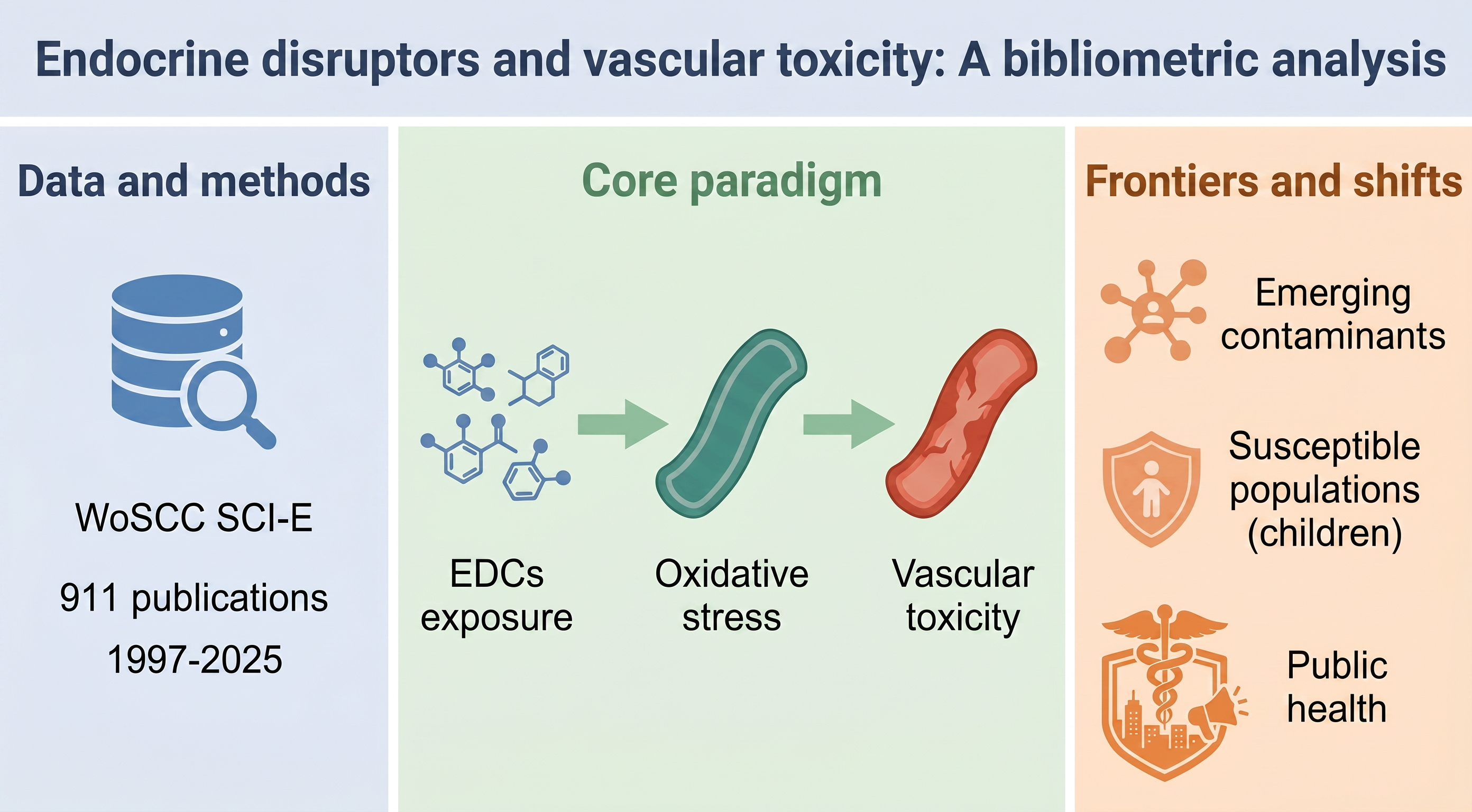
Methods: A total of 911 publications indexed in the Science Citation Index Expanded of the Web of Science Core Collection from 1997 to 2025 were analyzed using bibliometric and visualization methods to map collaboration patterns and knowledge evolution.
Results: The United States leads in both publication output and citation impact. Knowledge mapping indicates that an “exposure–mechanism–endpoint” framework is widely adopted in the literature, with “oxidative stress” appearing as a highly connected keyword linking environmental exposures and vascular outcomes. Trend analysis further suggests a shift in research focus from traditional persistent organic pollutants to emerging contaminants associated with modern lifestyles, alongside an increasing focus on early-life exposures and susceptible populations.
Conclusion: Research on EDC-related vascular toxicity is evolving toward greater emphasis on emerging contaminants, critical exposure windows, and mechanistic exploration. These trends may help inform future research directions and risk assessment strategies.
- Li Y, Cao GY, Jing WZ, Liu J, Liu M. Global trends and regional differences in incidence and mortality of cardiovascular disease, 1990-2019: findings from 2019 Global Burden of Disease Study. Eur J Prev Cardiol. 2023;30(3):276-286. doi: 10.1093/eurjpc/zwac285
- Wong ND, Sattar N. Cardiovascular risk in diabetes mellitus: epidemiology, assessment and prevention. Nat Rev Cardiol. 2023;20(10):685-695. doi: 10.1038/s41569-023-00877-z
- Kumar V, S H, Huligowda LKD, et al. Environmental pollutants as emerging concerns for cardiac diseases: a review on their impacts on cardiac health. Biomedicines. 2025;13(1):241. doi: 10.3390/biomedicines13010241
- Scimeca M, Palumbo V, Giacobbi E, et al. Impact of the environmental pollution on cardiovascular diseases: From epidemiological to molecular evidence. Heliyon. 2024;10(18):e38047. doi: 10.1016/j.heliyon.2024.e38047
- Annamalai J, Namasivayam V. Endocrine disrupting chemicals in the atmosphere: Their effects on humans and wildlife. Environ Int. 2015;76:78-97. doi: 10.1016/j.envint.2014.12.006
- Ahn C, Jeung EB. Endocrine-disrupting chemicals and disease endpoints. Int J Mol Sci. 2023;24(6):5342. doi: 10.3390/ijms24065342
- Lorigo M, Cairrao E. Regulation mechanisms of endocrine disruptors on vasodilation and vasoconstriction: Insights from ex vivo models. Biocell. 2022;46(6):1383-1389. doi: 10.32604/biocell.2022.018895
- Lemini C, Silveyra P, Segovia-Mendoza M. Cardiovascular disrupting effects of bisphenols, phthalates, and parabens related to endothelial dysfunction: review of toxicological and pharmacological mechanisms. Environ Toxicol Pharmacol. 2024;107:104407. doi: 10.1016/j.etap.2024.104407
- Morales-Grahl E, Hilz EN, Gore AC. Regrettable substitutes and the brain: what animal models and human studies tell us about the neurodevelopmental effects of bisphenol, per- and polyfluoroalkyl substances, and phthalate replacements. Int J Mol Sci. 2024;25(13):6887. doi: 10.3390/ijms25136887
- Macedo S, Teixeira E, Gaspar TB, et al. Endocrine-disrupting chemicals and endocrine neoplasia: A forty-year systematic review. Environ Res. 2023;218:114869. doi: 10.1016/j.envres.2022.114869
- Yang TC, Jovanovic N, Chong F, et al. Interventions to reduce exposure to synthetic phenols and phthalates from dietary intake and personal care products: a scoping review. Curr Environ Health Rep. 2023;10(2):184-214. doi: 10.1007/s40572-023-00394-8
- Roberts MS, Cheruvu HS, Mangion SE, et al. Topical drug delivery: history, percutaneous absorption, and product development. Adv Drug Deliv Rev. 2021;177:113929. doi: 10.1016/j.addr.2021.113929
- Ong HT, Samsudin H, Soto-Valdez H. Migration of endocrine-disrupting chemicals into food from plastic packaging materials: an overview of chemical risk assessment, techniques to monitor migration, and international regulations. Crit Rev Food Sci Nutr. 2022;62(4):957-979. doi: 10.1080/10408398.2020.1830747
- Muzeza C, Ngole-Jeme V, Msagati TAM. The mechanisms of plastic food-packaging monomers’ migration into food matrix and the implications on human health. Foods. 2023;12(18):3364. doi: 10.3390/foods12183364
- Rahaman MS, Rahman MM, Mise N, et al. Environmental arsenic exposure and its contribution to human diseases, toxicity mechanism and management. Environ Pollut. 2021;289:117940. doi: 10.1016/j.envpol.2021.117940
- Chen Y, Yang J, Yao B, Zhi D, Luo L, Zhou Y. Endocrine disrupting chemicals in the environment: Environmental sources, biological effects, remediation techniques, and perspective. Environ Pollut. 2022;310:119918. doi: 10.1016/j.envpol.2022.119918
- Thacharodi A, Hassan S, Hegde TA, Thacharodi DD, Brindhadevi K, Pugazhendhi A. Water a major source of endocrine-disrupting chemicals: An overview on the occurrence, implications on human health and bioremediation strategies. Environ Res. 2023;231(Pt 1):116097. doi: 10.1016/j.envres.2023.116097
- Münzel T, Steven S, Frenis K, Lelieveld J, Hahad O, Daiber A. Environmental factors such as noise and air pollution and vascular disease. Antioxid Redox Signal. 2020;33(9):581-601. doi: 10.1089/ars.2020.8090
- Amin A, Saadatakhtar M, Mohajerian A, et al. Mercury-mediated cardiovascular toxicity: mechanisms and remedies. Cardiovasc Toxicol. 2025;25(3):507-522. doi: 10.1007/s12012-025-09966-6
- Wei X, Hu Y, Zhu Q, Gao J, Liao C, Jiang G. Co-exposure and health risks of several typical endocrine disrupting chemicals in general population in eastern China. Environ Res. 2022;204(Pt D):112366. doi: 10.1016/j.envres.2021.112366
- Di D, Zhang R, Zhou H, et al. Exposure to phenols, chlorophenol pesticides, phthalate and PAHs and mortality risk: A prospective study based on 6 rounds of NHANES. Chemosphere. 2023;329:138650. doi: 10.1016/j.chemosphere.2023.138650
- Al-Dhfyan A, Alhoshani A, Korashy HM. Aryl hydrocarbon receptor/cytochrome P450 1A1 pathway mediates breast cancer stem cells expansion through PTEN inhibition and β-Catenin and Akt activation. Mol Cancer. 2017;16(1):14. doi: 10.1186/s12943-016-0570-y
- Vogel CFA, Van Winkle LS, Esser C, Haarmann-Stemmann T. The aryl hydrocarbon receptor as a target of environmental stressors – Implications for pollution mediated stress and inflammatory responses. Redox Biol. 2020;34:101530. doi: 10.1016/j.redox.2020.101530
- Omidi M, Ghafarian-Bahraman A, Mohammadi-Bardbori A. GSH/GSSG redox couple plays central role in aryl hydrocarbon receptor‐dependent modulation of cytochrome P450 1A1. J Biochem Mol Toxicol. 2018;32(7):e22164. doi: 10.1002/jbt.22164
- Stejskalova L, Pavek P. The function of cytochrome P450 1A1 enzyme (CYP1A1) and aryl hydrocarbon receptor (AhR) in the placenta. Curr Pharm Biotechnol. 2011;12(5):715-730. doi: 10.2174/138920111795470994
- Fenercioglu AK, Unal DO. The role of endocrine disrupting chemicals in the development of atherosclerosis. Cardiovasc Toxicol. 2025;25(11):1706-1717. doi: 10.1007/s12012-025-10054-y
- Ehrlich D, Krishna S, Kleinstreuer N. Data-driven derivation of an adverse outcome pathway linking vascular endothelial growth factor receptor (VEGFR), endocrine disruption, and atherosclerosis. ALTEX. 2024;41(4):617-632. doi: 10.14573/altex.2403211
- Incalza MA, D’Oria R, Natalicchio A, Perrini S, Laviola L, Giorgino F. Oxidative stress and reactive oxygen species in endothelial dysfunction associated with cardiovascular and metabolic diseases. Vascul Pharmacol. 2018;100:1-19. doi: 10.1016/j.vph.2017.05.005
- Chen H, Wang C, Li H, et al. A review of toxicity induced by persistent organic pollutants (POPs) and endocrine-disrupting chemicals (EDCs) in the nematode Caenorhabditis elegans. J Environ Manag. 2019;237:519-525. doi: 10.1016/j.jenvman.2019.02.102
- Badran A, Nasser SA, Mesmar J, et al. Reactive oxygen species: modulators of phenotypic switch of vascular smooth muscle cells. Int J Mol Sci. 2020;21(22):8764. doi: 10.3390/ijms21228764
- Joffre J, Hellman J. Oxidative stress and endothelial dysfunction in sepsis and acute inflammation. Antioxid Redox Signal. 2021;35(15):1291-1307. doi: 10.1089/ars.2021.0027
- Zhenyukh O, González-Amor M, Rodrigues-Diez RR, et al. Branched‐chain amino acids promote endothelial dysfunction through increased reactive oxygen species generation and inflammation. J Cell Mol Med. 2018;22(10):4948-4962. doi: 10.1111/jcmm.13759
- Grishanova AY, Perepechaeva ML. Aryl hydrocarbon receptor in oxidative stress as a double agent and its biological and therapeutic significance. Int J Mol Sci. 2022;23(12):6719. doi: 10.3390/ijms23126719
- Buoso E, Masi M, Racchi M, Corsini E. Endocrine-disrupting chemicals’ (EDCs) effects on tumour microenvironment and cancer progression: emerging contribution of RACK1. Int J Mol Sci. 2020;21(23):9229. doi: 10.3390/ijms21239229
- Pellegrini M, Bulzomi P, Lecis M, et al. Endocrine disruptors differently influence estrogen receptor β and androgen receptor in male and female rat VSMC. J Cell Physiol. 2014;229(8):1061-1068. doi: 10.1002/jcp.24530
- Kasal DA, Sena V, Huguenin GVB, De Lorenzo A, Tibirica E. Microvascular endothelial dysfunction in vascular senescence and disease. Front Cardiovasc Med. 2025;12:1505516. doi: 10.3389/fcvm.2025.1505516
- Grootaert MOJ, Bennett MR. Vascular smooth muscle cells in atherosclerosis: Time for a re-assessment. Cardiovasc Res. 2021;117(11):2326-2339. doi: 10.1093/cvr/cvab046
- Yu Y, Cai Y, Yang F, et al. Vascular smooth muscle cell phenotypic switching in atherosclerosis. Heliyon. 2024;10(18):e37727. doi: 10.1016/j.heliyon.2024.e37727
- Jarad S, Gill G, Amadi P, Gu HM, Zhang DW. VSMCs in atherosclerosis: Implications on the role of inflammation and extracellular matrix remodelling. Pharmacol Res. 2025;218:107833. doi: 10.1016/j.phrs.2025.107833
- Wu YL, Lin ZJ, Li CC, et al. Epigenetic regulation in metabolic diseases: mechanisms and advances in clinical study. Signal Transduct Target Ther. 2023;8(1):98. doi: 10.1038/s41392-023-01333-7
- Bianco-Miotto T, Craig JM, Gasser YP, Van Dijk SJ, Ozanne SE. Epigenetics and DOHaD: from basics to birth and beyond. J Dev Orig Health Dis. 2017;8(5):513-519. doi: 10.1017/s2040174417000733
- Vaiserman A. Epidemiologic evidence for association between adverse environmental exposures in early life and epigenetic variation: a potential link to disease susceptibility? Clin Epigenetics. 2015;7(1):96. doi: 10.1186/s13148-015-0130-0
- Chung FFL, Herceg Z. The promises and challenges of toxico-epigenomics: environmental chemicals and their impacts on the epigenome. Environ Health Perspect. 2020;128(1):15001. doi: 10.1289/ehp6104
- Baccarelli A, Bollati V. Epigenetics and environmental chemicals. Curr Opin Pediatr. 2009;21(2):243-251. doi: 10.1097/mop.0b013e32832925cc
- Kurita H, Ohuchi K, Inden M. Effects of environmental non-essential toxic heavy metals on epigenetics during development. Toxics. 2025;13(3):167. doi: 10.3390/toxics13030167
- Singh DD. Epigenetic mechanisms of endocrine-disrupting chemicals in breast cancer and their impact on dietary intake. J Xenobiot. 2024;15(1):1. doi: 10.3390/jox15010001
- Montjean D, Neyroud AS, Yefimova MG, Benkhalifa M, Cabry R, Ravel C. Impact of endocrine disruptors upon non-genetic inheritance. Int J Mol Sci. 2022;23(6):3350. doi: 10.3390/ijms23063350
- Akanbi CA, Rotimi DE, Ojo AB, Ojo OA. Endocrine-disrupting chemicals (EDCs) and epigenetic regulation in embryonic development: mechanisms, impacts, and emerging trends. Toxicol Rep. 2024;14:101885. doi: 10.1016/j.toxrep.2024.101885
- Fasolo F, Di Gregoli K, Maegdefessel L, Johnson JL. Non-coding RNAs in cardiovascular cell biology and atherosclerosis. Cardiovasc Res. 2019;115(12):1732-1756. doi: 10.1093/cvr/cvz203
- Meng Q, Pu L, Luo X, Wang B, Li F, Liu B. Regulatory roles of related long non-coding RNAs in the process of atherosclerosis. Front Physiol. 2020;11:564604. doi: 10.3389/fphys.2020.564604
- Tang Y, Li H, Chen C. Non-coding RNA-associated therapeutic strategies in atherosclerosis. Front Cardiovasc Med. 2022;9:889743. doi: 10.3389/fcvm.2022.889743
- Donthu N, Kumar S, Mukherjee D, Pandey N, Lim WM. How to conduct a bibliometric analysis: An overview and guidelines. J Bus Res. 2021;133:285-296. doi: 10.1016/j.jbusres.2021.04.070
- Mayr P, Scharnhorst A. Scientometrics and information retrieval: weak-links revitalized. Scientometrics. 2015;102:2193-2199. doi: 10.1007/s11192-014-1484-3
- Bukar UA, Sayeed MS, Razak SFA, Yogarayan S, Amodu OA, Mahmood RAR. A method for analyzing text using VOSviewer. MethodsX. 2023;11:102339. doi: 10.1016/j.mex.2023.102339
- Choudhary RK, Awasthi S. Bibliometric visualisation tools. Libr Prog. 2018;38(2):319. doi: 10.5958/2320-317x.2018.00034.x
- Coile RC. Lotka’s frequency distribution of scientific productivity. J Am Soc Inf Sci. 1977;28(6):366-370. doi: 10.1002/asi.4630280610
- Naranan S. Bradford’s law of bibliography of science: an interpretation. Nature. 1970;227(5258):631-632. doi: 10.1038/227631a0
- Brookes BC. Theory of the Bradford law. J Doc. 1977;33(3):180-209. doi: 10.1108/eb026641
- Mathews MT, Berk BC. PARP-1 inhibition prevents oxidative and nitrosative stress-induced endothelial cell death via transactivation of the VEGF receptor 2. Arter Thromb Vasc Biol. 2008;28(4):711-717. doi: 10.1161/atvbaha.107.156406
- Henning RJ, Bourgeois M, Harbison RD. Poly(ADP-ribose) polymerase (PARP) and PARP inhibitors: mechanisms of action and role in cardiovascular disorders. Cardiovasc Toxicol. 2018;18(6):493-506. doi: 10.1007/s12012-018-9462-2
- Yan R, Zhang X, Xu W, et al. ROS-induced endothelial dysfunction in the pathogenesis of atherosclerosis. Aging Dis. 2024;16(1):250-268. doi: 10.14336/ad.2024.0309
- Bu LL, Yuan HH, Xie LL, Guo MH, Liao DF, Zheng XL. New dawn for atherosclerosis: vascular endothelial cell senescence and death. Int J Mol Sci. 2023;24(20):15160. doi: 10.3390/ijms242015160
- Dominic A, Banerjee P, Hamilton DJ, Le NT, Abe JI. Time-dependent replicative senescence vs. disturbed flow-induced pre-mature aging in atherosclerosis. Redox Biol. 2020;37:101614. doi: 10.1016/j.redox.2020.101614
- Mikhed Y, Daiber A, Steven S. Mitochondrial oxidative stress, mitochondrial DNA damage and their role in age-related vascular dysfunction. Int J Mol Sci. 2015;16(7):15918-15953. doi: 10.3390/ijms160715918
- Wang R, Li C, Qiao P, et al. OGG1-initiated base excision repair exacerbates oxidative stress-induced parthanatos. Cell Death Dis. 2018;9(6):628. doi: 10.1038/s41419-018-0680-0
- Yu E, Calvert PA, Mercer JR, et al. Mitochondrial DNA damage can promote atherosclerosis independently of reactive oxygen species through effects on smooth muscle cells and monocytes and correlates with higher-risk plaques in humans. Circulation. 2013;128(7):702-712. doi: 10.1161/circulationaha.113.002271
- Nayak D, Adiga D, Khan NG, et al. Impact of bisphenol A on structure and function of mitochondria: a critical review. Rev Environ Contam Toxicol. 2022;260(1):10. doi: 10.1007/s44169-022-00011-z
68. Amir S, Shah STA, Mamoulakis C, et al. Endocrine disruptors acting on estrogen and androgen pathways cause reproductive disorders through multiple mechanisms: a review. Int J Environ Res Public Health. 2021;18(4):1464. doi: 10.3390/ijerph18041464