

Molecular and clinicopathological predictors of cervical lymph node metastasis in oral squamous cell carcinoma: A narrative review

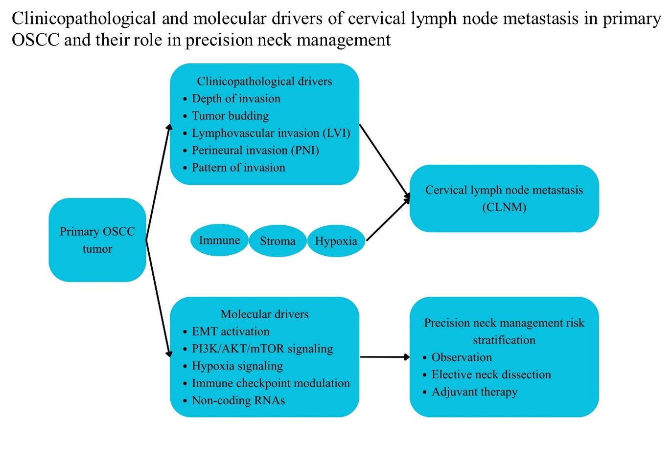
Cervical lymph node metastasis (CLNM) remains the principal determinant of survival in oral squamous cell carcinoma (OSCC), conferring significant reductions in disease-specific and overall survival even in early-stage disease. Despite advances in imaging and surgical staging, accurate identification of occult nodal involvement in clinically node-negative patients continues to represent a major clinical challenge. This narrative review synthesizes contemporary evidence on clinicopathological and molecular predictors of CLNM in OSCC, with emphasis on biological determinants that may refine risk-adapted neck management. A structured literature search of PubMed, Scopus, and Web of Science was conducted, prioritizing high-quality cohort studies, meta-analyses, and translational investigations evaluating histopathological parameters, molecular alterations, tumor microenvironment features, and integrative predictive models. Among clinicopathological factors, depth of invasion, lymphovascular invasion, perineural invasion, tumor budding, and infiltrative invasion patterns consistently demonstrate strong associations with nodal metastasis. Molecular drivers include epithelial–mesenchymal transition, activation of the phosphoinositide 3-kinase/protein kinase B/mechanistic target of rapamycin signaling pathway, hypoxia-mediated angiogenesis and lymphangiogenesis, immune checkpoint upregulation, and dysregulated non-coding RNAs. Emerging multivariable models that integrate pathological and molecular determinants demonstrate improved predictive performance compared to conventional tumor–node–metastasis staging. Collectively, CLNM reflects a coordinated biological process involving invasive tumor architecture and molecular reprogramming. Incorporation of validated biological predictors into routine pathological assessment may support precision-based neck management and enhance oncologic stratification in OSCC.
- Bray F, Ferlay J, Soerjomataram I, Siegel RL, Torre LA, Jemal A. Global cancer statistics 2018: GLOBOCAN estimates of incidence and mortality worldwide for 36 cancers in 185 countries. CA Cancer J Clin. 2018;68(6):394-424. doi: 10.3322/caac.21492
- Woolgar JA. Histopathological prognosticators in oral and oropharyngeal squamous cell carcinoma. Oral Oncol. 2006;42(3):229-239. doi: 10.1016/j.oraloncology.2005.05.008
- Huang SH, Hwang D, Lockwood G, Goldstein DP, O’Sullivan B. Predictive value of tumor thickness for cervical lymph-node involvement in squamous cell carcinoma of the oral cavity: a meta-analysis of reported studies. Cancer. 2009;115(7):1489-1497. doi: 10.1002/cncr.24161
- Almangush A, Pirinen M, Heikkinen I, et al. Tumour budding in oral squamous cell carcinoma: a meta-analysis. Br J Cancer. 2018;118(4):577-586. doi: 10.1038/bjc.2017.425
- Amin MB, Edge SB, Greene FL, et al., eds. AJCC Cancer Staging Manual. 8th ed. Springer; 2017.
- Huang SH, O’Sullivan B. Overview of the 8th edition TNM classification for head and neck cancer. Curr Treat Options Oncol. 2017;18(7):40. doi: 10.1007/s11864-017-0484-y
- Pentenero M, Gandolfo S, Carrozzo M. Importance of tumor thickness and depth of invasion in nodal involvement and prognosis of oral squamous cell carcinoma: a review of the literature. Head Neck. 2005;27(12):1080-1091. doi: 10.1002/hed.20275
- Seki M, Sano T, Yokoo S, Oyama T. Histologic assessment of tumor budding in preoperative biopsies to predict nodal metastasis in squamous cell carcinoma of the tongue and floor of the mouth. Head Neck. 2016;38(S1):E1582-E1590. doi: 10.1002/hed.24282
- Brandwein-Gensler M, Teixeira MS, Lewis CM, et al. Oral squamous cell carcinoma: histologic risk assessment, but not margin status, is strongly predictive of local disease-free and overall survival. Am J Surg Pathol. 2005;29(2):167-178. doi: 10.1097/01.pas.0000149687.90710.21
- Huang S, Zhu Y, Cai H, Zhang Y, Hou J. Impact of lymphovascular invasion in oral squamous cell carcinoma: a meta-analysis. Oral Surg Oral Med Oral Pathol Oral Radiol. 2021;131(3):319-328.e1. doi: 10.1016/j.oooo.2020.10.026
- Batsakis JG. Nerves and Neurotropic Carcinomas. Ann Otol Rhinol Laryngol. 1985;94(4):426-427. doi: 10.1177/000348948509400420
- Thiery JP, Acloque H, Huang RYJ, Nieto MA. Epithelial– mesenchymal transitions in development and disease. Cell. 2009;139(5):871-890. doi: 10.1016/j.cell.2009.11.007
- Leemans CR, Snijders PJF, Brakenhoff RH. The molecular landscape of head and neck cancer. Nat Rev Cancer. 2018;18(5):269-282. doi: 10.1038/nrc.2018.11
- Freudlsperger C, Burnett JR, Friedman J, Kannabiran VR, Chen Z, Van Waes C. EGFR–PI3K–AKT–mTOR signaling in head and neck squamous cell carcinomas: attractive targets for molecular-oriented therapy. Expert Opin Ther Targets. 2011;15(1):63-74. doi: 10.1517/14728222.2011.541440
- Molinolo AA, Amornphimoltham P, Squarize CH, et al. Dysregulated molecular networks in head and neck carcinogenesis. Oral Oncol. 2009;45(4-5):324-334. doi: 10.1016/j.oraloncology.2008.07.011
- Semenza GL. Hypoxia-inducible factor 1 and cancer pathogenesis. IUBMB Life. 2008;60(9):591-597. doi: 10.1002/iub.93
- Kyzas PA, Stefanou D, Batistatou A, Agnantis NJ. Prognostic significance of VEGF immunohistochemical expression and tumor angiogenesis in head and neck squamous cell carcinoma. J Cancer Res Clin Oncol. 2005;131(9):624-630. doi: 10.1007/s00432-005-0003-6
- Cho YA, Yoon HJ, Lee JI, Hong SP, Hong SD. Relationship between the expression of PD-L1 and tumor-infiltrating lymphocytes in oral squamous cell carcinoma. Oral Oncol. 2011;47(12):1148-1153. doi: 10.1016/j.oraloncology.2011.08.007
- Almangush A, Mäkitie AA, Triantafyllou A, et al. Staging and grading of oral squamous cell carcinoma: An update. Oral Oncol. 2020;107:104799. doi: 10.1016/j.oraloncology.2020.104799
- Almangush A, Bello IO, Keski-Säntti H, et al. Depth of invasion, tumor budding, and worst pattern of invasion: prognostic indicators in early-stage oral tongue cancer. Head Neck. 2014;36(6):811-818. doi: 10.1002/hed.23380
- Wong TS, Liu XB, Wong BYH, Ng RWM, Yuen APW, Wei WI. Mature miR-184 as potential oncogenic microRNA of squamous cell carcinoma of tongue. Clin Cancer Res. 2008;14(9):2588-2592. doi: 10.1158/1078-0432.CCR-07-0666
- Teng MWL, Ngiow SF, Ribas A, Smyth MJ. Classifying cancers based on T-cell infiltration and PD-L1. Cancer Res. 2015;75(11):2139-2145. doi: 10.1158/0008-5472.CAN-15-0255
- Kakaguchi W, Ashikaga Y, Yanagawa-Matsuda A, et al. Significant association of Yamamoto-Kohama classification and pathological depth of invasion with cervical lymph node metastasis in early-stage tongue squamous cell carcinoma (Stage I/II). J Dent Sci. 2023;18(4):1663-1668. doi: 10.1016/j.jds.2023.02.012
- Kumar D, Gupta A, Agrahari S, et al. Association of epithelial to mesenchymal transition markers on prognosis and clinicopathological characteristics in oral squamous cell carcinoma: a systematic review and meta-analysis. Head Neck Pathol. 2025;19(1):124. doi: 10.1007/s12105-025-01863-2
- Lu MY, Liao YW, Chen PY, et al. Targeting lncRNA HOTAIR suppresses cancer stemness and metastasis in oral carcinoma stem cells through modulation of EMT. Oncotarget. 2017;8(58):98542-98552. doi: 10.18632/oncotarget.21614
- Su YC, Lee WC, Wang CC, Yeh SA, Chen WH, Chen PJ. Targeting PI3K/AKT/mTOR signaling pathway as a radiosensitization in head and neck squamous cell carcinomas. Int J Mol Sci. 2022;23:15749. doi: 10.3390/ijms232415749
- Haider SP, Mahajan A, Zeevi T, et al. PET/CT radiomics signature of human papilloma virus association in oropharyngeal squamous cell carcinoma. Eur J Nucl Med Mol Imaging. 2020;47(13):2978-2991. doi: 10.1007/s00259-020-04839-2