

Time saving with artificial intelligence-assisted lumbar spine magnetic resonance imaging reporting: A preliminary study

Introduction: Lumbar spine magnetic resonance imaging (MRI) is a high-volume diagnostic examination, yet increasing caseloads and reporting complexity continue to strain radiology workflows. Emerging artificial intelligence (AI)-assisted reading tools may help streamline interpretation and reduce report turnaround times, but their real-world impact on efficiency remains insufficiently quantified.
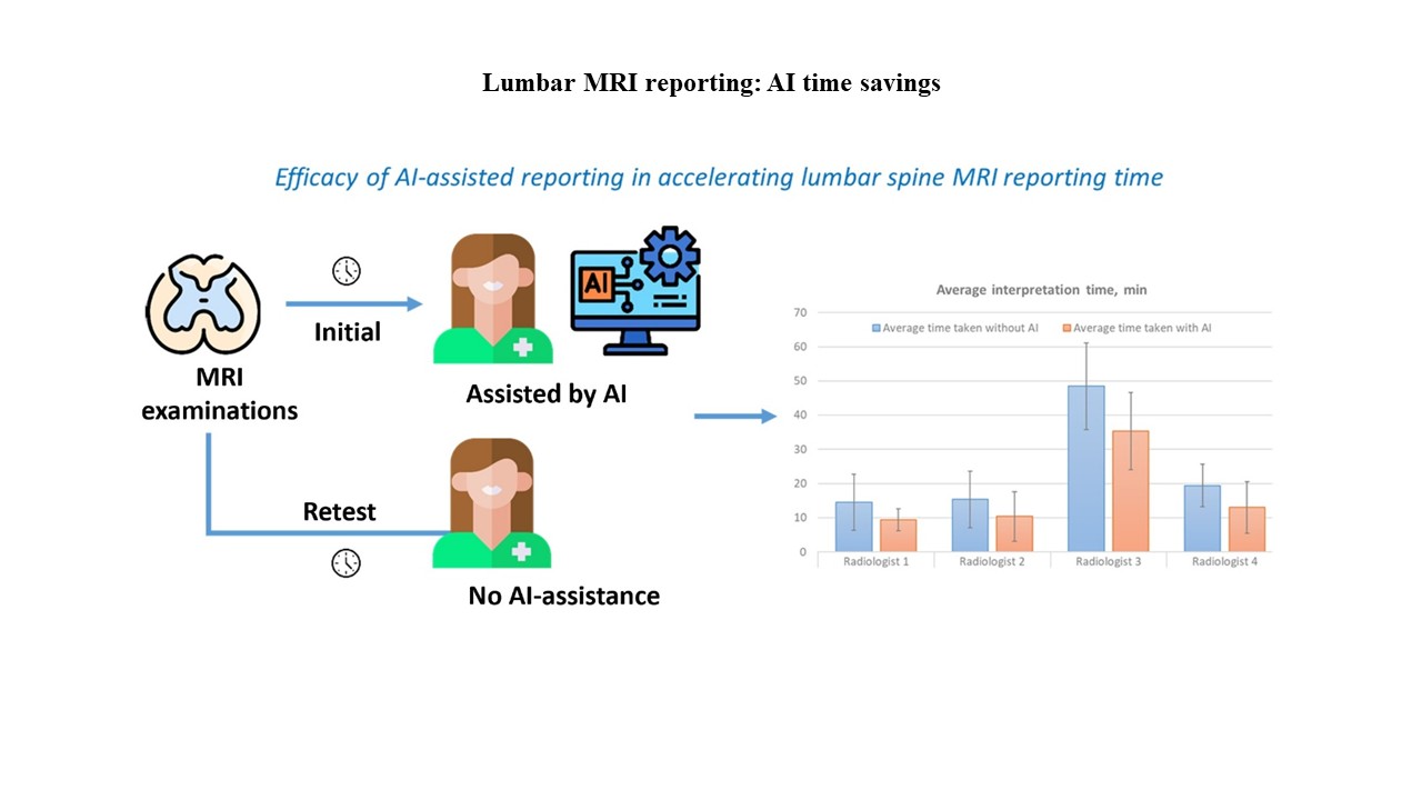
Objective: To evaluate the impact of an AI-based reading tool on lumbar spine MRI interpretation and reporting time.
Methods: We randomly selected 236 lumbar spine MRI examinations performed between 2018 and 2023 in patients aged 18 and older. Cases with prior lumbar surgery or scoliosis were excluded. Digital imaging and communications in medicine (DICOM) data were processed using a commercial deep-learning software package, and outputs were reviewed in a standard DICOM viewer. Five radiologists participated. Studies 1 and 2 assessed the effect of AI on interpretation time using a within-reader design: radiologists interpreted each examination with AI support and then reinterpreted the same examinations 2 months later without AI, enabling direct comparison of interpretation times. Study 3 evaluated the effect of AI by comparing AI-assisted and unassisted interpretations in 146 randomly selected examinations.
Results: AI assistance significantly accelerated report generation. Across the full dataset, AI-supported interpretation reduced time by approximately 52% compared with unassisted reading. AI-assisted generation of preliminary reports reduced radiologists’ overall time by nearly 30%. Linear mixed-effects modeling indicated that these reductions were statistically significant. The smaller reduction observed in Study 3 (9.21%) may reflect limited familiarity with the software’s reporting style and occasional instances in which the AI outputs did not fully support the radiologists’ findings.
Conclusion: AI assistance improves the efficiency of lumbar spine MRI reporting and shortens reporting time.
- Jeong J, Kim S, Pan L, et al. Reducing the workload of medical diagnosis through artificial intelligence: A narrative review. Medicine (Baltimore). 2025;104(6):e41470. doi: 10.1097/MD.0000000000041470
- Gill A, Rainey C, McLaughlin L, et al. Artificial intelligence user interface preferences in radiology: A scoping review. J Med Imaging Radiat Sci. 2025;56(3):101866. doi: 10.1016/j.jmir.2025.101866
- Sim JZT, Bhanu Prakash KN, Huang WM, Tan CH. Harnessing artificial intelligence in radiology to augment population health. Front Med Technol. 2023;5:1281500. doi: 10.3389/fmedt.2023.1281500
- Obuchowicz R, Lasek J, Wodzinski M, Piorkowski A, Strzelecki M, Nurzynska K. Artificial intelligence-empowered radiology-current status and critical review. Diagnostics (Basel). 2025;15(3):282. doi: 10.3390/diagnostics15030282
- Melancia JL, Francisco AF, Antunes JL. Spinal stenosis. Handb Clin Neurol. 2014;119:541-549. doi: 10.1016/b978-0-7020-4086-3.00035-7
- Stroman PW, Wheeler-Kingshott C, Bacon M, et al. The current state-of-the-art of spinal cord imaging: Methods. Neuroimage. 2014;84:1070-1081. doi: 10.1016/j.neuroimage.2013.04.124
- Wahid G, Ammara H, Mehreen S, Naila T. Causes of delay in radiological reporting and ways to reduce them. J Saidu Med Coll Swat. 2022;12(3):133-137. doi: 10.52206/jsmc.2022.12.3.697
- Taylor-Phillips S, Stinton C. Fatigue in radiology: A fertile area for future research. Br J Radiol. 2019;92(1099):20190043. doi: 10.1259/bjr.20190043
- Roller BL, Boutin RD, O’Gara TJ, et al. Accurate prediction of lumbar microdecompression level with an automated MRI grading system. Skeletal Radiol. 2021;50(1):69-78. doi: 10.1007/s00256-020-03505-w
- Georgiev R, Novakova M, Bliznakova K. Clinical assessment of columbo deep learning system for central canal stenosis diagnostics. Euras J Med Oncol. 2023;7(1):42-48. doi: 10.14744/ejmo.2023.59207
- Rathmann E, Hemkemeier P, Raths S, et al. Changes in MRI workflow of multiple sclerosis after introduction of an AI-software: A qualitative study. Healthcare (Basel). 2024;12(10):978 doi: 10.3390/healthcare12100978
- Yang YXC, Yee SY, Tan TSE, et al. An artificial intelligence boost to MRI lumbar spine reporting. Eur J Radiol. 2024;179:111636. doi: 10.1016/j.ejrad.2024.111636
- Georgiev N, Asenov A. Automatic Segmentation of Lumbar Spine MRI Using Ensemble of 2D Algorithms. Berlin: Springer International Publishing; 2019. p. 154-162.
- Lu JT, Pedemonte S, Bizzo B, et al. DeepSPINE: Automated Lumbar Vertebral Segmentation, Disc-level Designation, and Spinal Stenosis Grading Using Deep Learning. United States: Cornell University; 2018. doi: 10.48550/arXiv.1807.10215 abs/1807.10215
- Granata V, Fusco R, Coluccino S, et al. Preliminary data on artificial intelligence tool in magnetic resonance imaging assessment of degenerative pathologies of lumbar spine. Radiol Med. 2024;129(4):623-630. doi: 10.1007/s11547-024-01791-1
- Lehnen NC, Haase R, Faber J, et al. Detection of degenerative changes on MR images of the lumbar spine with a convolutional neural network: A feasibility study. Diagnostics (Basel). 2021;11(5):902 doi: 10.3390/diagnostics11050902
- Herzog R, Elgort DR, Flanders AE, Moley PJ. Variability in diagnostic error rates of 10 MRI centers performing lumbar spine MRI examinations on the same patient within a 3-week period. Spine J. 2017;17(4):554-561. doi: 10.1016/j.spinee.2016.11.009
- Al Meslamani AZ. Beyond implementation: The long-term economic impact of AI in healthcare. J Med Econ. 2023;26(1):1566-1569. doi: 10.1080/13696998.2023.2285186
- Alowais SA, Alghamdi SS, Alsuhebany N, et al. Revolutionizing healthcare: The role of artificial intelligence in clinical practice. Bmc Med Educ. 2023;23(1):689. doi: 10.1186/s12909-023-04698-z
- Bajwa J, Munir U, Nori A, Williams B. Artificial intelligence in healthcare: Transforming the practice of medicine. Future Healthc J. 2021;8(2):e188-e194. doi: 10.7861/fhj.2021-0095
- Cartolovni A, Malesevic A, Poslon L. Critical analysis of the AI impact on the patient-physician relationship: A multi-stakeholder qualitative study. Digit Health. 2023;9:20552076231220833. doi: 10.1177/20552076231220833
- Kutsarova D. Innovations in Imaging Diagnostics. Bulgaria: Master of Science. Medical University of Varna; 2025.