

Application of CRISPR–Cas9 technology in the treatment of chronic lymphocytic leukemia with TP53 mutations

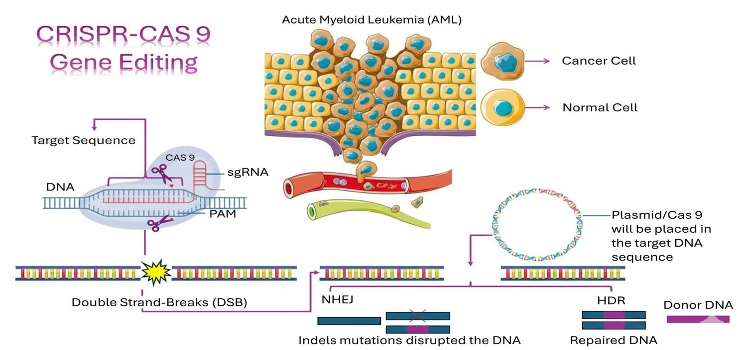
This study proposes the implementation of clustered regularly interspaced short palindromic repeats (CRISPR)–CRISPR-associated protein 9 (Cas9) technology for gene therapy targeting genetic mutations in human lymphocytes affected by chronic lymphocytic leukemia (CLL), offering new opportunities for effective treatment of this heterogeneous disease. It focuses on the application of CRISPR–Cas9-mediated targeted sequencing to systematically characterize the biological effects of monoallelic and biallelic TP53 gene lesions, aiming to replace mutant TP53 genes in CLL cells through this technology. CRISPR–Cas9 technology employs a specific enzyme guided by a designed guide RNA (gRNA) to a DNA target. The enzyme first introduces a cut at the target site, and following this cleavage event, it can further disrupt the TP53 gene. The gRNA plays a crucial role by directing the Cas9 protein to the DNA sequence of interest. The gRNA consists of CRISPR RNA (crRNA) and trans-activating CRISPR RNA (tracrRNA) sequences, responsible for target recognition and Cas9 binding, respectively. Examination of the predicted secondary structure of the tracrRNA–crRNA duplex suggests that the features required for Cas9-catalyzed DNA cleavage at specific sites can be captured within a single chimeric RNA. Although the natural tracrRNA–crRNA mechanism operates efficiently, the use of a single RNA-guided Cas9 system is particularly attractive due to its potential for programmed DNA cleavage and genome editing. Importantly, Cas9 can bind and cleave a target sequence only if it is adjacent to a protospacer adjacent motif. Once the gRNA–Cas9 complex binds to the target DNA, Cas9 induces a double-strand break at the specified site. In conclusion, CRISPR–Cas9 technology represents a powerful genetic engineering tool capable of inserting, deleting, or replacing DNA within an organism’s genome using these “molecular scissors.”
- Ishino Y, Shinagawa H, Makino K, Amemura M, Nakata A. Nucleotide sequence of the iap gene, responsible for alkaline phosphatase isozyme conversion in Escherichia coli, and identification of the gene product. J Bacteriol. 1987;169(12):5429-5433. doi: 10.1128/jb.169.12.5429-5433.1987
- Lander ES. The heroes of CRISPR. Cell. 2016;164(1-2):18-28. doi: 10.1016/j.cell.2015.12.041
- Hille F, Charpentier E. CRISPR-Cas: Biology, mechanisms and relevance. Philos Trans R Soc Lond B Biol Sci. 2016;371(1707):20150496. doi: 10.1098/rstb.2015.0496
- Chang HH, Pannunzio NR, Adachi N, Lieber MR. Non-homologous DNA end joining and alternative pathways to double-strand break repair. Nat Rev Mol Cell Biol. 2017;18(8):495-506. doi: 10.1038/nrm.2017.48.
- Sallmyr A, Tomkinson AE. Repair of DNA double-strand breaks by mammalian alternative end-joining pathways. J Biol Chem. 2018;293(27):10536-10546. doi: 10.1074/jbc.TM117.000375
- Bhargava R, Onyango DO, Stark JM. Regulation of single-strand annealing and its role in genome maintenance. Trends Genet. 2016;2(9):566-575. doi: 10.1016/j.tig.2016.06.007
- Chen JM, Férec C, Cooper DN. Gene conversion in human genetic disease. Genes (Basel). 2010;1(3):550-563. doi: 10.3390/genes1030550
- Verma P, Greenberg RA. Noncanonical views of homology-directed DNA repair. Genes Dev. 2016;30(10):1138-1154. doi: 10.1101/gad.280545.116
- Savic N, Ringnalda FC, Lindsay H, et al. Covalent linkage of the DNA repair template to the CRISPR-Cas9 nuclease enhances homology-directed repair. Elife. 2018;7:e33761. doi: 10.7554/eLife.33761
- Zaboikin M, Zaboikina T, Freter C, Srinivasakumar N. Non-homologous end joining and homology directed DNA repair frequency of double-stranded breaks introduced by genome editing reagents. PLoS One. 2017;12(1):e0169931. doi: 10.1371/journal.pone.0169931
- Qi LS, Larson MH, Gilbert LA, et al. Repurposing CRISPR as an RNA-guided platform for sequence-specific control of gene expression. Cell. 2013;152(5):1173-1183. doi: 10.1016/j.cell.2013.02.022. Erratum in: Cell. 2021;184(3):844. doi: 10.1016/j.cell.2021.01.019
- Konermann S, Brigham MD, Trevino AE, et al. Genome-scale transcriptional activation by an engineered CRISPR-Cas9 complex. Nature. 2015;517(7536):583-588. doi: 10.1038/nature14136
- Polstein LR, Gersbach CA. A light-inducible CRISPR-Cas9 system for control of endogenous gene activation. Nat Chem Biol. 2015;11(3):198-200. doi: 10.1038/nchembio.1753
- Gilbert LA, Larson MH, Morsut L, et al. CRISPR-mediated modular RNA-guided regulation of transcription in eukaryotes. Cell. 2013;154(2):442-451. doi: 10.1016/j.cell.2013.06.044
- Zhang T, Zhu Z, Xun G, Zhao H. ScienceDirect. Curr Opin Biomed Eng. 2023;28:10048.
- Tadić V, Josipović G, Zoldoš V, Vojta A. CRISPR/Cas9-based epigenome editing: An overview of dCas9-based tools with special emphasis on off-target activity. Methods. 2019;164- 165:109-119. doi: 10.1016/j.ymeth.2019.05.003
- Hong Y, Lu G, Duan J Liu W, Yu Z. Comparison and optimization of CRISPR/dCas9/gRNA genome-labeling systems for live cell imaging. Genome Biol. 2018;19:39. doi: 10.1186/s13059-018-1413-5
- Schubert MS, Cedrone E, Neun B, Behlke MA, Dobrovolskaia MA. Chemical modification of CRISPR gRNAs eliminate type I interferon responses in human peripheral blood mononuclear cells. J Cytokine Biol. 2018;3(1):121. doi: 10.4172/2576-3881
- Anzalone AV, Randolph PB, Davis JR, et al. Search-and-replace genome editing without double-strand breaks or donor DNA. Nature. 2019;576(7785):149-157. doi: 10.1038/s41586-019-1711-4
- Zetsche B, Gootenberg JS, Abudayyeh OO, et al. Cpf1 is a single RNA-guided endonuclease of a class 2 CRISPR-Cas system. Cell. 2015;163(3):759-771. doi: 10.1016/j.cell.2015.09.038
- Gregg C, Ohtsuka M, Gurumurthy CB, Behlke MA. Simplified CRISPR tools for efficient genome editing and streamlined protocols for their delivery into mammalian cells and mouse zygotes. Methods. 2017;121-122:16-28. doi: 10.1016/j.ymeth.2017.03.021
- Miura H, Gurumurthy CB, Sato T, Sato M, Ohtsuka M. CRISPR/Cas9-based generation of knockdown mice by intronic insertion of artificial microRNA using longer single-stranded DNA. Sci Rep. 2015;5:12799. doi: 10.1038/srep12799
- Yoshimi K, Kunihiro Y, Kaneko T, Nagahora H, Voigt B, Mashimo T. ssODN-mediated knock-in with CRISPR-Cas for large genomic regions in zygotes. Nat Commun. 2016;7:10431. doi: 10.1038/ncomms10431
- Crosetto N, Mitra A, Silva MJ, et al. Nucleotide-resolution DNA double-strand break mapping by next-generation sequencing. Nat Methods. 2013;10(4):361-365. doi: 10.1038/nmeth.2408
- Kim D, Bae S, Park J, et al. Digenome-seq: Genome-wide profiling of CRISPR-Cas9 off-target effects in human cells. Nat Methods. 2015;12(3):237-43, 1 p following 243. doi: 10.1038/nmeth.3284
- Tsai SQ, Nguyen NT, Malagon-Lopez J, Topkar VV, Aryee MJ, Joung JK. CIRCLE-seq: A highly sensitive in vitro screen for genome-wide CRISPR-Cas9 nuclease off-targets. Nat Methods. 2017;14(6):607-614. doi: 10.1038/nmeth.4278
- Cameron P, Fuller CK, Donohoue PD, et al. Mapping the genomic landscape of CRISPR-Cas9 cleavage. Nat Methods. 2017;14(6):600-606. doi: 10.1038/nmeth.4284
- Tsai SQ, Zheng Z, Nguyen NT, et al. GUIDE-seq enables genome-wide profiling of off-target cleavage by CRISPR-Cas nucleases. Nat Biotechnol. 2015;33(2):187-197. doi: 10.1038/nbt.3117
- Wienert B, Wyman SK, Richardson CD, et al. Unbiased detection of CRISPR off-targets in vivo using DISCOVER-Seq. Science. 2019;A364(6437):286-289. doi: 10.1126/science.aav9023
- Barbu D, Popescu DM. Immunophenotypic diagnosis in images. In: Clinical Hematology Laboratory Notebooks. Bucharest: Medical Publishing House; 2018.
- Udristioiu A, Gheorghe IG, Nica-Badea D. Book “Isoform p53 Protein’s Major Role in the Pathophysiology of Malignant Hematologic Diseases. Ch. 2. Cambridge, UK: Cambridge Scholars Publishing; 2024.
- Lefter M, Vis JK, Vermaat M, den Dunnen JT, Taschner PE, Laros JFJ. Mutalyzer 2: Next generation HGVS nomenclature checker. Bioinformatics. 2021;37:2811-28117. doi: 10.1093/bioinformatics/btab051